- Phare
- Validé par KD/KO
Anticorps Polyclonal de lapin anti-YTHDF1
YTHDF1 Polyclonal Antibody for WB, IHC, IF/ICC, IF-P, IP, ELISA
Hôte / Isotype
Lapin / IgG
Réactivité testée
Humain, rat, souris et plus (4)
Applications
WB, IHC, IF/ICC, IF-P, IP, CoIP, RIP, ELISA
Conjugaison
Non conjugué
N° de cat : 17479-1-AP
Synonymes
Galerie de données de validation
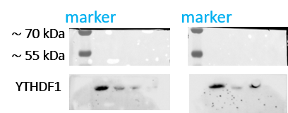
 with sh-Control and sh-YTHDF1 transfected HepG2 cells.")
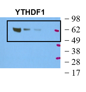
 at dilution of 1:4000 incubated at room temperature for 1.5 hours.")
 at dilution of 1:8000 incubated at room temperature for 1.5 hours.")
 at dilution of 1:15000 incubated at room temperature for 1.5 hours.")
 with mouse brain tissue lysate 2640ug.")
 at dilution of 1:500 (under 10x lens). Heat mediated antigen retrieval with Tris-EDTA buffer (pH 9.0).")
 at dilution of 1:500 (under 40x lens). Heat mediated antigen retrieval with Tris-EDTA buffer (pH 9.0).")
 at dilution of 1:200 (under 40x lens). Heat mediated antigen retrieval with Tris-EDTA buffer (pH 9.0).")
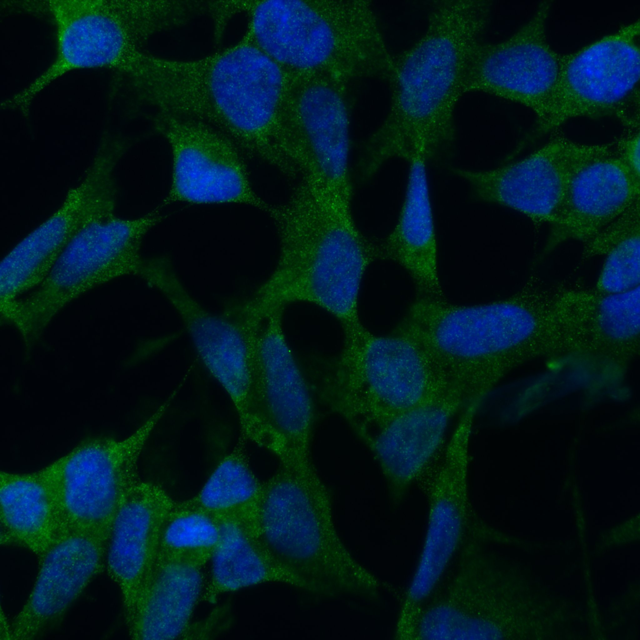
 fixed mouse brain tissue using YTHDF1 antibody (17479-1-AP) at dilution of 1:400 and CoraLite®488-Conjugated AffiniPure Goat Anti-Rabbit IgG(H+L).")
 fixed mouse brain tissue using 17479-1-AP (YTHDF1 antibody) at dilution of 1:50 and Alexa Fluor 488-Conjugated AffiniPure Goat Anti-Rabbit IgG(H+L).")
 fixed U2OS cells using YTHDF1 antibody (17479-1-AP) at dilution of 1:400 and Multi-rAb CoraLite ® Plus 488-Goat Anti-Rabbit Recombinant Secondary Antibody (H+L) (RGAR002).")
Applications testées
| Résultats positifs en WB | cellules HeLa, cellules A549, cellules HEK-293, cellules HepG2, cellules Jurkat, cellules MCF-7, tissu cérébral de rat, tissu cérébral de souris |
| Résultats positifs en IP | tissu cérébral de souris |
| Résultats positifs en IHC | tissu rénal humain, tissu d'hypothalamus humain il est suggéré de démasquer l'antigène avec un tampon de TE buffer pH 9.0; (*) À défaut, 'le démasquage de l'antigène peut être 'effectué avec un tampon citrate pH 6,0. |
| Résultats positifs en IF-P | tissu cérébral de souris, |
| Résultats positifs en IF/ICC | cellules U2OS, |
Dilution recommandée
| Application | Dilution |
|---|---|
| Western Blot (WB) | WB : 1:1000-1:8000 |
| Immunoprécipitation (IP) | IP : 0.5-4.0 ug for 1.0-3.0 mg of total protein lysate |
| Immunohistochimie (IHC) | IHC : 1:250-1:1000 |
| Immunofluorescence (IF)-P | IF-P : 1:200-1:800 |
| Immunofluorescence (IF)/ICC | IF/ICC : 1:200-1:800 |
| It is recommended that this reagent should be titrated in each testing system to obtain optimal results. | |
| Sample-dependent, check data in validation data gallery | |
Informations sur le produit
17479-1-AP cible YTHDF1 dans les applications de WB, IHC, IF/ICC, IF-P, IP, CoIP, RIP, ELISA et montre une réactivité avec des échantillons Humain, rat, souris
| Réactivité | Humain, rat, souris |
| Réactivité citée | rat, Humain, poisson-zèbre, porc, poulet, singe, souris |
| Hôte / Isotype | Lapin / IgG |
| Clonalité | Polyclonal |
| Type | Anticorps |
| Immunogène | YTHDF1 Protéine recombinante Ag11509 |
| Nom complet | YTH domain family, member 1 |
| Masse moléculaire calculée | 559 aa, 61 kDa |
| Poids moléculaire observé | 60 kDa |
| Numéro d’acquisition GenBank | BC050284 |
| Symbole du gène | YTHDF1 |
| Identification du gène (NCBI) | 54915 |
| Conjugaison | Non conjugué |
| Forme | Liquide |
| Méthode de purification | Purification par affinité contre l'antigène |
| Tampon de stockage | PBS with 0.02% sodium azide and 50% glycerol |
| Conditions de stockage | Stocker à -20°C. Stable pendant un an après l'expédition. L'aliquotage n'est pas nécessaire pour le stockage à -20oC Les 20ul contiennent 0,1% de BSA. |
Informations générales
YTHDF1, also named YTH domain-containing family protein 1 or C20orf21, is a 559 amino acid protein, which localizes in the cytoplasm. YTHDF1 specifically recognizes and binds N6-methyladenosine (m6A)-containing mRNAs, and promotes mRNA translation efficiency. M6A is a modification present at the internal sites of mRNAs and some non-coding RNAs and plays a role in the efficiency of mRNA splicing, processing, and stability. YTHDF1 acts as a regulator of mRNA translation efficiency: promotes ribosome loading to m6A-containing mRNAs and interacts with translation initiation factors eIF3 (EIF3A or EIF3B) to facilitate translation initiation. YTHDF1 exists two isoforms and the calculated molecular weight of isoforms are 61 kDa and 21 kDa.
Protocole
| Product Specific Protocols | |
|---|---|
| WB protocol for YTHDF1 antibody 17479-1-AP | Download protocol |
| IHC protocol for YTHDF1 antibody 17479-1-AP | Download protocol |
| IF protocol for YTHDF1 antibody 17479-1-AP | Download protocol |
| IP protocol for YTHDF1 antibody 17479-1-AP | Download protocol |
| Standard Protocols | |
|---|---|
| Click here to view our Standard Protocols |
Publications
| Species | Application | Title |
|---|---|---|
Nature m6A facilitates hippocampus-dependent learning and memory through YTHDF1.
| ||
Cell G3BP1 Is a Tunable Switch that Triggers Phase Separation to Assemble Stress Granules. | ||
Cell A Unified Model for the Function of YTHDF Proteins in Regulating m6A-Modified mRNA.
| ||
Cell The Mammalian Ribo-interactome Reveals Ribosome Functional Diversity and Heterogeneity. |
Avis
The reviews below have been submitted by verified Proteintech customers who received an incentive for providing their feedback.
FH Vikas (Verified Customer) (12-23-2024) | Works very good. used for WB.
|
FH RASHMI (Verified Customer) (11-27-2024) | Works great in WB
|
FH Fanpeng (Verified Customer) (03-14-2024) | Good for immunostaining and WB analysis using brain tissues and multiple brain cells.
|
FH Tatyana (Verified Customer) (12-04-2023) | Works well in WB and also gives a cytoplasmic signal in ICC, although it is fairly week. Also works in PLA.
 |
FH Shinford (Verified Customer) (10-26-2023) | We conducted tests on various m6A-related modulators, and the antibodies related to the YTHDF family consistently performed well. Even at a lower concentration of 1:3000, they yielded satisfactory results.
 |
FH Sarah (Verified Customer) (01-05-2021) | Works great in WB at 1/1000 (5% Milk TBS Tween). Tested on HeLa and 293T lysates;
|
FH Hannah (Verified Customer) (08-14-2020) | Band observed by WB of correct size that is reduced with siRNA knockdown but required long exposure or Pico supersignal ECL for a good signal from fibroblast lysates
|
FH Biao (Verified Customer) (03-11-2020) | This antibody is very specific and good quality.
|
FH Zizhao (Verified Customer) (12-03-2018) |
 |

