- Phare
- Validé par KD/KO
Anticorps Polyclonal de lapin anti-Lamin A/C
Lamin A/C Polyclonal Antibody for WB, IHC, IF/ICC, FC (Intra), IP, ELISA
Hôte / Isotype
Lapin / IgG
Réactivité testée
Humain, rat, souris et plus (2)
Applications
WB, IHC, IF/ICC, FC (Intra), IP, CoIP, ELISA
Conjugaison
Non conjugué
N° de cat : 10298-1-AP
Synonymes
 at dilution of 1:6000 incubated at room temperature for 1.5 hours.")
 at dilution of 1:35000 incubated at room temperature for 1.5 hours.")
 with sh-Control and sh-Lamin A/C transfected HeLa cells.")
 at dilution of 1:600 incubated at room temperature for 1.5 hours.")
 at dilution of 1:1000 incubated at room temperature for 1.5 hours.")
 at dilution of 1:800 incubated at room temperature for 1.5 hours.")
 at dilution of 1:800 incubated at room temperature for 1.5 hours.")
 at dilution of 1:1000 incubated at room temperature for 1.5 hours.")
 with HeLa cells lysate 1360 ug.")
 with A375 cells lysate 800ug.")
 at dilution of 1:7000 (under 20x lens). Heat mediated antigen retrieval with Tris-EDTA buffer (pH 9.0).")
 at dilution of 1:4000 (under 10x lens). Heat mediated antigen retrieval with Tris-EDTA buffer (pH 9.0).")
 at dilution of 1:4000 (under 40x lens). Heat mediated antigen retrieval with Tris-EDTA buffer (pH 9.0).")
 fixed HepG2 cells using 10298-1-AP (Lamin A/C antibody) at dilution of 1:200 and Alexa Fluor 488-conjugated AffiniPure Goat Anti-Rabbit IgG(H+L).")
 fixed HepG2 cells using Lamin A/C antibody (10298-1-AP) at dilution of 1:800 and CoraLite®488-Conjugated AffiniPure Goat Anti-Rabbit IgG(H+L), CL594-Phalloidin (red).")
 fixed HepG2 cells using 10298-1-AP (Lamin A/C antibody) at dilution of 1:100 and Alexa Fluor 488-conjugated AffiniPure Goat Anti-Rabbit IgG(H+L).")
 fixed HeLa cells using 10298-1-AP (Lamin A/C antibody) at dilution of 1:100 and CL594-66467 (CL594-Mouse anti-Rabbit IgG heavy chain) as secondary antibody with dilution 1:400. .")
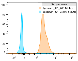
 and CoraLite®488-Conjugated AffiniPure Goat Anti-Rabbit IgG(H+L) at dilution 1:1000 (red), or 0.4 ug Isotype Control. Cells were fixed and permeabilized with Transcription Factor Staining Buffer Kit (PF00011).")
"Lamin A/C Antibodies" Comparison
View side-by-side comparison of Lamin A/C antibodies from other vendors to find the one that best suits your research needs.
Applications testées
| Résultats positifs en WB | cellules A431, cellules A375, cellules C6, cellules HEK-293, cellules HeLa, cellules HUVEC, cellules NIH/3T3, cellules SKOV-3, tissu ovarien de souris |
| Résultats positifs en IP | cellules HeLa, cellules A375 |
| Résultats positifs en IHC | tissu cardiaque de souris, human normal colon il est suggéré de démasquer l'antigène avec un tampon de TE buffer pH 9.0; (*) À défaut, 'le démasquage de l'antigène peut être 'effectué avec un tampon citrate pH 6,0. |
| Résultats positifs en IF/ICC | cellules HepG2, cellules HeLa |
| Résultats positifs en FC (Intra) | cellules HEK-293T |
Dilution recommandée
| Application | Dilution |
|---|---|
| Western Blot (WB) | WB : 1:5000-1:50000 |
| Immunoprécipitation (IP) | IP : 0.5-4.0 ug for 1.0-3.0 mg of total protein lysate |
| Immunohistochimie (IHC) | IHC : 1:2000-1:8000 |
| Immunofluorescence (IF)/ICC | IF/ICC : 1:400-1:1600 |
| Flow Cytometry (FC) (INTRA) | FC (INTRA) : 0.40 ug per 10^6 cells in a 100 µl suspension |
| It is recommended that this reagent should be titrated in each testing system to obtain optimal results. | |
| Sample-dependent, check data in validation data gallery | |
Applications publiées
| KD/KO | See 4 publications below |
| WB | See 242 publications below |
| IHC | See 2 publications below |
| IF | See 18 publications below |
| IP | See 4 publications below |
| CoIP | See 1 publications below |
Informations sur le produit
10298-1-AP cible Lamin A/C dans les applications de WB, IHC, IF/ICC, FC (Intra), IP, CoIP, ELISA et montre une réactivité avec des échantillons Humain, rat, souris
| Réactivité | Humain, rat, souris |
| Réactivité citée | rat, Humain, singe, souris, duck |
| Hôte / Isotype | Lapin / IgG |
| Clonalité | Polyclonal |
| Type | Anticorps |
| Immunogène | Lamin A/C Protéine recombinante Ag0408 |
| Nom complet | lamin A/C |
| Masse moléculaire calculée | 65 kDa |
| Poids moléculaire observé | 65 kDa, 70 kDa |
| Numéro d’acquisition GenBank | BC003162 |
| Symbole du gène | Lamin A/C |
| Identification du gène (NCBI) | 4000 |
| Conjugaison | Non conjugué |
| Forme | Liquide |
| Méthode de purification | Purification par affinité contre l'antigène |
| Tampon de stockage | PBS with 0.02% sodium azide and 50% glycerol |
| Conditions de stockage | Stocker à -20°C. Stable pendant un an après l'expédition. L'aliquotage n'est pas nécessaire pour le stockage à -20oC Les 20ul contiennent 0,1% de BSA. |
Informations générales
Lamin A/C is also named as LMNA or LMN1. The lamin family of proteins make up the matrix and are highly conserved in evolution. During mitosis, the lamina matrix is reversibly disassembled as the lamin proteins are phosphorylated. Lamin proteins are thought to be involved in nuclear stability, chromatin structure, and gene expression. The lack of lamin A/C can be as a novel marker for undifferentiated embryonic stem cells and lamin A/C expression is an early indicator of differentiation (PMID: 16179429). Mutations in this gene lead to several diseases: Emery-Dreifuss muscular dystrophy, familial partial lipodystrophy, limb-girdle muscular dystrophy, dilated cardiomyopathy, Charcot-Marie-Tooth disease, and Hutchinson-Gilford progeria syndrome. This protein has 4 isoforms produced by alternative splicing with the molecular weight of 74 kDa, 65 kDa, 70 kDa, and 64 kDa. This antibody can recognize 4 isoforms of Lamin A/C.
Protocole
| Product Specific Protocols | |
|---|---|
| WB protocol for Lamin A/C antibody 10298-1-AP | Download protocol |
| IHC protocol for Lamin A/C antibody 10298-1-AP | Download protocol |
| IF protocol for Lamin A/C antibody 10298-1-AP | Download protocol |
| IP protocol for Lamin A/C antibody 10298-1-AP | Download protocol |
| Standard Protocols | |
|---|---|
| Click here to view our Standard Protocols |
Publications
| Species | Application | Title |
|---|---|---|
Mol Cancer Cell surface CD55 traffics to the nucleus leading to cisplatin resistance and stemness by inducing PRC2 and H3K27 trimethylation on chromatin in ovarian cancer | ||
Nat Microbiol Nuclear pore blockade reveals that HIV-1 completes reverse transcription and uncoating in the nucleus. | ||
Mol Cell Lactylation-driven METTL3-mediated RNA m6A modification promotes immunosuppression of tumor-infiltrating myeloid cells. | ||
J Extracell Vesicles Extracellular vesicles derived from oesophageal cancer containing P4HB promote muscle wasting via regulating PHGDH/Bcl-2/caspase-3 pathway. | ||
Nat Chem Biol E2-Ub-R74G strategy reveals E2-specific ubiquitin conjugation profiles in live cells | ||
Cancer Cell SET1A-Mediated Mono-Methylation at K342 Regulates YAP Activation by Blocking Its Nuclear Export and Promotes Tumorigenesis. |
Avis
The reviews below have been submitted by verified Proteintech customers who received an incentive for providing their feedback.
FH Zahida (Verified Customer) (04-15-2025) | Easy to reveal
|
FH Alejandro (Verified Customer) (08-22-2022) | Shows clear staining in IF and well staining using FACS
 |
FH Charlotte (Verified Customer) (07-29-2022) | Cell fraction performed on NIH-3T3 cells to show the nucleus part. Blot super clean. Antibody specific. Easy to reveal.
 |
FH S (Verified Customer) (12-31-2021) | good antibody.
 |
FH Azita (Verified Customer) (06-16-2021) | It was used as a marker in WB to confirm proper nuclear fractionation of cell lysate.
|
FH Declan (Verified Customer) (11-29-2018) |
|

