Anticorps Polyclonal de lapin anti-mCherry
mCherry Polyclonal Antibody for WB, IF/ICC, IP, ELISA
Hôte / Isotype
Lapin / IgG
Réactivité testée
Protéine recombinante et plus (7)
Applications
WB, IHC, IF/ICC, IP, CoIP, ELISA
Conjugaison
Non conjugué
N° de cat : 26765-1-AP
Synonymes
Galerie de données de validation
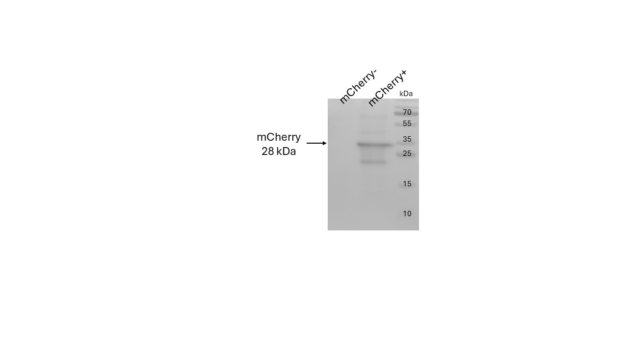
 at dilution of 1:8000 incubated at room temperature for 1.5 hours.")
 with negative control and mCherry overexpressed Transfected HEK-293 cells.")
 with negative control and mCherry overexpressed Transfected HEK-293 cells.")
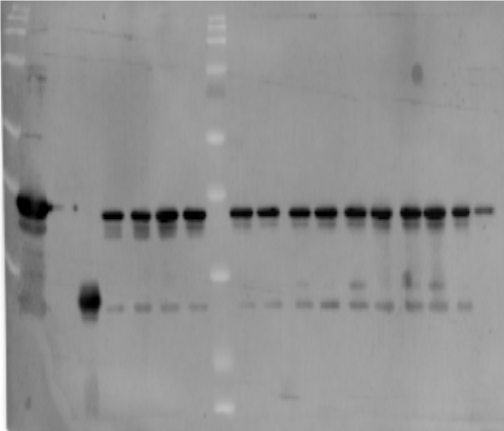
 with Transfected HEK-293T cells lysate 1280 ug.")
 fixed Transfected HEK-293 cells using mCherry antibody (26765-1-AP) at dilution of 1:800 and CoraLite®488-Conjugated AffiniPure Goat Anti-Rabbit IgG(H+L).")
Applications testées
| Résultats positifs en WB | cellules HEK-293 transfectées, Protéine recombinante |
| Résultats positifs en IP | Transfected HEK-293T cells, |
| Résultats positifs en IF/ICC | cellules HEK-293 transfectées, |
Dilution recommandée
| Application | Dilution |
|---|---|
| Western Blot (WB) | WB : 1:1000-1:4000 |
| Immunoprécipitation (IP) | IP : 0.5-4.0 ug for 1.0-3.0 mg of total protein lysate |
| Immunofluorescence (IF)/ICC | IF/ICC : 1:400-1:1600 |
| It is recommended that this reagent should be titrated in each testing system to obtain optimal results. | |
| Sample-dependent, check data in validation data gallery | |
Applications publiées
| WB | See 67 publications below |
| IHC | See 4 publications below |
| IF | See 39 publications below |
| IP | See 10 publications below |
| CoIP | See 4 publications below |
Informations sur le produit
26765-1-AP cible mCherry dans les applications de WB, IHC, IF/ICC, IP, CoIP, ELISA et montre une réactivité avec des échantillons Protéine recombinante
| Réactivité | Protéine recombinante |
| Réactivité citée | Humain, Lapin, levure, plante, poisson-zèbre, singe, souris |
| Hôte / Isotype | Lapin / IgG |
| Clonalité | Polyclonal |
| Type | Anticorps |
| Immunogène | mCherry Protéine recombinante Ag25320 |
| Nom complet | mCherry |
| Masse moléculaire calculée | 27 kDa |
| Symbole du gène | |
| Identification du gène (NCBI) | |
| Conjugaison | Non conjugué |
| Forme | Liquide |
| Méthode de purification | Purification par affinité contre l'antigène |
| Tampon de stockage | PBS with 0.02% sodium azide and 50% glycerol |
| Conditions de stockage | Stocker à -20°C. Stable pendant un an après l'expédition. L'aliquotage n'est pas nécessaire pour le stockage à -20oC Les 20ul contiennent 0,1% de BSA. |
Informations générales
Red fluorescent proteins (RFPs) is a collective term referring to a heterogenous group of red chromophore-carrying proteins, originating from various species and forming different protein lineages.
The original RFP (dsRed) is a 225 amino acid fluorescent protein (25.9 kDa) derived from Discosoma sp.. It emits red light with a peak wavelength of 593 nm upon excitation by green light (excitation peak at 558 nm).
When fused with other proteins, RFP serves as a versatile reporter protein e.g. for quantifying expression levels or facilitates visualization of subcellular localization through fluorescence microscopy.
This antibody is a rabbit polyclonal antibody raised against mCherry. It can be used to detect mCherry, dsRed, tdTomato, and mScarlet.
Protocole
| Product Specific Protocols | |
|---|---|
| WB protocol for mCherry antibody 26765-1-AP | Download protocol |
| IF protocol for mCherry antibody 26765-1-AP | Download protocol |
| IP protocol for mCherry antibody 26765-1-AP | Download protocol |
| Standard Protocols | |
|---|---|
| Click here to view our Standard Protocols |
Publications
| Species | Application | Title |
|---|---|---|
Nat Struct Mol Biol Aurora kinase A-mediated phosphorylation triggers structural alteration of Rab1A to enhance ER complexity during mitosis | ||
Nat Commun Stalled translation by mitochondrial stress upregulates a CNOT4-ZNF598 ribosomal quality control pathway important for tissue homeostasis | ||
J Extracell Vesicles Identification of the SNARE complex that mediates the fusion of multivesicular bodies with the plasma membrane in exosome secretion | ||
Cell Death Differ RING1 dictates GSDMD-mediated inflammatory response and host susceptibility to pathogen infection |
Avis
The reviews below have been submitted by verified Proteintech customers who received an incentive for providing their feedback.
FH Christine (Verified Customer) (09-23-2025) | Very strong and clean western blot on cells transfected with a mCHERRY tagged protein. Very sensitive antibody, as good as the home made one we were trying to replace. The monoclonal rabbit one sold by Proteintech (81202-2-RR) is also excellent and clean (but a slightly less strong), but would another great choice.
|
FH Alexandra (Verified Customer) (07-02-2024) | Not the strongest antibody I've ever used but it works fine, the maximum recomended dilution is 1000x, but possibly 500x would be more effective.
 |
FH Verdiana (Verified Customer) (10-12-2023) | Nice and clear bands!
|
FH Andrea (Verified Customer) (10-05-2023) | Good and strong signal for the mCherry fusion protein. In my case it was a 1:1000 dilution.
 |
FH Tatyana (Verified Customer) (01-09-2023) | Excellent antibody, very good bright signal, no background or non-specific bands. HeLa cells were transfected with plasmids for expression of mCherry tagged proteins, lysed after 24 hours, followed by WB (semi-dry transfer, blocking in 4% milk). Antibody was diluted in 4% milk.
 |
FH Laura (Verified Customer) (01-14-2020) | Antibody was used in 5% TBST overnight, gave a clean blot.
|
FH Benjamin (Verified Customer) (12-10-2019) | This antibody has been used to image mCherry fusion proteins expressed in HEK293T cells via western blot with high rates of success.Antibody was diluted in 3% milk TBST and incubated overnight at 4 degrees.
|
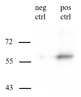
FH Liang (Verified Customer) (05-27-2019) | The antibody works perfect. According the attached image, The 2nd from the left is the negative control protein (mGFP) and the 3rd lane is the mCherry protein as the positive control, the others are the target protein.
 |

