- Phare
- Validé par KD/KO
Anticorps Polyclonal de lapin anti-NF-κB p65
NF-κB p65 Polyclonal Antibody for WB, IHC, IF/ICC, IP, ELISA
Hôte / Isotype
Lapin / IgG
Réactivité testée
Humain, rat, souris et plus (6)
Applications
WB, IHC, IF/ICC, IP, CoIP, ChIP, ELISA
Conjugaison
Non conjugué
N° de cat : 10745-1-AP
Synonymes
Galerie de données de validation
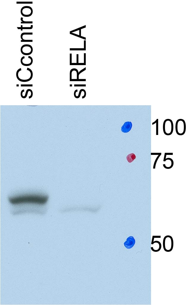
 with si-Control and si-RELA,p65 transfected HeLa cells.")
 at dilution of 1:3000 incubated at room temperature for 1.5 hours.")
 at dilution of 1:1000 incubated at room temperature for 1.5 hours.")
 at dilution of 1:800 incubated at room temperature for 1.5 hours.")
 at dilution of 1:800 incubated at room temperature for 1.5 hours.")
 with HeLa cells lysate 1280 ug.")
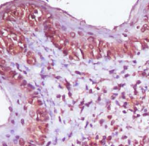
 at dilution of 1:200 (under 0x lens). Heat mediated antigen retrieval with Tris-EDTA buffer (pH 9.0).")
 at dilution of 1:500 (under 40x lens). Heat mediated antigen retrieval with Tris-EDTA buffer (pH 9.0).")
 at dilution of 1:500 (under 20x lens). Heat mediated antigen retrieval with Sodium Citrate buffer (pH 6.0).")
 at dilution of 1:100 (under 10x lens).")
 at dilution of 1:50 (under 10x lens).")
 at dilution of 1:50 (under 10x lens).")
 at dilution of 1:50 (under 40x lens).")
 at dilution of 1:50 (under 40x lens).")
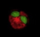
 fixed HepG2 cells using 10745-1-AP (p65; RELA antibody) at dilution of 1:100 and Alexa Fluor 488-conjugated AffiniPure Goat Anti-Rabbit IgG(H+L).")
 fixed TNF alpha treated HT-1080 cells using NF-κB p65 antibody (10745-1-AP) at dilution of 1:400 and Multi-rAb CoraLite ® Plus 488-Goat Anti-Rabbit Recombinant Secondary Antibody (H+L) (RGAR002), CL594-phalloidin (red).")
Applications testées
| Résultats positifs en WB | cellules A431, cellules HEK-293, cellules HeLa, cellules Jurkat, cellules K-562, cellules MCF-7, cellules NIH/3T3, cellules Raji |
| Résultats positifs en IP | cellules HeLa, |
| Résultats positifs en IHC | tissu de cancer du sein humain, tissu de cancer du foie humain, tissu de côlon de souris, tissu de côlon humain, tissu d'estomac humain il est suggéré de démasquer l'antigène avec un tampon de TE buffer pH 9.0; (*) À défaut, 'le démasquage de l'antigène peut être 'effectué avec un tampon citrate pH 6,0. |
| Résultats positifs en IF/ICC | cellules HepG2, TNF alpha treated HT-1080 cells |
Dilution recommandée
| Application | Dilution |
|---|---|
| Western Blot (WB) | WB : 1:1000-1:6000 |
| Immunoprécipitation (IP) | IP : 0.5-4.0 ug for 1.0-3.0 mg of total protein lysate |
| Immunohistochimie (IHC) | IHC : 1:50-1:500 |
| Immunofluorescence (IF)/ICC | IF/ICC : 1:50-1:500 |
| It is recommended that this reagent should be titrated in each testing system to obtain optimal results. | |
| Sample-dependent, check data in validation data gallery | |
Informations sur le produit
10745-1-AP cible NF-κB p65 dans les applications de WB, IHC, IF/ICC, IP, CoIP, ChIP, ELISA et montre une réactivité avec des échantillons Humain, rat, souris
| Réactivité | Humain, rat, souris |
| Réactivité citée | rat, Chèvre, Humain, Lapin, poisson-zèbre, poulet, singe, souris, fish |
| Hôte / Isotype | Lapin / IgG |
| Clonalité | Polyclonal |
| Type | Anticorps |
| Immunogène | NF-κB p65 Protéine recombinante Ag1199 |
| Nom complet | v-rel reticuloendotheliosis viral oncogene homolog A (avian) |
| Masse moléculaire calculée | 65 kDa |
| Poids moléculaire observé | 65 kDa |
| Numéro d’acquisition GenBank | BC011603 |
| Symbole du gène | NF-κB p65 |
| Identification du gène (NCBI) | 5970 |
| Conjugaison | Non conjugué |
| Forme | Liquide |
| Méthode de purification | Purification par affinité contre l'antigène |
| Tampon de stockage | PBS with 0.02% sodium azide and 50% glycerol |
| Conditions de stockage | Stocker à -20°C. Stable pendant un an après l'expédition. L'aliquotage n'est pas nécessaire pour le stockage à -20oC Les 20ul contiennent 0,1% de BSA. |
Informations générales
Nuclear factor k B (NF-kB) is a sequence-specific DNA-binding protein complex which regulates the expression of viral genomes, including the human immunodeficiency virus, and a variety of cellular genes, particularly those involved in immune and inflammatory responses. The members of the NF-kB family in mammalian cells include the proto-oncogene c-Rel,p50/p105 (NFkB1), p65 (RelA), p52/p100 (NFkB2), and RelB. All of these proteins share a conserved 300-amino acid region known as the Rel homology domain which is responsible for DNA binding, dimerization, and nuclear translocation of NF-kB. The p65 subunit is a major component of NF-kB complexes and is responsible for trans-activation. NF-kB heterodimeric p65-p50 and p65-c-Rel complexes are transcriptional activators. The NF-kB p65-p65 complex appears to be involved in invasin-mediated activation of IL-8 expression. The inhibitory effect of IkB upon NF-kB the cytoplasm is exerted primarily through the interaction with p65. p65 shows a weak DNA-binding site which could contribute directly to DNA binding in the NF-kB complex. It associates with chromatin at the NF-kB promoter region via association with DDX1. This antibody is a rabbit polyclonal antibody raised against residues near the N terminus of human RELA.
Protocole
| Product Specific Protocols | |
|---|---|
| WB protocol for NF-κB p65 antibody 10745-1-AP | Download protocol |
| IHC protocol for NF-κB p65 antibody 10745-1-AP | Download protocol |
| IF protocol for NF-κB p65 antibody 10745-1-AP | Download protocol |
| IP protocol for NF-κB p65 antibody 10745-1-AP | Download protocol |
| Standard Protocols | |
|---|---|
| Click here to view our Standard Protocols |
Publications
| Species | Application | Title |
|---|---|---|
Bioact Mater Silicate ions as soluble form of bioactive ceramics alleviate aortic aneurysm and dissection | ||
Mol Cell Serine synthesis sustains macrophage IL-1β production via NAD+-dependent protein acetylation | ||
Nat Microbiol IFI16 directly senses viral RNA and enhances RIG-I transcription and activation to restrict influenza virus infection. | ||
Adv Sci (Weinh) Sirtuin 5-Mediated Desuccinylation of ALDH2 Alleviates Mitochondrial Oxidative Stress Following Acetaminophen-Induced Acute Liver Injury |
Avis
The reviews below have been submitted by verified Proteintech customers who received an incentive for providing their feedback.
FH Maximilian (Verified Customer) (06-30-2023) | Quite a bit of unspecific binding. Did not work in mouse cell lines.
|
FH Xie (Verified Customer) (09-29-2022) | NF-κB p65 Antibody staining.
 |
FH Murali (Verified Customer) (01-24-2022) | Very nice antibody for IHC
 |
FH Azita (Verified Customer) (05-31-2021) | NSC-34 cells (motor neuron-like cells) strong labelling in WB
|
FH Zehua (Verified Customer) (12-17-2019) | After siRNA treatment for 48 hours, specific signal shown here. Works excellent!
 |
FH Feng-qian (Verified Customer) (12-07-2018) |
|
FH Fraser (Verified Customer) (06-06-2018) |
|

