Tested Applications
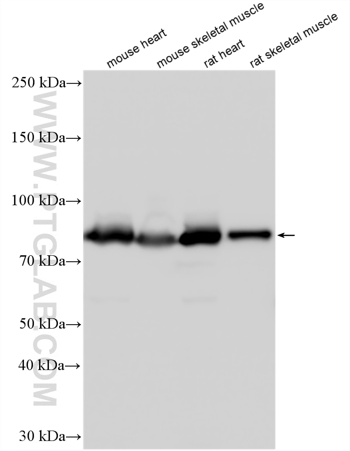
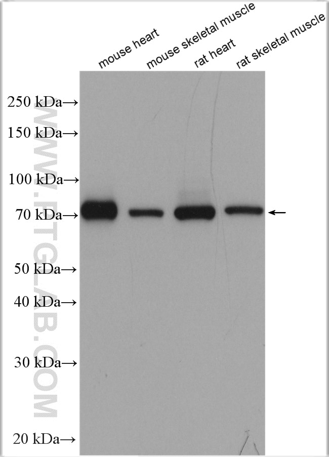
| Positive WB detected in | mouse heart tissue, mouse skeletal muscle tissue, rat heart tissue, rat skeletal muscle tissue |
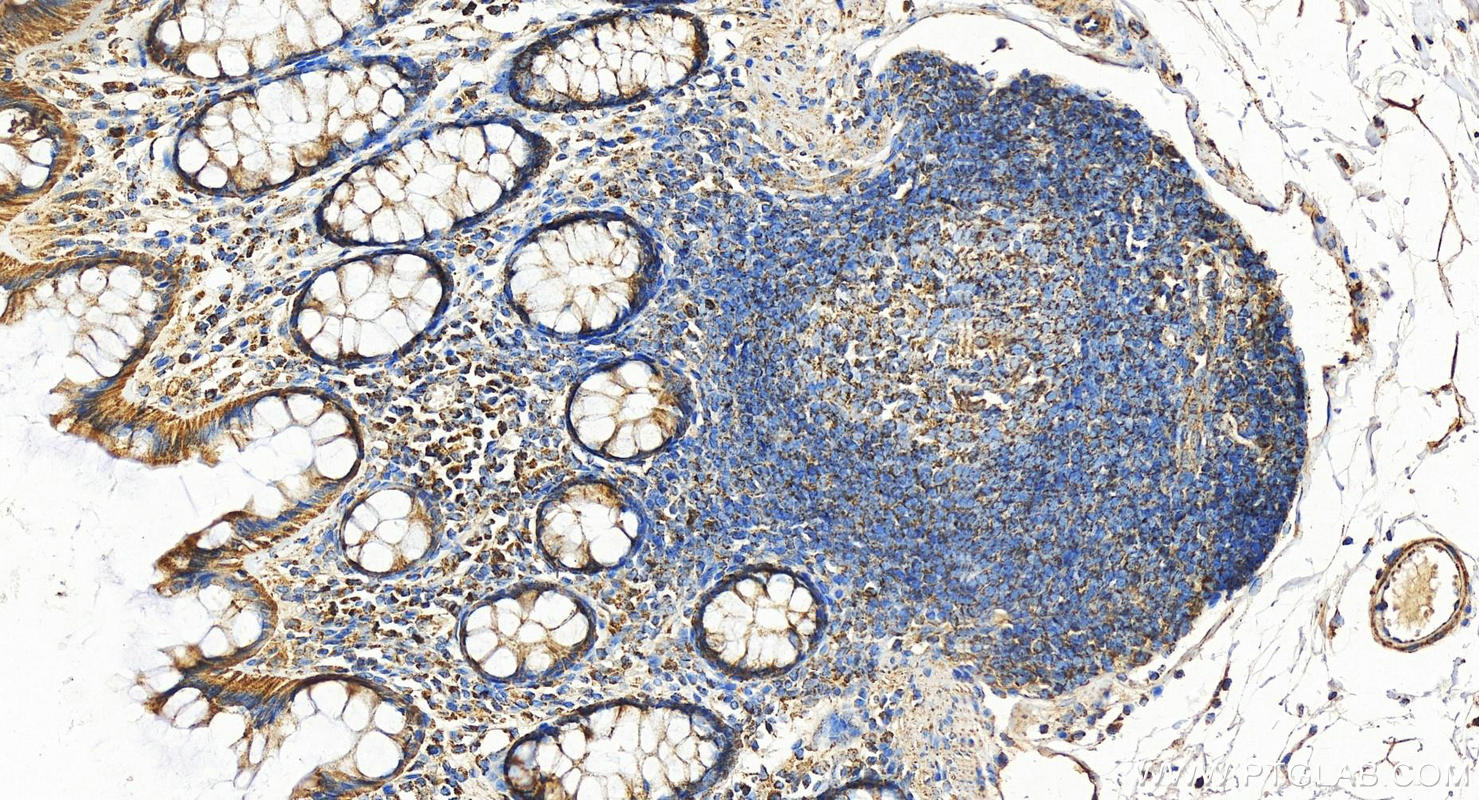
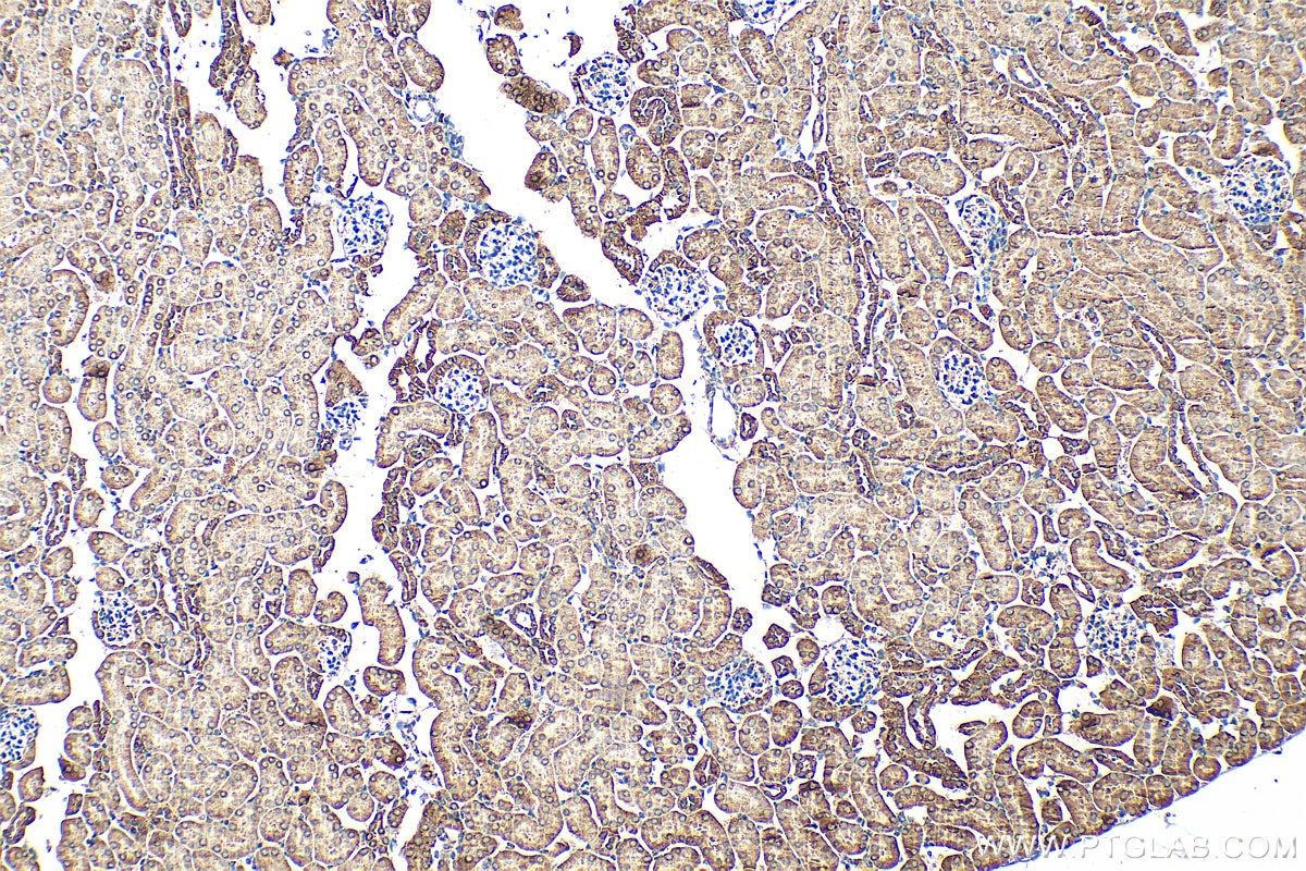
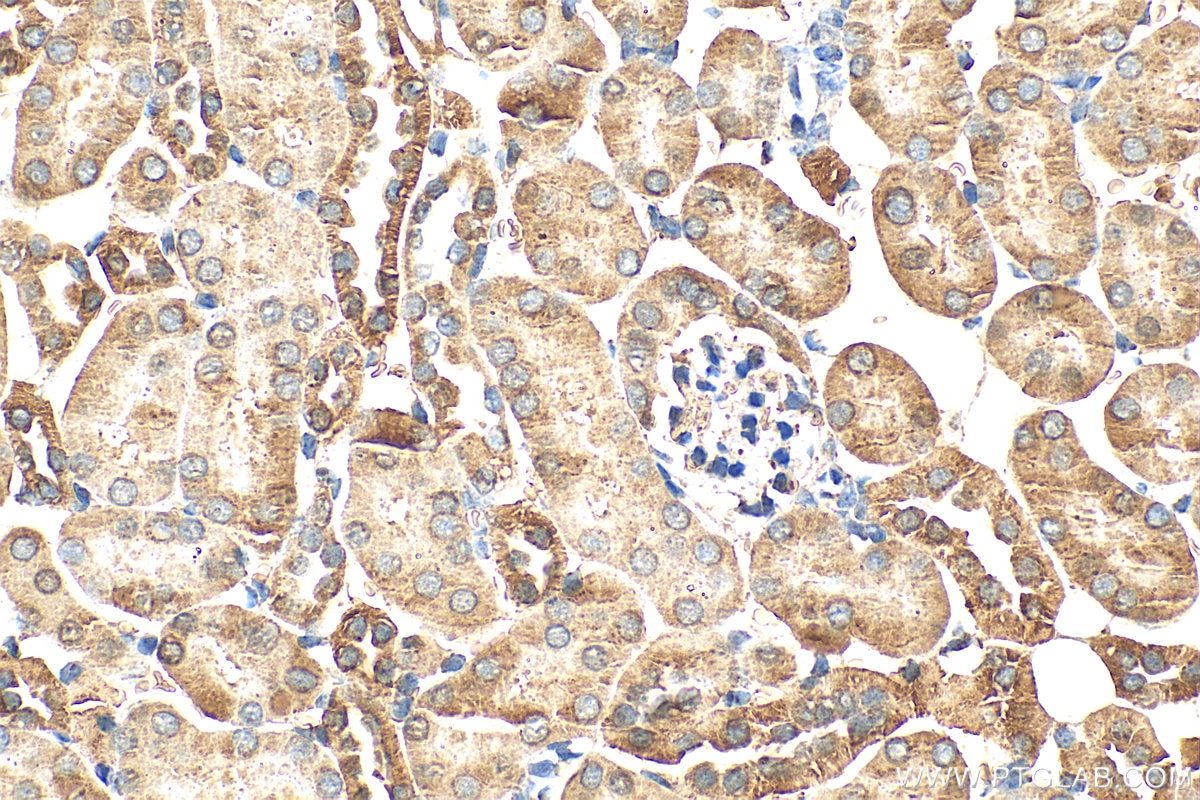
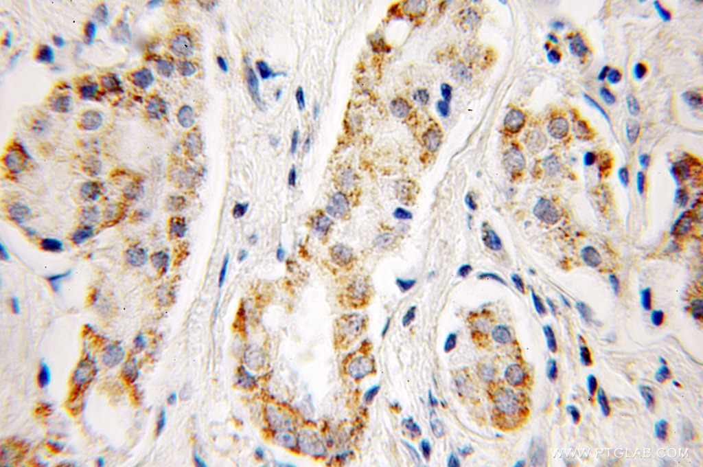
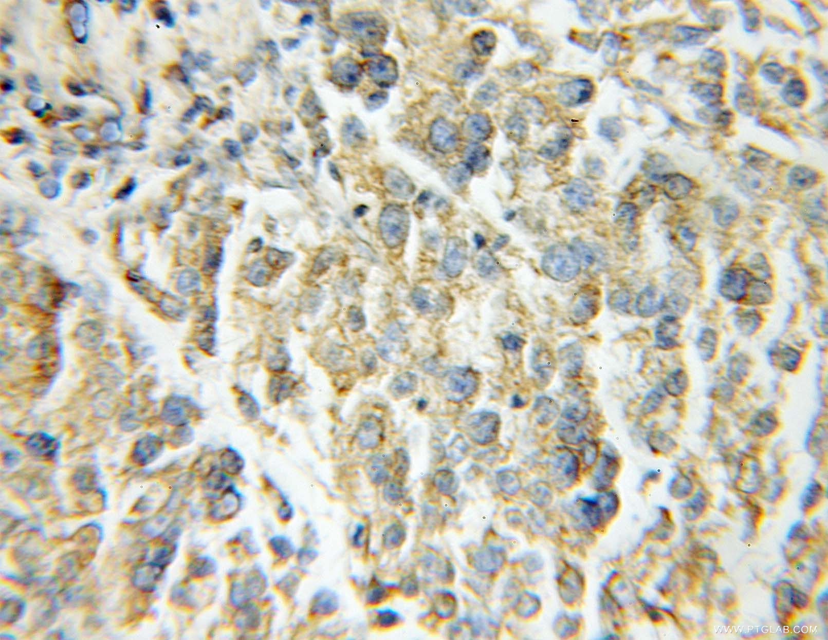
| Positive IHC detected in | mouse kidney tissue, human colon tissue, human lung cancer tissue, human prostate cancer tissue Note: suggested antigen retrieval with TE buffer pH 9.0; (*) Alternatively, antigen retrieval may be performed with citrate buffer pH 6.0 |
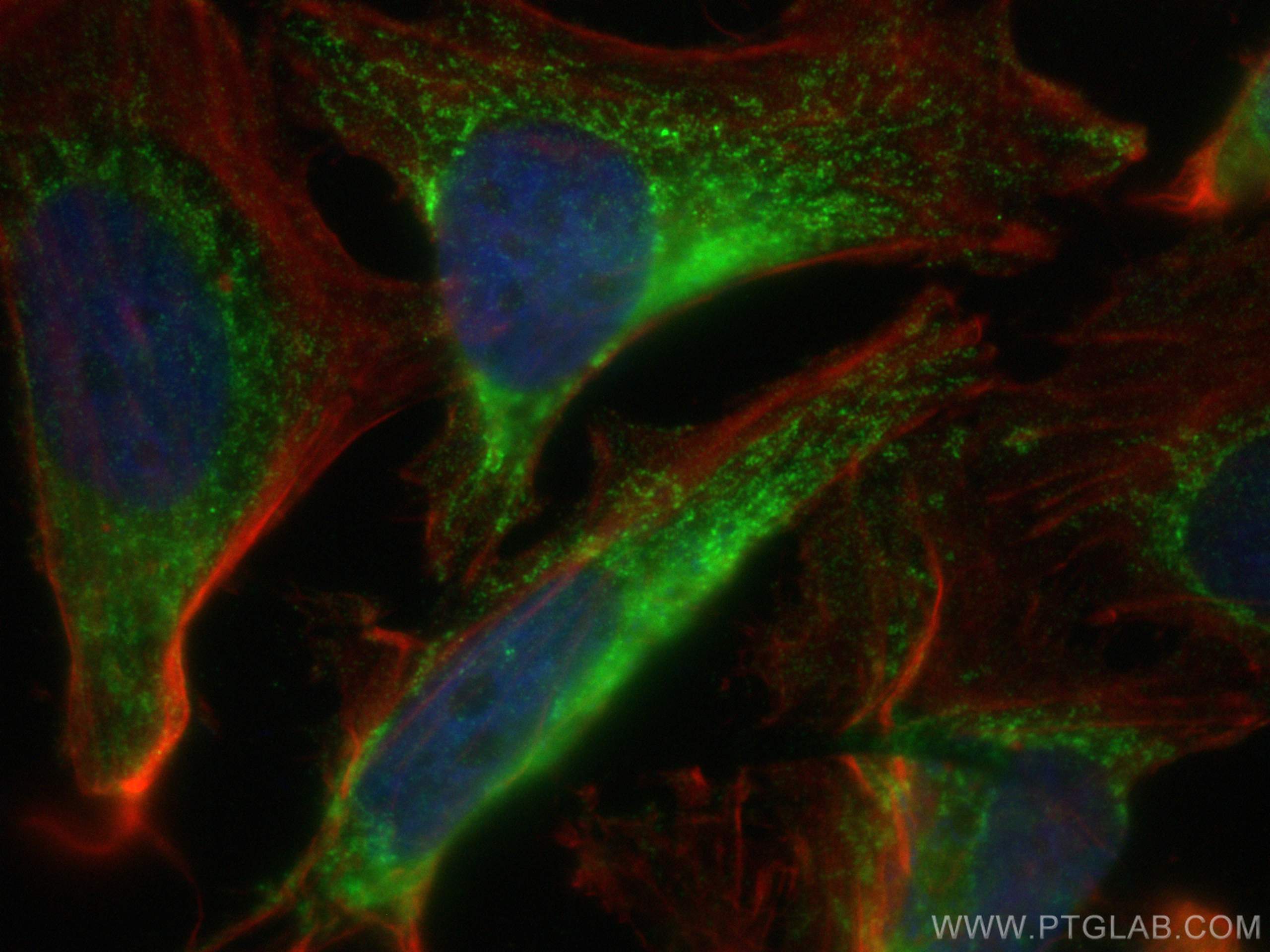
| Positive IF/ICC detected in | HeLa cells |
Recommended dilution
| Application | Dilution |
|---|---|
| Western Blot (WB) | WB : 1:5000-1:50000 |
| Immunohistochemistry (IHC) | IHC : 1:200-1:1200 |
| Immunofluorescence (IF)/ICC | IF/ICC : 1:300-1:1200 |
| It is recommended that this reagent should be titrated in each testing system to obtain optimal results. | |
| Sample-dependent, Check data in validation data gallery. | |
Published Applications
| KD/KO | See 1 publications below |
| WB | See 29 publications below |
| IHC | See 2 publications below |
| IF | See 6 publications below |
Product Information
11134-1-AP targets Aconitase 2 in WB, IHC, IF/ICC, ELISA applications and shows reactivity with human, mouse, rat samples.
| Tested Reactivity | human, mouse, rat |
| Cited Reactivity | human, mouse, rat |
| Host / Isotype | Rabbit / IgG |
| Class | Polyclonal |
| Type | Antibody |
| Immunogen |
CatNo: Ag1590 Product name: Recombinant human ACO2 protein Source: e coli.-derived, PGEX-4T Tag: GST Domain: 1-306 aa of BC014092 Sequence: MAPYSLLVTRLQKALGVRQYHVASVLCQRAKVAMSHFEPNEYIHYDLLEKNINIVRKRLNRPLTLSEKIVYGHLDDPASQEIERGKSYLRLRPDRVAMQDATAQMAMLQFISSGLSKVAVPSTIHCDHLIEAQVGGEKDLRRAKDINQEVYNFLATAGAKYGVGFWKPGSGIIHQIILENYAYPGVLLIGTDSHTPNGGGLGGICIGVGGADAVDVMAGIPWELKCPKVIGVKLTGSLSGWSSPKDVILKVAGILTVKGGTGAIVEYHGPGVDSISCTGMATICNMGAEIGATTSVFPYNHRMKKY Predict reactive species |
| Full Name | aconitase 2, mitochondrial |
| Calculated Molecular Weight | 85 kDa |
| Observed Molecular Weight | 85 kDa |
| GenBank Accession Number | BC014092 |
| Gene Symbol | ACO2 |
| Gene ID (NCBI) | 50 |
| RRID | AB_2289288 |
| Conjugate | Unconjugated |
| Form | Liquid |
| Purification Method | Antigen affinity purification |
| UNIPROT ID | Q99798 |
| Storage Buffer | PBS with 0.02% sodium azide and 50% glycerol, pH 7.3. |
| Storage Conditions | Store at -20°C. Stable for one year after shipment. Aliquoting is unnecessary for -20oC storage. 20ul sizes contain 0.1% BSA. |
Background Information
ACO2(aconitate hydratase, mitochondrial) is also named as citrate hydro-lyase and belongs to the aconitase/IPM isomerase family. It plays a key function in cellular energy production, and loss of its activity has a major impact on cellular and organismal survival. Western blot shows two bands of 83 kDa and 40 kDa. The 40 kDa fragment decreases with age and oxidative stress, whereas other fragmentation products with molecular weights between 40 and 83 kDa increased with age and MnSOD(mitochondrial manganese superoxide dismutase) deficiency(PMID:12459471). Defects in ACO2 are the cause of infantile cerebellar-retinal degeneration (ICRD).
Protocols
| Product Specific Protocols | |
|---|---|
| IF protocol for Aconitase 2 antibody 11134-1-AP | Download protocol |
| IHC protocol for Aconitase 2 antibody 11134-1-AP | Download protocol |
| WB protocol for Aconitase 2 antibody 11134-1-AP | Download protocol |
| Standard Protocols | |
|---|---|
| Click here to view our Standard Protocols |
Publications
| Species | Application | Title |
|---|---|---|
Cell Metab Malic enzyme 2 connects the Krebs cycle intermediate fumarate to mitochondrial biogenesis. | ||
Aging Dis Dietary Salt Disrupts Tricarboxylic Acid Cycle and Induces Tau Hyperphosphorylation and Synapse Dysfunction during Aging | ||
Sci Total Environ TRIM24-DTNBP1-ATP7A mediated astrocyte cuproptosis in cognition and memory dysfunction caused by Y2O3 NPs | ||
Cell Rep A LON-ClpP Proteolytic Axis Degrades Complex I to Extinguish ROS Production in Depolarized Mitochondria. | ||
EMBO Rep UBXN1 maintains ER proteostasis and represses UPR activation by modulating translation | ||
Cell Biosci Hinokitiol-iron complex is a ferroptosis inducer to inhibit triple-negative breast tumor growth |
Reviews
The reviews below have been submitted by verified Proteintech customers who received an incentive for providing their feedback.
FH Tanusree (Verified Customer) (12-18-2019) | Product worked well in WB at 1:500 dilution
|
FH Svitlana (Verified Customer) (03-19-2019) | SDS-PAGE: mitochondrial lysate 10 ug protein/well, 4-12% Bis-Tris.Transfer: Immobilon-FL transfer membranes O/N at 30V, 4CBlocking: SEA Block Blocking Buffer 1h at room temperature.Secondary Ab: IRDye 800CW Goat anti-Rabbit 1 h at room temperature.Lines on WB:1. BioRad Precision Plus Protein standard2. Lysate of mitochondrial fraction.
 |

