Tested Applications
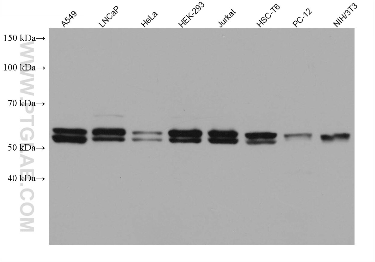
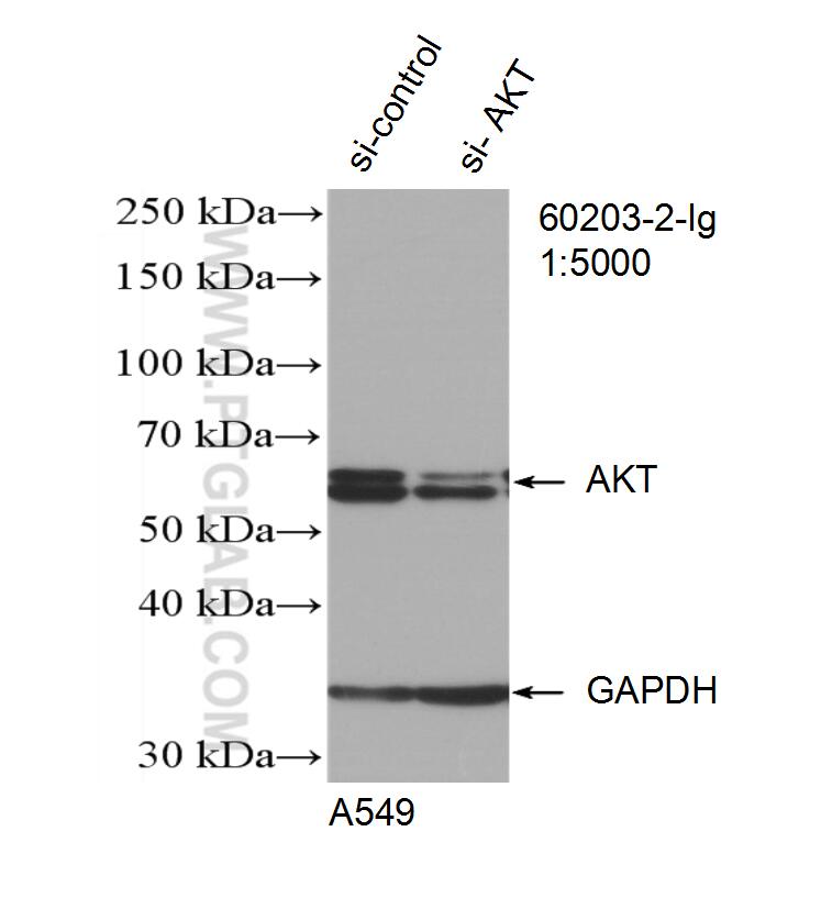
| Positive WB detected in | A549 cells, HEK-293 cells, mouse brain tissue, ROS1728 cells, LNCaP cells, Hela cells, Jurkat cells, HSC-T6 cells, PC-12 cells, NIH/3T3 cells |
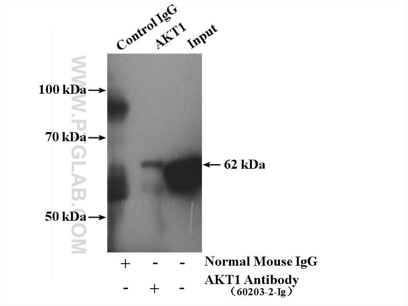
| Positive IP detected in | mouse brain tissue |
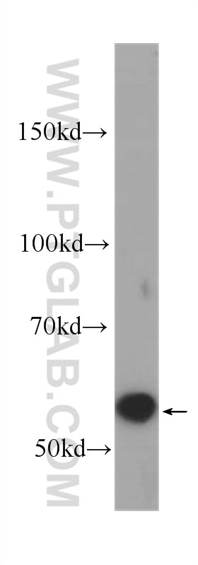
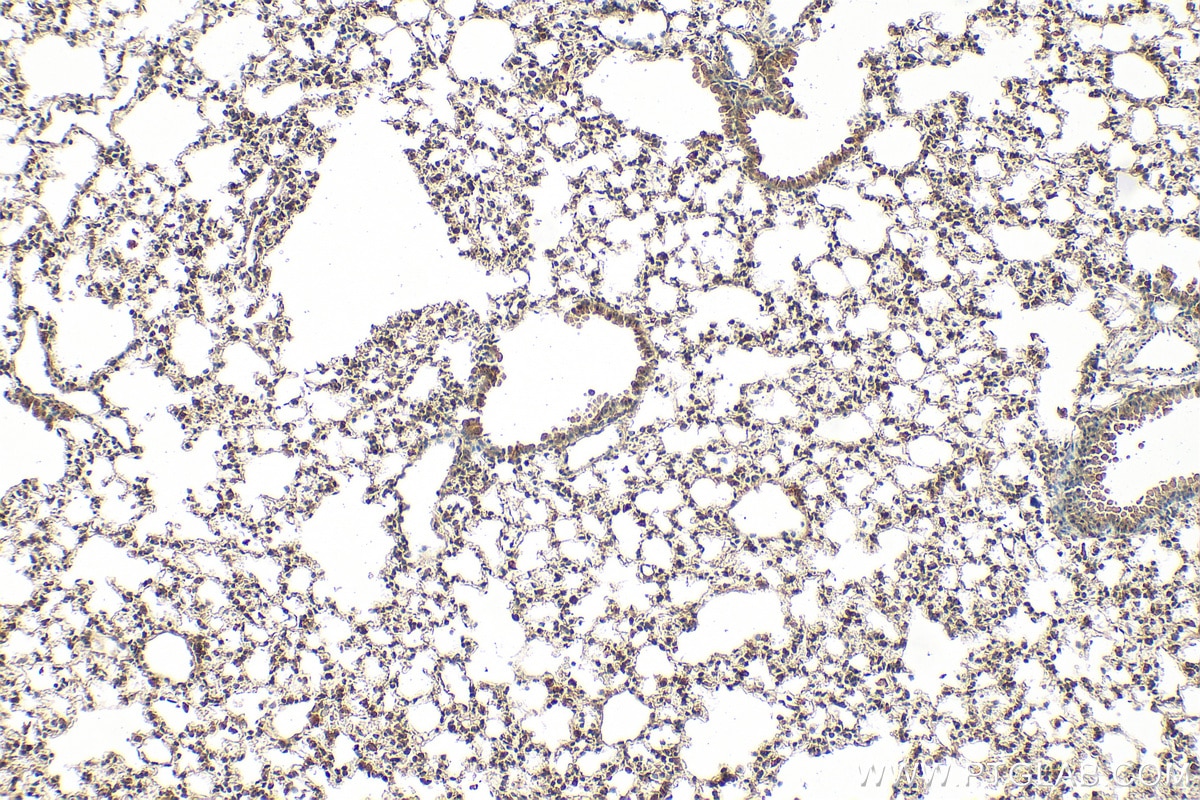
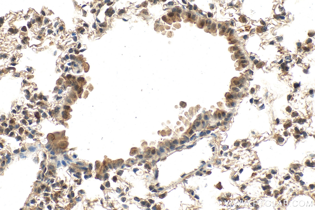
| Positive IHC detected in | mouse lung tissue Note: suggested antigen retrieval with TE buffer pH 9.0; (*) Alternatively, antigen retrieval may be performed with citrate buffer pH 6.0 |
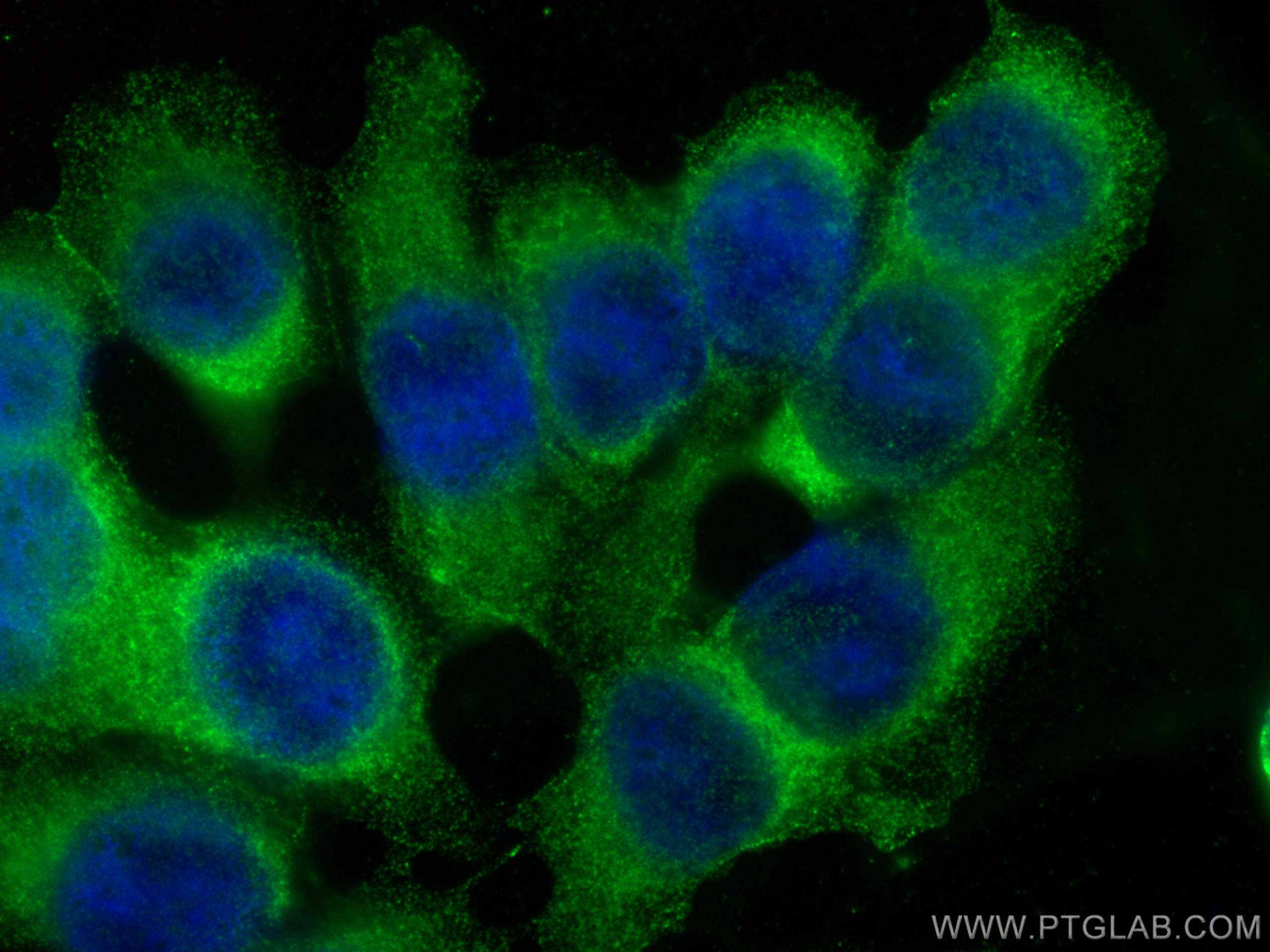
| Positive IF/ICC detected in | MCF-7 cells |
Recommended dilution
| Application | Dilution |
|---|---|
| Western Blot (WB) | WB : 1:5000-1:50000 |
| Immunoprecipitation (IP) | IP : 0.5-4.0 ug for 1.0-3.0 mg of total protein lysate |
| Immunohistochemistry (IHC) | IHC : 1:250-1:1000 |
| Immunofluorescence (IF)/ICC | IF/ICC : 1:200-1:800 |
| It is recommended that this reagent should be titrated in each testing system to obtain optimal results. | |
| Sample-dependent, Check data in validation data gallery. | |
Published Applications
| KD/KO | See 3 publications below |
| WB | See 789 publications below |
| IHC | See 24 publications below |
| IF | See 27 publications below |
| IP | See 5 publications below |
| CoIP | See 4 publications below |
Product Information
60203-2-Ig targets AKT in WB, IHC, IF/ICC, IP, CoIP, ELISA applications and shows reactivity with human, mouse, rat samples.
| Tested Reactivity | human, mouse, rat |
| Cited Reactivity | human, mouse, rat, pig, rabbit, chicken, hamster, sheep, goat, duck |
| Host / Isotype | Mouse / IgG1 |
| Class | Monoclonal |
| Type | Antibody |
| Immunogen |
CatNo: Ag16695 Product name: Recombinant human AKT protein Source: e coli.-derived, PET28a Tag: 6*His Domain: 1-224 aa of BC000479 Sequence: MSDVAIVKEGWLHKRGEYIKTWRPRYFLLKNDGTFIGYKERPQDVDQREAPLNNFSVAQCQLMKTERPRPNTFIIRCLQWTTVIERTFHVETPEEREEWTTAIQTVADGLKKQEEEEMDFRSGSPSDNSGAEEMEVSLAKPKHRVTMNEFEYLKLLGKGTFGKVILVKEKATGRYYAMKILKKEVIVAKDEVAHTLTENRVLQNSRHPFLTALKYSFQTHDRLC Predict reactive species |
| Full Name | v-akt murine thymoma viral oncogene homolog 1 |
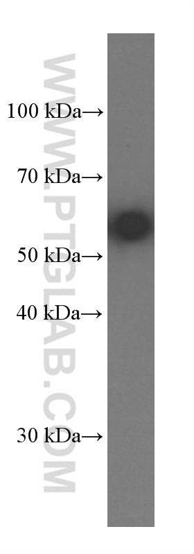
| Calculated Molecular Weight | 56 kDa |
| Observed Molecular Weight | 56-62 kDa |
| GenBank Accession Number | BC000479 |
| Gene Symbol | AKT1 |
| Gene ID (NCBI) | 207 |
| RRID | AB_10912803 |
| Conjugate | Unconjugated |
| Form | Liquid |
| Purification Method | Protein G purification |
| UNIPROT ID | P31749 |
| Storage Buffer | PBS with 0.02% sodium azide and 50% glycerol, pH 7.3. |
| Storage Conditions | Store at -20°C. Stable for one year after shipment. Aliquoting is unnecessary for -20oC storage. 20ul sizes contain 0.1% BSA. |
Background Information
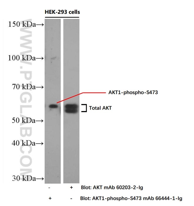
The serine-threonine protein kinase AKT1 is catalytically inactive in serum-starved primary and immortalized fibroblasts. Survival factors can suppress apoptosis in a transcription-independent manner by activating the serine/threonine kinase AKT1, which then phosphorylates and inactivates components of the apoptotic machinery. This antibody detects all the members of AKT with/without phospho- modification.
Protocols
| Product Specific Protocols | |
|---|---|
| IF protocol for AKT antibody 60203-2-Ig | Download protocol |
| IHC protocol for AKT antibody 60203-2-Ig | Download protocol |
| IP protocol for AKT antibody 60203-2-Ig | Download protocol |
| WB protocol for AKT antibody 60203-2-Ig | Download protocol |
| Standard Protocols | |
|---|---|
| Click here to view our Standard Protocols |
Publications
| Species | Application | Title |
|---|---|---|
Signal Transduct Target Ther Circulating tumor cells shielded with extracellular vesicle-derived CD45 evade T cell attack to enable metastasis | ||
Cell Metab Acetate enables metabolic fitness and cognitive performance during sleep disruption | ||
Nat Commun FOXP3+ regulatory T cell perturbation mediated by the IFNγ-STAT1-IFITM3 feedback loop is essential for anti-tumor immunity | ||
Cell Rep Med Targeting phenylpyruvate restrains excessive NLRP3 inflammasome activation and pathological inflammation in diabetic wound healing | ||
Cell Rep Med Reprograming immunosuppressive microenvironment by eIF4G1 targeting to eradicate pancreatic ductal adenocarcinoma | ||
Adv Healthc Mater A ROS-Responsive Liposomal Composite Hydrogel Integrating Improved Mitochondrial Function and Pro-Angiogenesis for Efficient Treatment of Myocardial Infarction. |
Reviews
The reviews below have been submitted by verified Proteintech customers who received an incentive for providing their feedback.
FH Mounika (Verified Customer) (01-08-2026) | Antibodies are very wonderful, works very amazingly!
|
FH Sai Sindhura (Verified Customer) (01-08-2026) | AKT works very good for IHC
|
FH Areen (Verified Customer) (12-15-2025) | VERY GOOD ANTIBODY
|
FH A (Verified Customer) (07-15-2025) | Used for caco2 cells and animal colon tissue
|
FH C (Verified Customer) (06-25-2025) | A reliable Ab to detect endo Akt.
 |
FH Janine (Verified Customer) (02-18-2019) | Yielded clear and distinguishable bands at the expected size of ~60kDa after 10 sec exposure using enhanced chemiluminescence.
|