Tested Applications
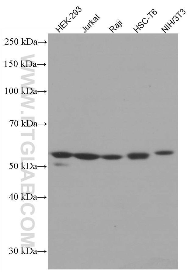
| Positive WB detected in | HEK-293 cells, HepG2 cells, HeLa cells, Jurkat cells, Raji cells, HSC-T6 cells, NIH/3T3 cells |
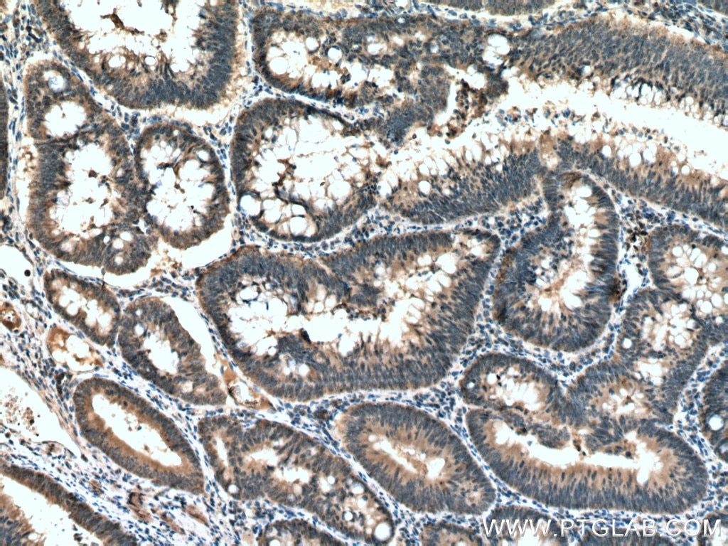
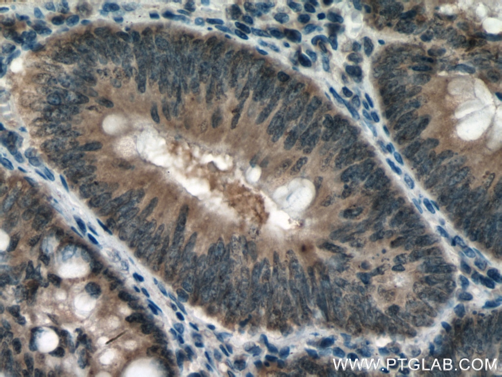
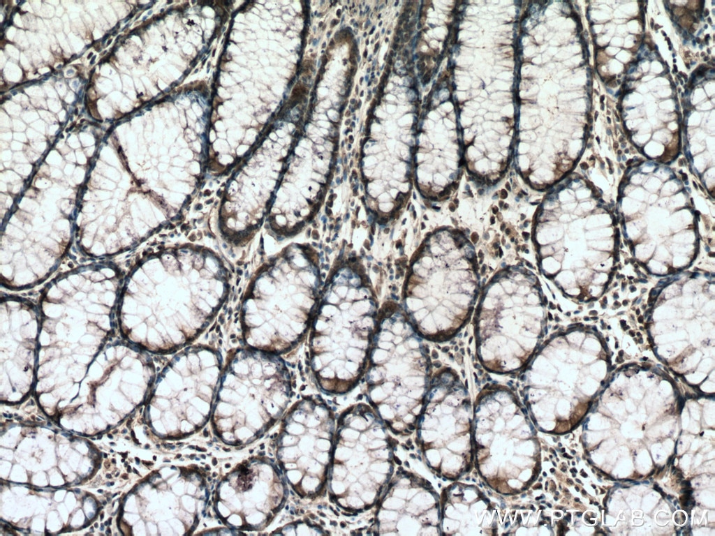
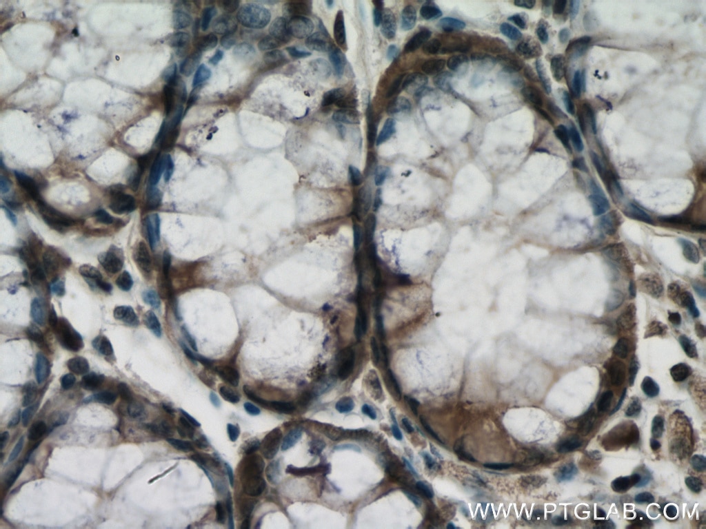
| Positive IHC detected in | human colon cancer tissue Note: suggested antigen retrieval with TE buffer pH 9.0; (*) Alternatively, antigen retrieval may be performed with citrate buffer pH 6.0 |
Recommended dilution
| Application | Dilution |
|---|---|
| Western Blot (WB) | WB : 1:1000-1:6000 |
| Immunohistochemistry (IHC) | IHC : 1:150-1:600 |
| It is recommended that this reagent should be titrated in each testing system to obtain optimal results. | |
| Sample-dependent, Check data in validation data gallery. | |
Published Applications
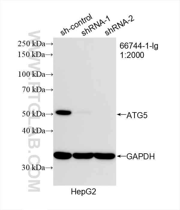
| KD/KO | See 6 publications below |
| WB | See 52 publications below |
| IHC | See 2 publications below |
| IF | See 3 publications below |
| IP | See 1 publications below |
Product Information
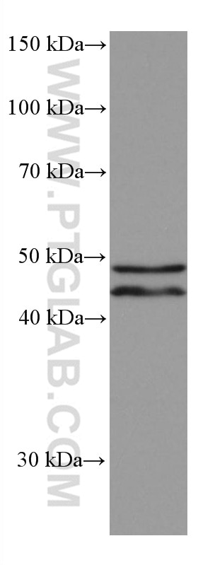
66744-1-Ig targets ATG5 in WB, IHC, IF, IP, ELISA applications and shows reactivity with human, mouse, rat samples.
| Tested Reactivity | human, mouse, rat |
| Cited Reactivity | human, mouse, rat, pig, bovine |
| Host / Isotype | Mouse / IgG2a |
| Class | Monoclonal |
| Type | Antibody |
| Immunogen |
CatNo: Ag16685 Product name: Recombinant human ATG5 protein Source: e coli.-derived, PET28a Tag: 6*His Domain: 28-275 aa of BC002699 Sequence: TEREAEPYYLLLPRVSYLTLVTDKVKKHFQKVMRQEDISEIWFEYEGTPLKWHYPIGLLFDLLASSSALPWNITVHFKSFPEKDLLHCPSKDAIEAHFMSCMKEADALKHKSQVINEMQKKDHKQLWMGLQNDRFDQFWAINRKLMEYPAEENGFRYIPFRIYQTTTERPFIQKLFRPVAADGQLHTLGDLLKEVCPSAIDPEDGEKKNQVMIHGIEPMLETPLQWLSEHLSYPDNFLHISIIPQPTD Predict reactive species |
| Full Name | ATG5 autophagy related 5 homolog (S. cerevisiae) |
| Calculated Molecular Weight | 32 kDa |
| Observed Molecular Weight | 50-55 and 40-45 kDa |
| GenBank Accession Number | BC002699 |
| Gene Symbol | ATG5 |
| Gene ID (NCBI) | 9474 |
| RRID | AB_2882092 |
| Conjugate | Unconjugated |
| Form | Liquid |
| Purification Method | Protein A purification |
| UNIPROT ID | Q9H1Y0 |
| Storage Buffer | PBS with 0.02% sodium azide and 50% glycerol, pH 7.3. |
| Storage Conditions | Store at -20°C. Stable for one year after shipment. Aliquoting is unnecessary for -20oC storage. 20ul sizes contain 0.1% BSA. |
Background Information
ATG5, also named as APG5L and ASP, belongs to the ATG5 family. It is required for autophagy. It plays an important role in the apoptotic process, possibly within the modified cytoskeleton. Its expression is a relatively late event in the apoptotic process, occurring downstream of caspase activity. Autophagy is a catabolic process for the autophagosomic-lysosomal degradation of bulk cytoplasmic contents. Formation of the autophagosome involves a ubiquitin-like conjugation system in which ATG12 is covalently bound to ATG5 and targeted to autophagosome vesicles. It mediates autophagosome-independent host protection. 66744-1-Ig antibody can recognize the ATG5-ATG12 complex (50-55 kDa) which can be truncated and generate a 40-45 kDa band. (PMID: 19783656, 25032862)
Protocols
| Product Specific Protocols | |
|---|---|
| IHC protocol for ATG5 antibody 66744-1-Ig | Download protocol |
| WB protocol for ATG5 antibody 66744-1-Ig | Download protocol |
| Standard Protocols | |
|---|---|
| Click here to view our Standard Protocols |
Publications
| Species | Application | Title |
|---|---|---|
Gastroenterology Pancreatic acinar cells-derived sphingosine-1-phosphate contributes to fibrosis of chronic pancreatitis via inducing autophagy and activation of pancreatic stellate cells | ||
Adv Sci (Weinh) Mitochondrial tRNAGlu 14693A>G Mutation, an "Enhancer" to the Phenotypic Expression of Leber's Hereditary Optic Neuropathy | ||
Dev Cell ATG5 attenuates inflammatory signaling in mouse embryonic stem cells to control differentiation
| ||
J Nanobiotechnology Cold exposure protects against medial arterial calcification development via autophagy | ||
J Nanobiotechnology Reciprocal regulation of NRF2 by autophagy and ubiquitin-proteasome modulates vascular endothelial injury induced by copper oxide nanoparticles.
| ||
Biochim Biophys Acta Mol Basis Dis Hydroxysafflor yellow A inhibits neuronal ferroptosis and ferritinophagy in ischemic stroke |
Reviews
The reviews below have been submitted by verified Proteintech customers who received an incentive for providing their feedback.
FH Marina (Verified Customer) (11-22-2021) | ATG 5 and the complex ATG5-ATG12 are visible, almost no background.
|