
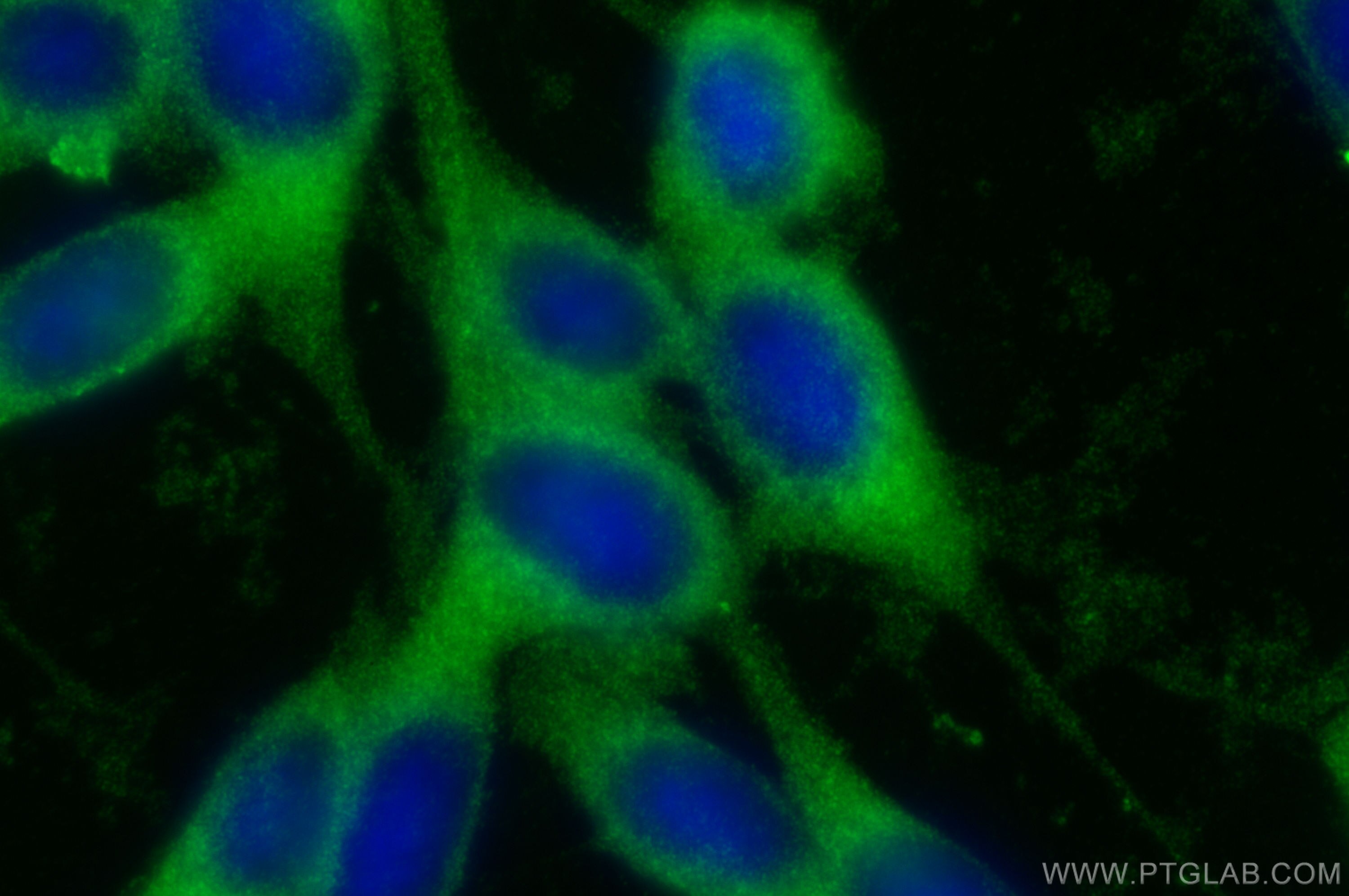

Tested Applications
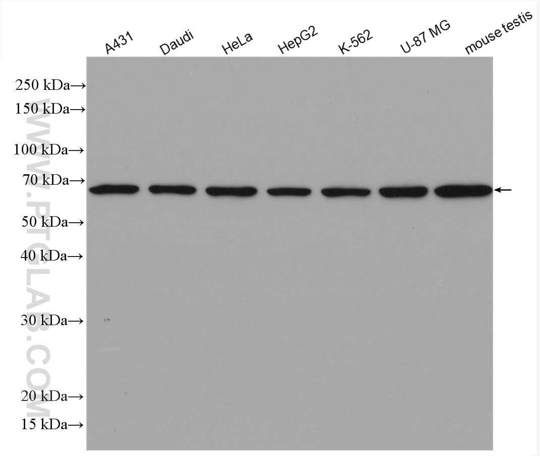
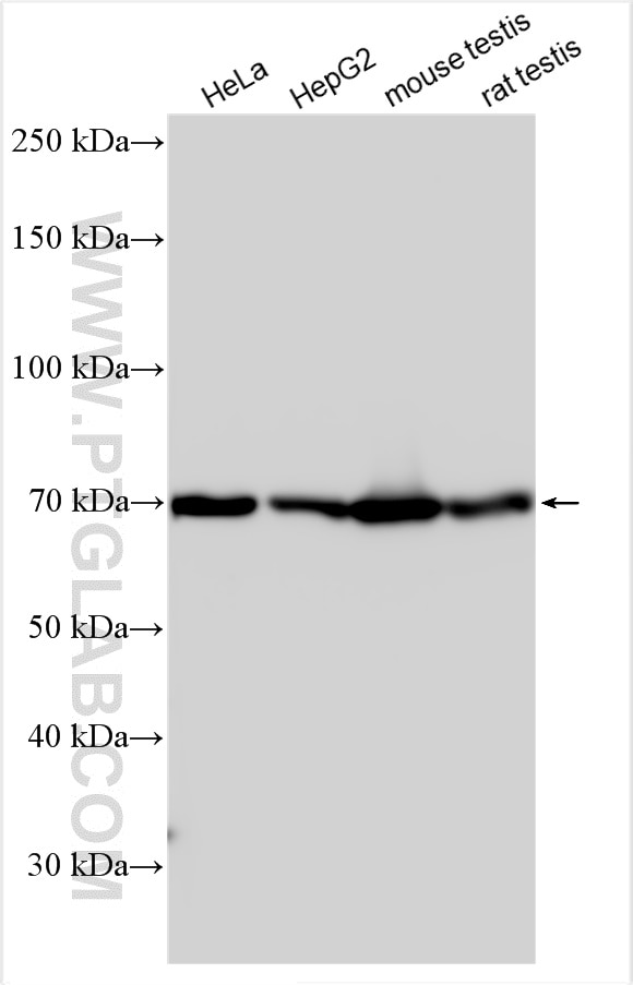
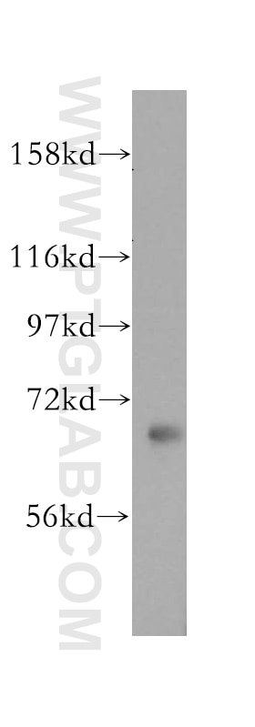
| Positive WB detected in | A431 cells, human placenta tissue, mouse kidney tissue, HeLa cells, Daudi cells, HepG2 cells, K-562 cells, U-87 MG cells, mouse testis tissue, rat testis tissue |
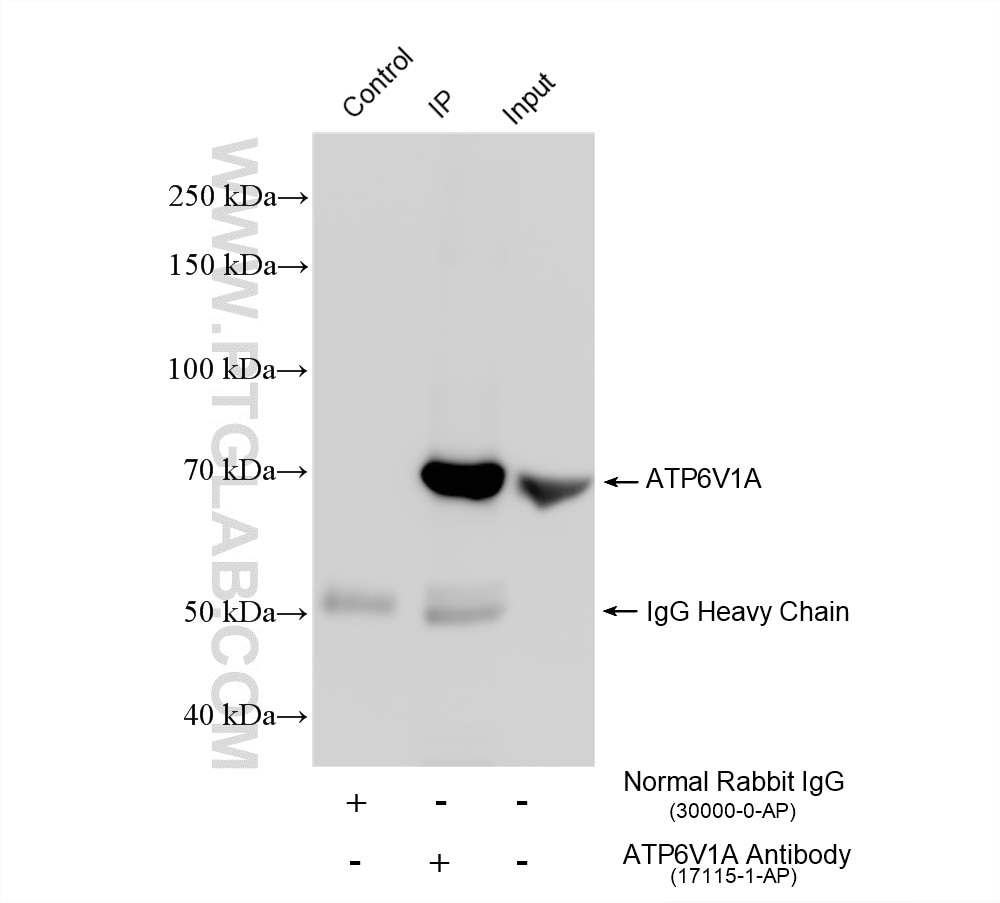
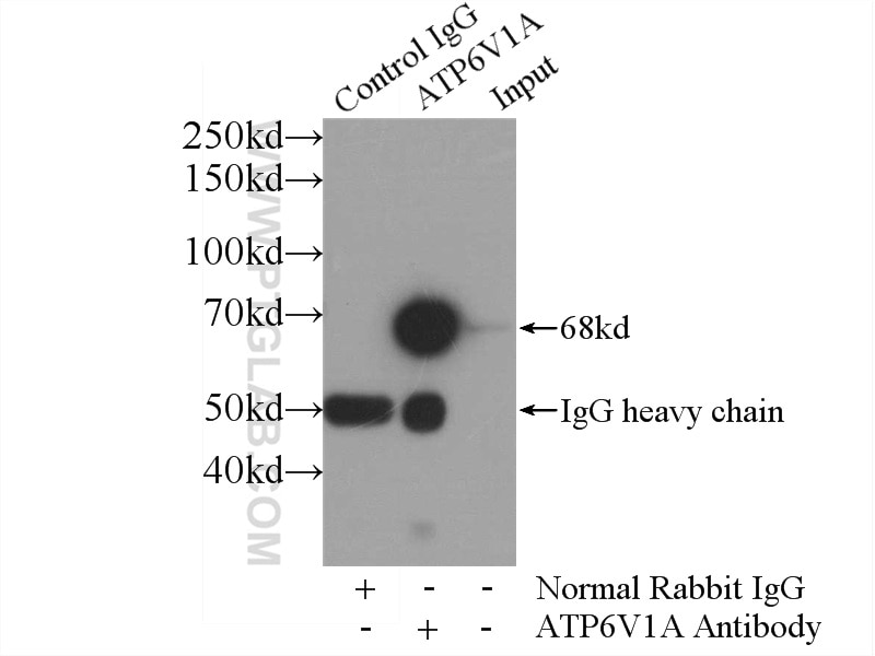
| Positive IP detected in | HeLa cells, RAW 264.7 cells |
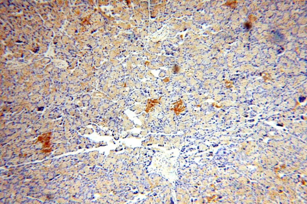
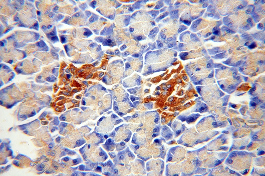
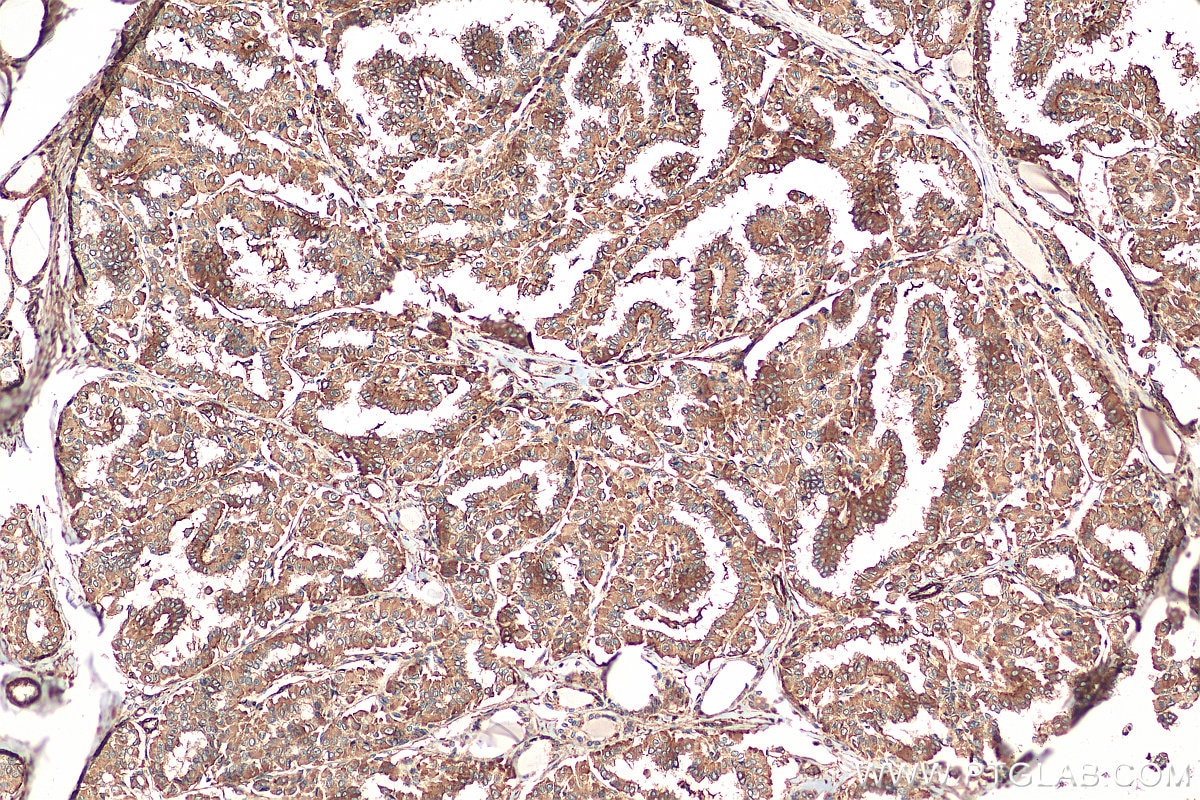
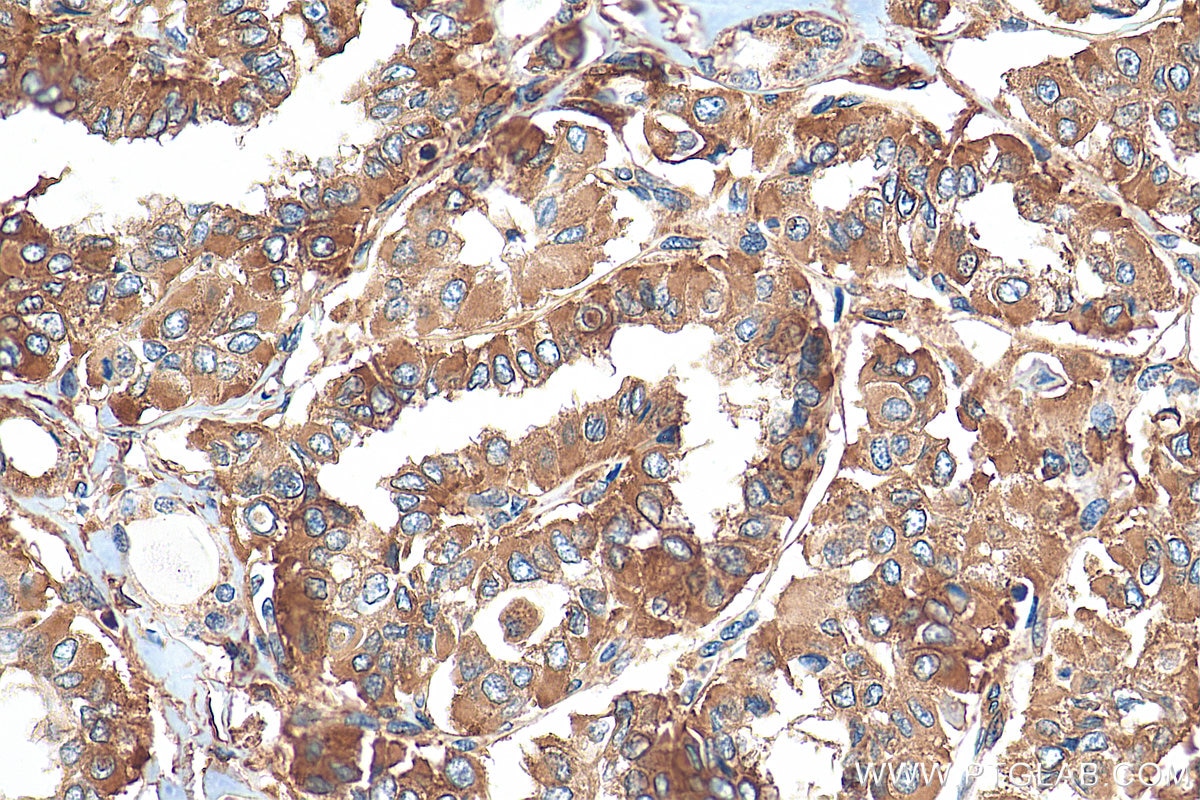
| Positive IHC detected in | human pancreas tissue, human thyroid cancer tissue Note: suggested antigen retrieval with TE buffer pH 9.0; (*) Alternatively, antigen retrieval may be performed with citrate buffer pH 6.0 |
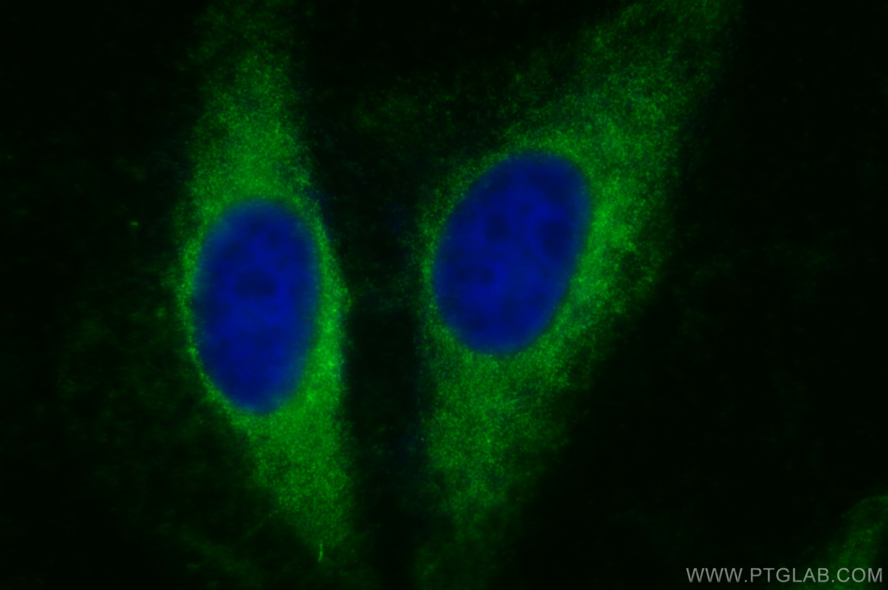
| Positive IF/ICC detected in | NIH/3T3 cells, HeLa cells |
Recommended dilution
| Application | Dilution |
|---|---|
| Western Blot (WB) | WB : 1:2000-1:16000 |
| Immunoprecipitation (IP) | IP : 0.5-4.0 ug for 1.0-3.0 mg of total protein lysate |
| Immunohistochemistry (IHC) | IHC : 1:20-1:200 |
| Immunofluorescence (IF)/ICC | IF/ICC : 1:200-1:800 |
| It is recommended that this reagent should be titrated in each testing system to obtain optimal results. | |
| Sample-dependent, Check data in validation data gallery. | |
Product Information
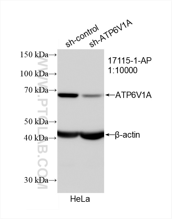
17115-1-AP targets ATP6V1A in WB, IHC, IF/ICC, IP, ELISA applications and shows reactivity with human, mouse, rat samples.
| Tested Reactivity | human, mouse, rat |
| Cited Reactivity | human, mouse, rat, pig, monkey, zebrafish, c.elegans |
| Host / Isotype | Rabbit / IgG |
| Class | Polyclonal |
| Type | Antibody |
| Immunogen |
CatNo: Ag10801 Product name: Recombinant human ATP6V1A protein Source: e coli.-derived, PGEX-4T Tag: GST Domain: 268-617 aa of BC013138 Sequence: NSDVIIYVGCGERGNEMSEVLRDFPELTMEVDGKVESIMKRTALVANTSNMPVAAREASIYTGITLSEYFRDMGYHVSMMADSTSRWAEALREISGRLAEMPADSGYPAYLGARLASFYERAGRVKCLGNPEREGSVSIVGAVSPPGGDFSDPVTSATLGIVQVFWGLDKKLAQRKHFPSVNWLISYSKYMRALDEYYDKHFTEFVPLRTKAKEILQEEEDLAEIVQLVGKASLAETDKITLEVAKLIKDDFLQQNGYTPYDRFCPFYKTVGMLSNMIAFYDMARRAVETTAQSDNKITWSIIREHMGDILYKLSSMKFKDPLKDGEAKIKSDYAQLLEDMQNAFRSLED Predict reactive species |
| Full Name | ATPase, H+ transporting, lysosomal 70kDa, V1 subunit A |
| Calculated Molecular Weight | 617 aa, 68 kDa |
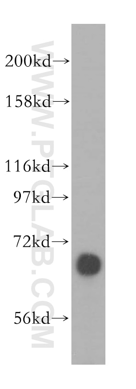
| Observed Molecular Weight | 68 kDa |
| GenBank Accession Number | BC013138 |
| Gene Symbol | ATP6V1A |
| Gene ID (NCBI) | 523 |
| RRID | AB_2290195 |
| Conjugate | Unconjugated |
| Form | Liquid |
| Purification Method | Antigen affinity purification |
| UNIPROT ID | P38606 |
| Storage Buffer | PBS with 0.02% sodium azide and 50% glycerol, pH 7.3. |
| Storage Conditions | Store at -20°C. Stable for one year after shipment. Aliquoting is unnecessary for -20oC storage. 20ul sizes contain 0.1% BSA. |
Background Information
The vacuolar-type H(+)-ATPase (V-ATPase) is responsible for the acidification of endosomes, lysosomes, and other intracellular organelles. It is also involved in hydrogen ion transport across the plasma membrane into the extracellular space. The V-ATPase is a multisubunit complex with cytosolic and transmembrane domains. The cytosolic catalytic domain consists of 3 A subunits and 3 B subunits, which bind and hydrolyze ATP, as well as regulatory accessory subunits.ATP6V1A is V-type proton ATPase catalytic subunit A.
Protocols
| Product Specific Protocols | |
|---|---|
| IF protocol for ATP6V1A antibody 17115-1-AP | Download protocol |
| IHC protocol for ATP6V1A antibody 17115-1-AP | Download protocol |
| IP protocol for ATP6V1A antibody 17115-1-AP | Download protocol |
| WB protocol for ATP6V1A antibody 17115-1-AP | Download protocol |
| Standard Protocols | |
|---|---|
| Click here to view our Standard Protocols |
Publications
What published studies show
The ATP6V1A Polyclonal antibody (Cat# 17115-1-AP) has accumulated 65 citations published in high-impact journals including Cell, Nature Immunology, Nature Cell Biology, Nature Microbiology, Autophagy, Cell Death & Differentiation, Cell Death & Disease, Journal of Clinical Investigation, EMBO Journal, and ACS Nano. Associated research fields encompass lysosomal biology and V-ATPase function, autophagy and lipophagy, multiple cancer types (hepatocellular, prostate, thyroid, breast), neurodegenerative diseases (Parkinson's, Alzheimer's), infectious diseases (SARS-CoV-2, tuberculosis, Zika), metabolic disorders (NAFLD, diabetes), cardiology, ferroptosis, and developmental biology in zebrafish and other model organisms.
| Species | Application | Title |
|---|---|---|
Nat Immunol Heme drives hemolysis-induced susceptibility to infection via disruption of phagocyte functions. | ||
Cell Mol Immunol Mycobacterium tuberculosis MEM39 (Rv1977) hijacks host aldolase A (ALDOA) to subvert immunometabolism to facilitate bacterial intracellular survival. | ||
Nat Cell Biol Membrane receptors cluster phosphatidylserine to activate LC3-associated phagocytosis | ||
Nat Microbiol The interferon-inducible isoform of NCOA7 inhibits endosome-mediated viral entry. | ||
Autophagy LAMP2-FLOT2 interaction enhances autophagosome-lysosome fusion to protect the septic heart in response to ILC2 |
Reviews
What customers say
The single review for this product is a 5-star rating for immunofluorescence. The user worked with MDA-MB-231 cells, fixing them in 2% PFA for 20 minutes and permeabilizing with 0.05% saponin plus 5% horse serum in PBS. The primary antibody was applied at a 1:100 dilution in saponin solution for one hour at room temperature, followed by an anti-rabbit AlexaFluor 488 secondary at 1:200 for one hour. The protocol was reported to work well, and an image was included to support the result.
The reviews below have been submitted by verified Proteintech customers who received an incentive for providing their feedback.
Paulina (Verified Customer) (03-26-2025) | Fixation: 2% PFA for 20min. Permeabilization/antibody dilution solution: 0.05% saponin, 5% horse serum in PBS. Primary antibody diluted 1:100 in saponin solution for 1hr at room temperature, followed by anti-rabbit conjugated to AlexaFluor 488 (1:200 in saponin solution) for 1 hr at room temperature.
 |














