

Tested Applications
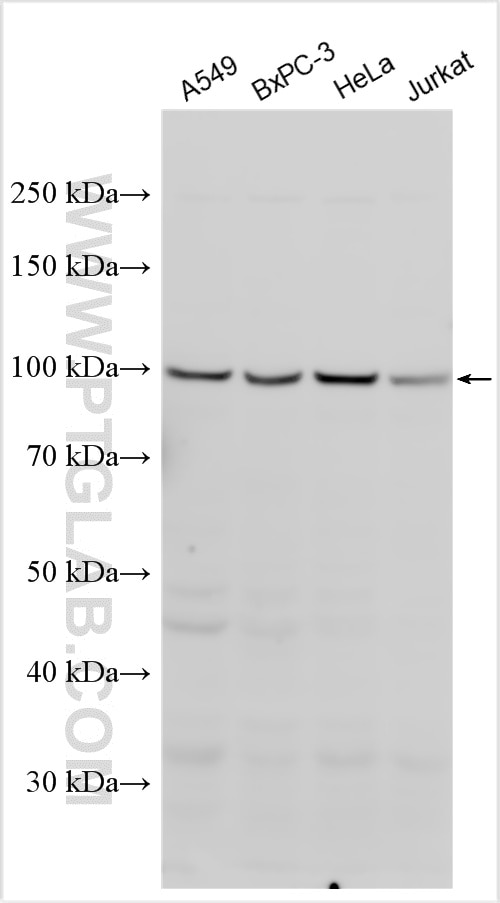
| Positive WB detected in | A549 cells, BxPC-3 cells, HeLa cells, Jurkat cells |
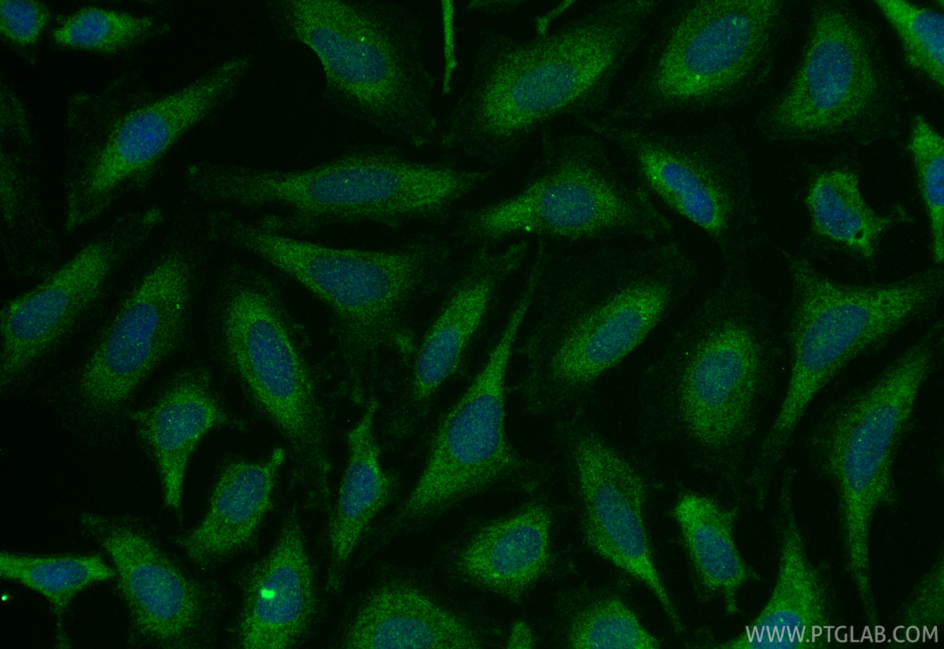
| Positive IF/ICC detected in | HeLa cells |
Recommended dilution
| Application | Dilution |
|---|---|
| Western Blot (WB) | WB : 1:1000-1:8000 |
| Immunofluorescence (IF)/ICC | IF/ICC : 1:200-1:800 |
| It is recommended that this reagent should be titrated in each testing system to obtain optimal results. | |
| Sample-dependent, Check data in validation data gallery. | |
Product Information
20540-1-AP targets AXIN2 in WB, IHC, IF/ICC, IP, ELISA applications and shows reactivity with human, mouse, rat samples.
| Tested Reactivity | human, mouse, rat |
| Cited Reactivity | human, mouse, rat |
| Host / Isotype | Rabbit / IgG |
| Class | Polyclonal |
| Type | Antibody |
| Immunogen |
CatNo: Ag14574 Product name: Recombinant human AXIN2 protein Source: e coli.-derived, PGEX-4T Tag: GST Domain: 30-83 aa of BC006295 Sequence: EGETPPCQPGVGKGQVTKPMPVSSNTRRNEDGLGEPEGRASPDSPLTRWTKSLH Predict reactive species |
| Full Name | axin 2 |
| Calculated Molecular Weight | 843 aa, 94 kDa |
| Observed Molecular Weight | 94-100 kDa |
| GenBank Accession Number | BC006295 |
| Gene Symbol | AXIN2 |
| Gene ID (NCBI) | 8313 |
| RRID | AB_10694569 |
| Conjugate | Unconjugated |
| Form | Liquid |
| Purification Method | Antigen affinity purification |
| UNIPROT ID | Q9Y2T1 |
| Storage Buffer | PBS with 0.02% sodium azide and 50% glycerol, pH 7.3. |
| Storage Conditions | Store at -20°C. Stable for one year after shipment. Aliquoting is unnecessary for -20oC storage. 20ul sizes contain 0.1% BSA. |
Background Information
Axis inhibition protein2 (AXIN2), also known as Coductin or Axil, is a multidomain scaffold protein that negatively regulates Wnt signaling. AXIN2 can directly interact with beta-catenin and GSK3B. AXIN2 has a number of phosphorylation sites and can also undergo poly(ADP-ribosylation)lation by tankyrase TNKS and TNKS2. Poly(ADP-ribosylated)lated AXIN2 then gets ubiquitinated by RNF146 and leads to its degradation and subsequent activation of Wnt signaling. AXIN2 is localized in the cytoplasm, and its mutation is involved in colorectal cancer. Catalgo#20540-1-AP is a rabbit polyclonal antibody raised against the 34-amino-acid N-terminus of human AXIN2. The observed MW of AXIN2 is from 93-100 kDa (PMID: 11809809; 24607787).
Protocols
| Product Specific Protocols | |
|---|---|
| IF protocol for AXIN2 antibody 20540-1-AP | Download protocol |
| WB protocol for AXIN2 antibody 20540-1-AP | Download protocol |
| Standard Protocols | |
|---|---|
| Click here to view our Standard Protocols |
Publications
What published studies show
The AXIN2 Polyclonal antibody (catalog no. 20540-1-AP) has 81 citations. These appear across journals including Signal Transduction and Targeted Therapy, Nature Communications, Nature Cell Biology, Molecular Cancer, Cell Research, Autophagy, Oncogene, Theranostics, Cell Death & Disease, Cancer Letters, EMBO Reports, and Science Reports. Research fields center on Wnt/β-catenin signaling, spanning cancer biology (pancreatic, colorectal, liver, gastric, lung, breast, ovarian, and bladder cancers), stem cell biology, autophagy, inflammation, and tissue regeneration. Applications are predominantly Western blot, with some IHC, IF, and IP.
| Species | Application | Title |
|---|---|---|
Signal Transduct Target Ther B-lymphoid tyrosine kinase-mediated FAM83A phosphorylation elevates pancreatic tumorigenesis through interacting with β-catenin | ||
Signal Transduct Target Ther Serine/threonine/tyrosine kinase 1 drives pancreatic carcinogenesis via GSK3β sequestration-mediated Wnt/β-catenin pathway hyperactivation. | ||
Cell Res Hierarchical activation of compartmentalized pools of AMPK depends on severity of nutrient or energy stress. | ||
Nat Cell Biol LncGata6 maintains stemness of intestinal stem cells and promotes intestinal tumorigenesis. | ||
Autophagy TSPAN1 promotes autophagy flux and mediates cooperation between WNT-CTNNB1 signaling and autophagy via the MIR454-FAM83A-TSPAN1 axis in pancreatic cancer. |
Reviews
What customers say
Both reviews are for Western Blot applications. One user reported a clean, specific band at 100 kDa in RKO cells at a 1:2000 dilution, rating the antibody 5 stars. Another user observed a band at 95 kDa in MCF7 cells at 1:1000 but noted significant nonspecific bands, giving 4 stars. Overall, the antibody detects the expected target size, though some users may need to optimize conditions to reduce background.
The reviews below have been submitted by verified Proteintech customers who received an incentive for providing their feedback.
Aurélie (Verified Customer) (10-06-2023) | This antibody gives a specific band at 100 kDa.
 |
Guannan (Verified Customer) (01-31-2019) | This antibody gives me a band at 95 kd, but also a lot of nonspecific bands.
|



