Tested Applications
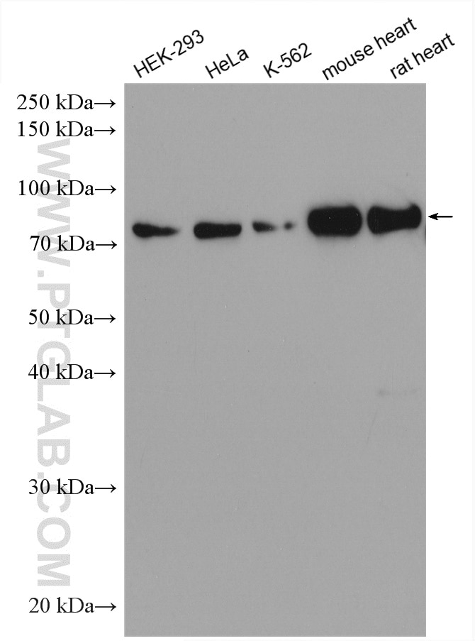
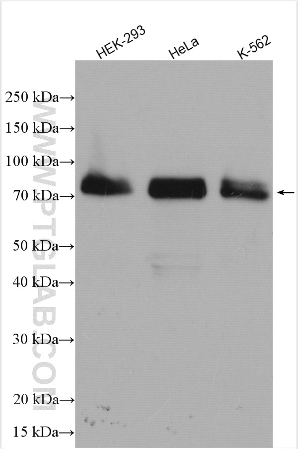
| Positive WB detected in | HEK-293 cells, HeLa cells, K-562 cells, mouse heart, rat heart |
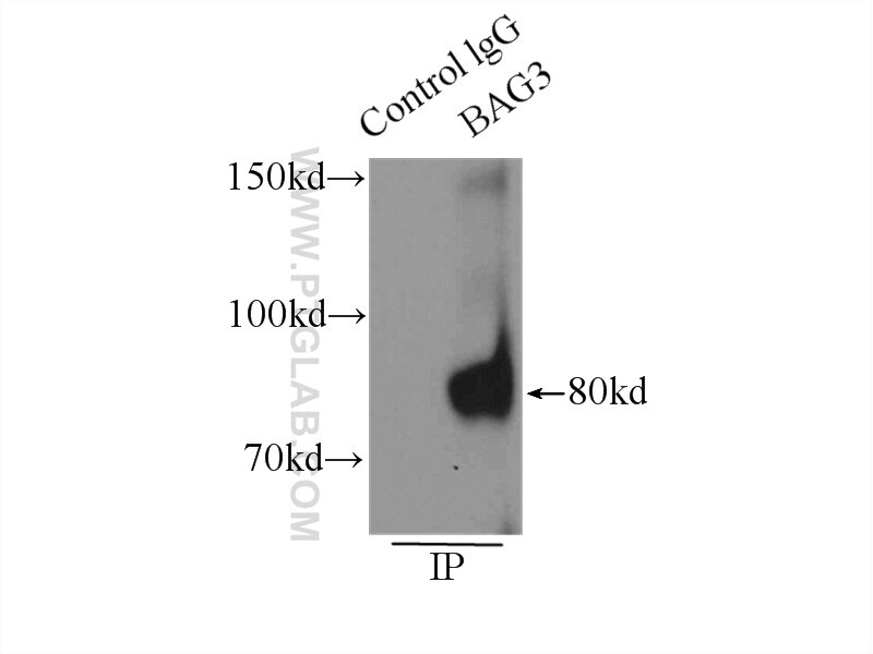
| Positive IP detected in | K-562 cells |
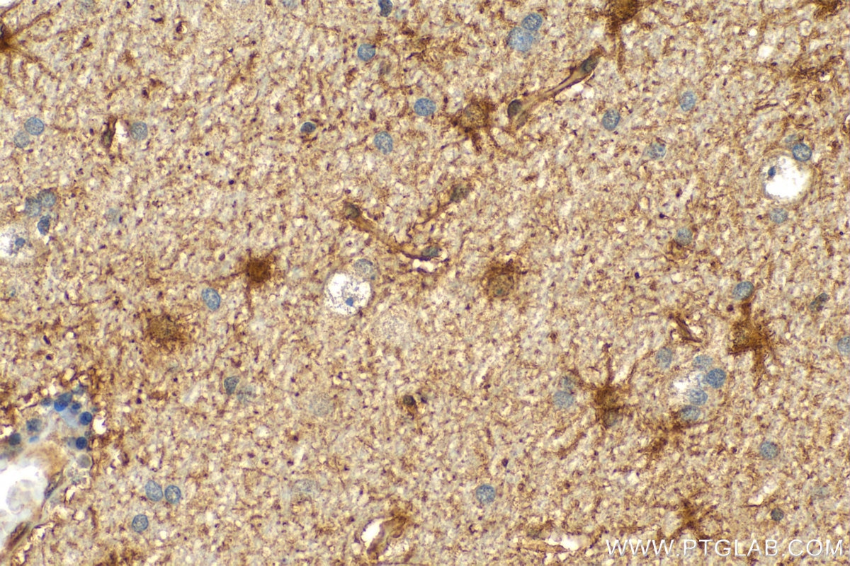
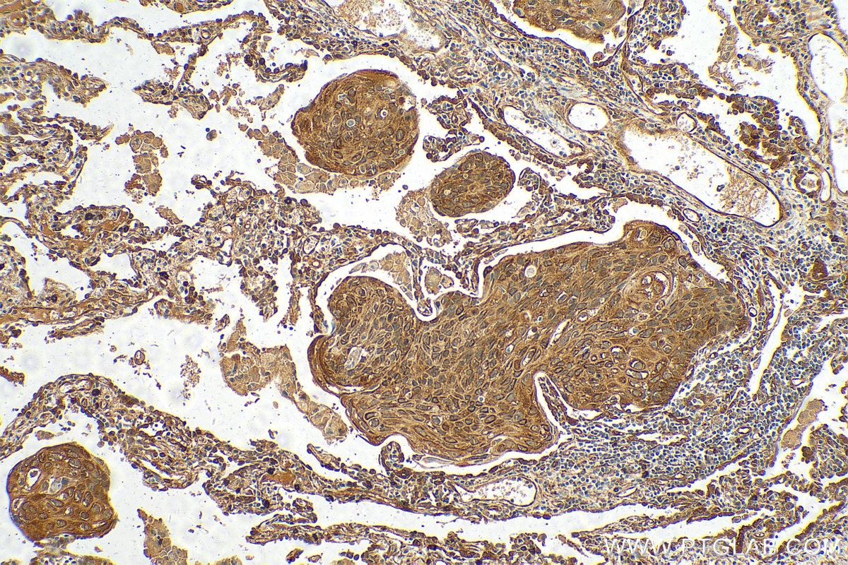
| Positive IHC detected in | human lung cancer tissue, human gliomas tissue Note: suggested antigen retrieval with TE buffer pH 9.0; (*) Alternatively, antigen retrieval may be performed with citrate buffer pH 6.0 |
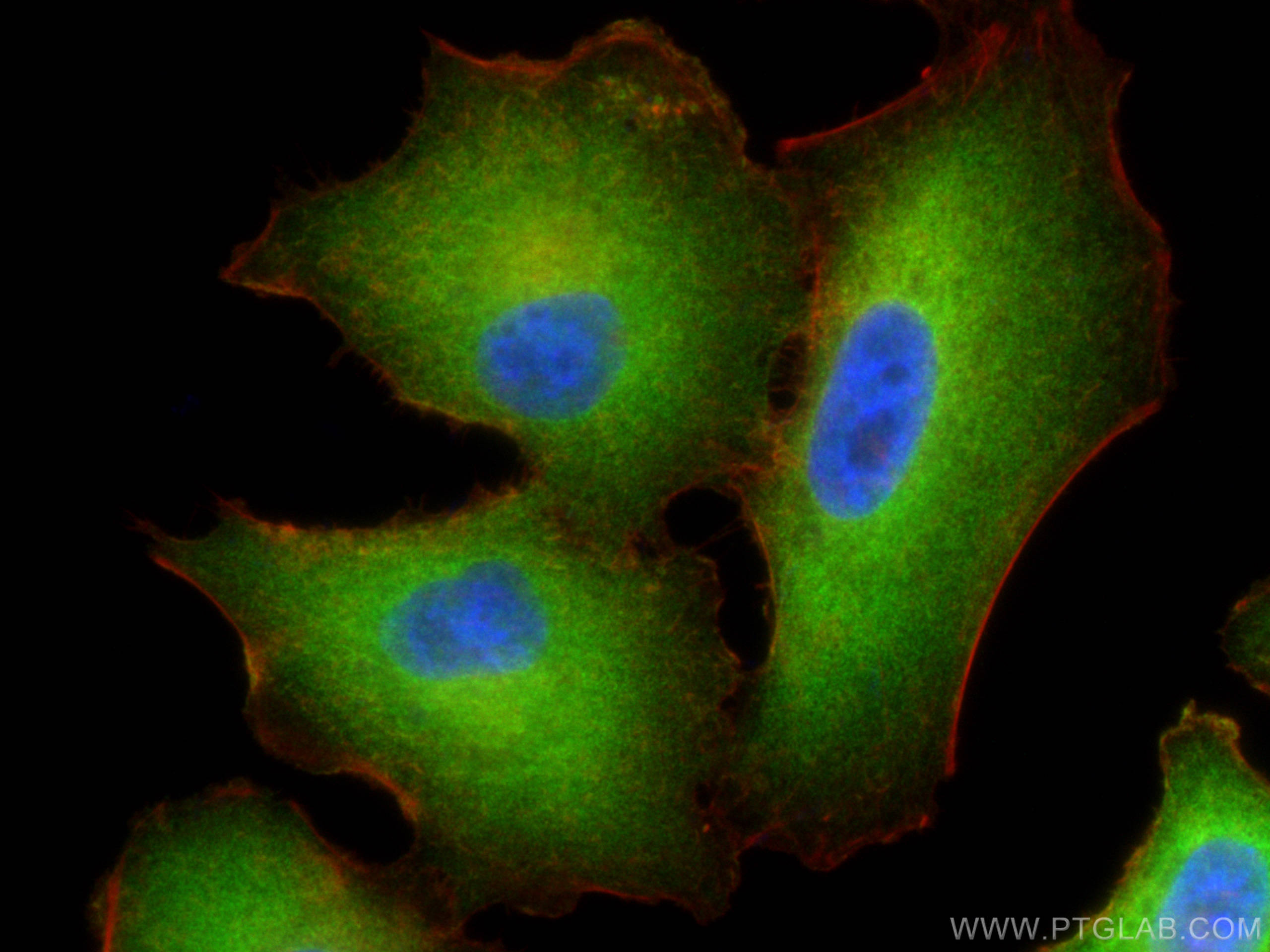
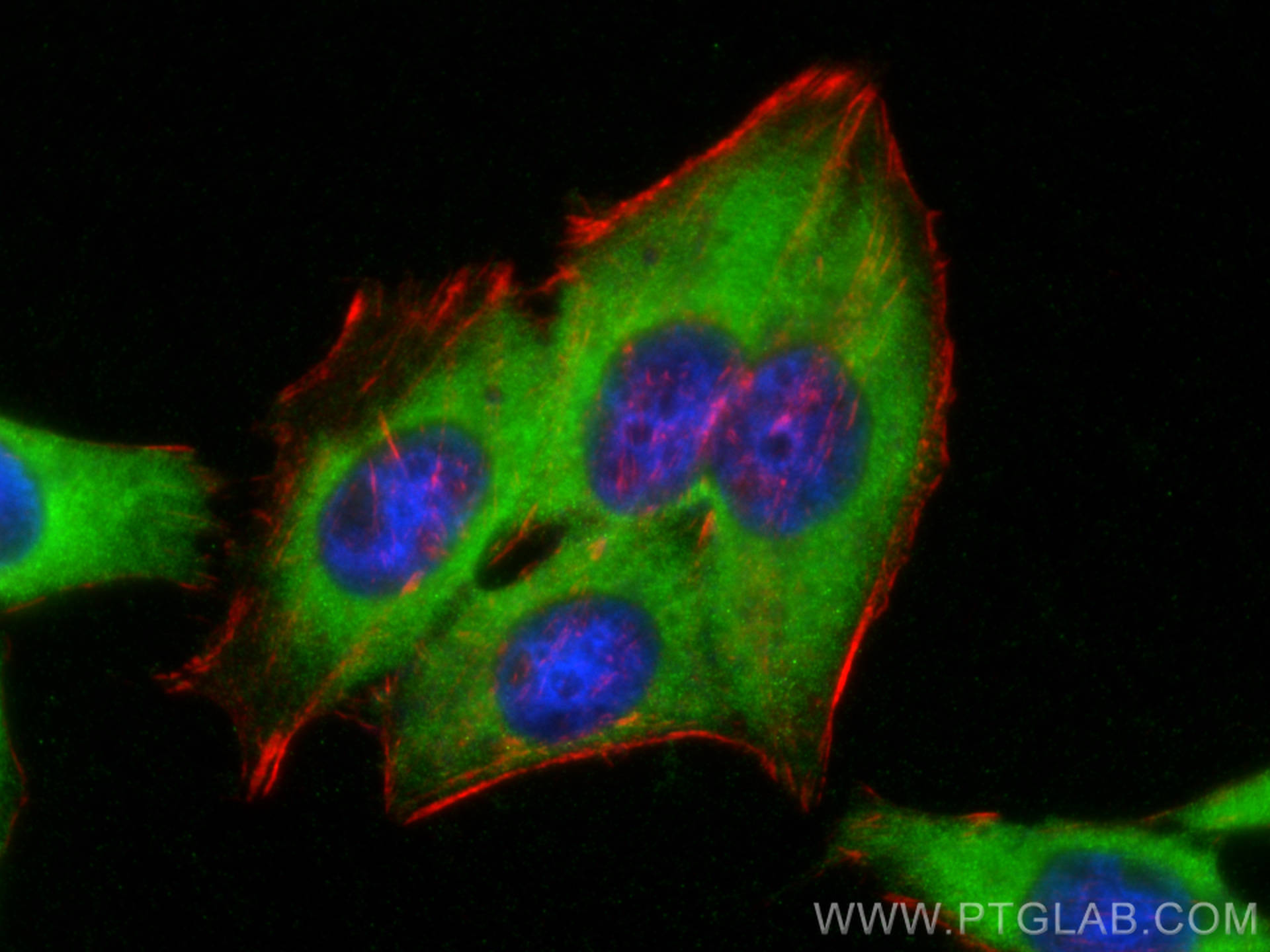
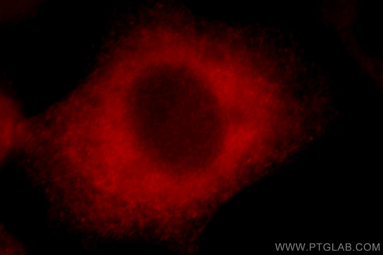
| Positive IF/ICC detected in | A549 cells, HeLa cells, HepG2 cells |
Recommended dilution
| Application | Dilution |
|---|---|
| Western Blot (WB) | WB : 1:30000-1:60000 |
| Immunoprecipitation (IP) | IP : 0.5-4.0 ug for 1.0-3.0 mg of total protein lysate |
| Immunohistochemistry (IHC) | IHC : 1:500-1:2000 |
| Immunofluorescence (IF)/ICC | IF/ICC : 1:50-1:500 |
| It is recommended that this reagent should be titrated in each testing system to obtain optimal results. | |
| Sample-dependent, Check data in validation data gallery. | |
Published Applications
| KD/KO | See 34 publications below |
| WB | See 125 publications below |
| IHC | See 7 publications below |
| IF | See 53 publications below |
| IP | See 8 publications below |
| CoIP | See 6 publications below |
Product Information
10599-1-AP targets BAG3 in WB, IHC, IF/ICC, IP, CoIP, ELISA applications and shows reactivity with human, mouse, rat samples.
| Tested Reactivity | human, mouse, rat |
| Cited Reactivity | human, mouse, rat, monkey, hamster |
| Host / Isotype | Rabbit / IgG |
| Class | Polyclonal |
| Type | Antibody |
| Immunogen |
CatNo: Ag0956 Product name: Recombinant human BAG3 protein Source: e coli.-derived, PGEX-4T Tag: GST Domain: 275-575 aa of BC006418 Sequence: SREGSPARSSTPLHSPSPIRVHTVVDRPQQPMTHRETAPVSQPENKPESKPGPVGPELPPGHIPIQVIRKEVDSKPVSQKPPPPSEKVEVKVPPAPVPCPPPSPGPSAVPSSPKSVATEERAAPSTAPAEATPPKPGEAEAPPKHPGVLKVEAILEKVQGLEQAVDNFEGKKTDKKYLMIEEYLTKELLALDSVDPEGRADVRQARRDGVRKVQTILEKLEQKAIDVPGQVQVYELQPSNLEADQPLQAIMEMGAVAADKGKKNAGNAEDPHTETQQPEATAAATSNPSSMTDTPGNPAAP Predict reactive species |
| Full Name | BCL2-associated athanogene 3 |
| Calculated Molecular Weight | 61 kDa |
| Observed Molecular Weight | 74-80 kDa |
| GenBank Accession Number | BC006418 |
| Gene Symbol | BAG3 |
| Gene ID (NCBI) | 9531 |
| RRID | AB_2062602 |
| Conjugate | Unconjugated |
| Form | Liquid |
| Purification Method | Antigen affinity purification |
| UNIPROT ID | O95817 |
| Storage Buffer | PBS with 0.02% sodium azide and 50% glycerol, pH 7.3. |
| Storage Conditions | Store at -20°C. Stable for one year after shipment. Aliquoting is unnecessary for -20oC storage. 20ul sizes contain 0.1% BSA. |
Background Information
BAG3 (Bcl2-associated athanogene 3) belongs to the BAG protein family, the co-chaperone that binds to Hsc70/Hsp70 through the BAG domain and modulates their activity in polypeptide folding. BAG3 contains also a WW domain and a proline-rich (PXXP) repeat, that mediate binding to partners different from Hsp70. Through interacting with different molecular partner, BAG3 influences several cell processes, such as apoptosis, autophagy and cell motility. BAG3 protein has been reported to sustain cell survival, resistance to therapy, and/or motility and metastatization in several tumor types, thus being identified as a potential target for anticancer therapies. In addition, defects in BAG3 are the cause of some myopathy. BAG3 normally migrates around 74-80 kDa; a slightly different molecular weight or a doublet form can be observed in some cell types and/or following cell exposure to stressors. A synaptosome associated form of 40 kDa has recently been described.
Protocols
| Product Specific Protocols | |
|---|---|
| IF protocol for BAG3 antibody 10599-1-AP | Download protocol |
| IHC protocol for BAG3 antibody 10599-1-AP | Download protocol |
| IP protocol for BAG3 antibody 10599-1-AP | Download protocol |
| WB protocol for BAG3 antibody 10599-1-AP | Download protocol |
| Standard Protocols | |
|---|---|
| Click here to view our Standard Protocols |
Publications
| Species | Application | Title |
|---|---|---|
Nat Genet Mutations affecting the cytoplasmic functions of the co-chaperone DNAJB6 cause limb-girdle muscular dystrophy. | ||
Nat Neurosci A tau homeostasis signature is linked with the cellular and regional vulnerability of excitatory neurons to tau pathology.
| ||
Acta Neuropathol Missense mutations in small muscle protein X-linked (SMPX) cause distal myopathy with protein inclusions. | ||
J Intern Med Transglutaminase type 2 plays a key role in the pathogenesis of Mycobacterium tuberculosis infection. | ||
Nat Commun Misfolded polypeptides are selectively recognized and transported toward aggresomes by a CED complex. |
Reviews
The reviews below have been submitted by verified Proteintech customers who received an incentive for providing their feedback.
FH Andrea (Verified Customer) (01-03-2023) | Strong, clear band on the membrane using a 1:500 ratio.
|
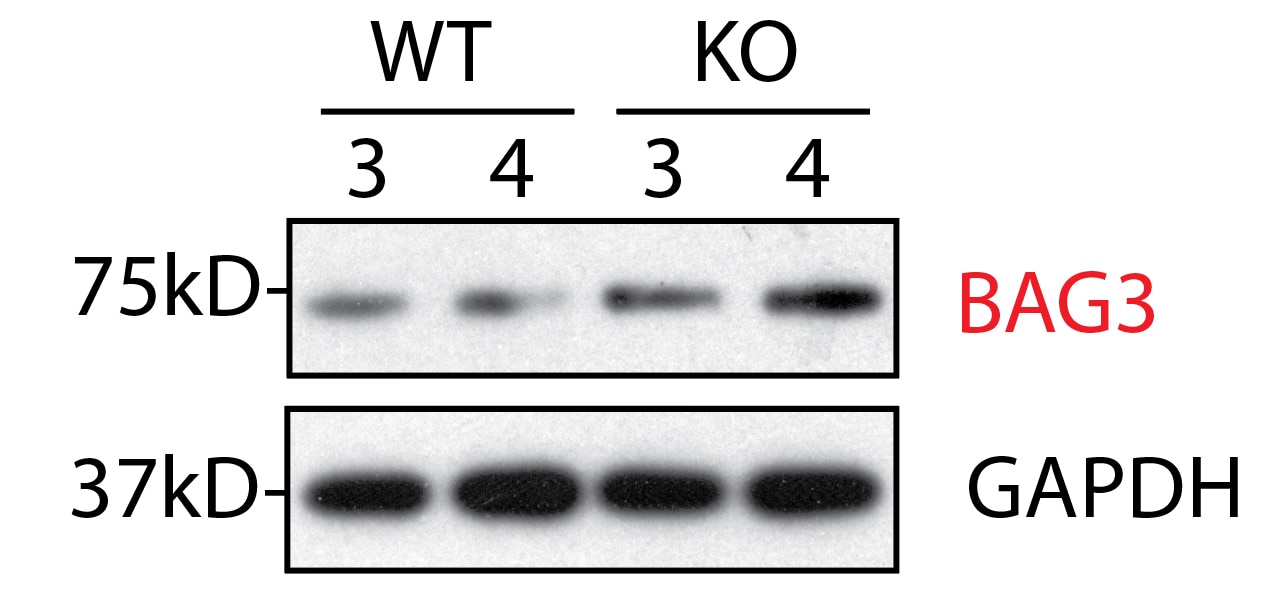
FH WEI (Verified Customer) (03-03-2022) | Very strong antibody, confirmed by KO tissue
|
FH Jane (Verified Customer) (02-03-2022) | One specific band at 80kDa in mouse heart tissue lysate
|
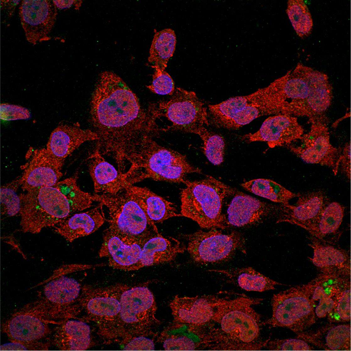
FH Eric (Verified Customer) (01-25-2021) | The red stain is 1:100 BAG3 under laser confocal microscopy in human microglia cells.
 |
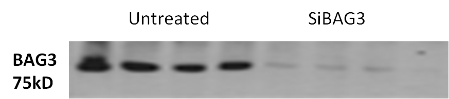
FH Tongbin (Verified Customer) (08-25-2020) | This bag3 antibody is very specific without any non-specific bands. It is the best Bag3 antibody out there for western blots.
 |
FH Afaque (Verified Customer) (10-17-2019) | I have used this antibody to study autophagy. It has worked very well for my Western blots. I also tried for IF and got very good results.
 |
FH SHUBHAM (Verified Customer) (02-25-2019) | excellent Antibody
 |
FH Praveen (Verified Customer) (12-03-2018) |
|