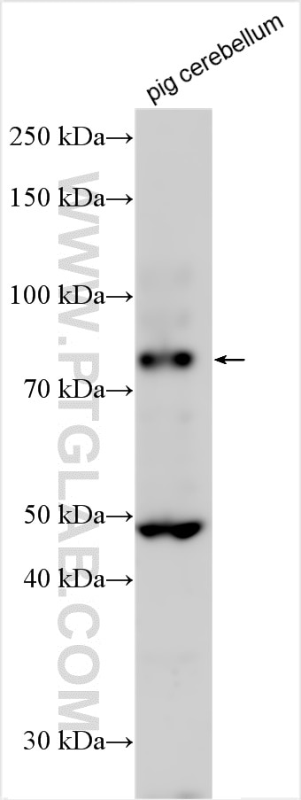
Various lysates were subjected to SDS PAGE followed by western blot with 31459-1-AP (DAB1 antibody) at dilution of 1:2000 incubated at room temperature for 1.5 hours.
Various lysates were subjected to SDS PAGE followed by western blot with 31459-1-AP (DAB1 antibody) at dilution of 1:2000 incubated at room temperature for 1.5 hours.
WB analysis using 31459-1-AP
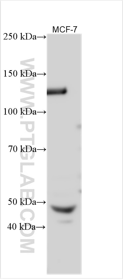
Various lysates were subjected to SDS PAGE followed by western blot with 31459-1-AP (DAB1 antibody) at dilution of 1:1000 incubated at room temperature for 1.5 hours.
Various lysates were subjected to SDS PAGE followed by western blot with 31459-1-AP (DAB1 antibody) at dilution of 1:1000 incubated at room temperature for 1.5 hours.
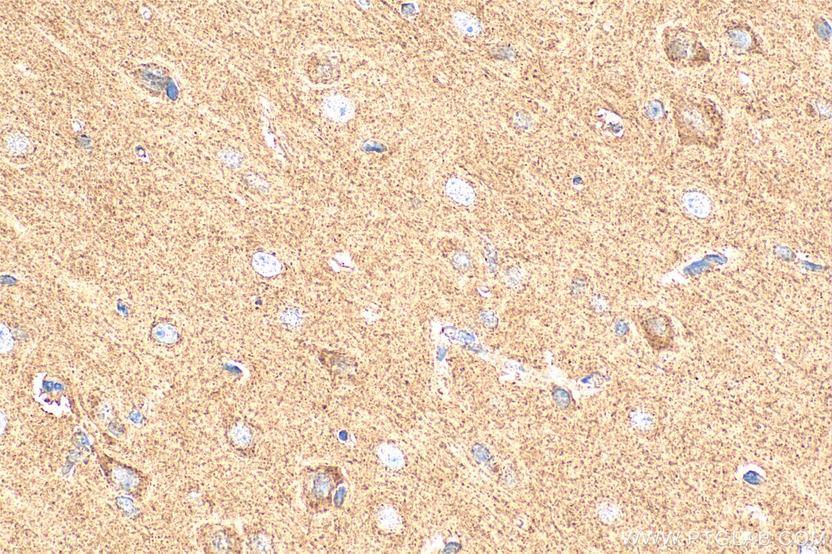
IHC staining of rat brain using 31459-1-AP
Immunohistochemical analysis of paraffin-embedded rat brain tissue slide using 31459-1-AP (DAB1 antibody) at dilution of 1:200 (under 40x lens). Heat mediated antigen retrieval with Tris-EDTA buffer (pH 9.0).
Immunohistochemical analysis of paraffin-embedded rat brain tissue slide using 31459-1-AP (DAB1 antibody) at dilution of 1:200 (under 40x lens). Heat mediated antigen retrieval with Tris-EDTA buffer (pH 9.0).
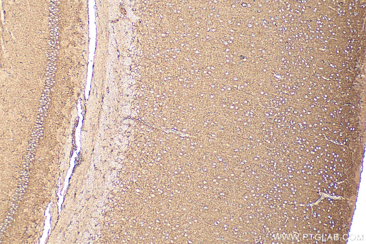
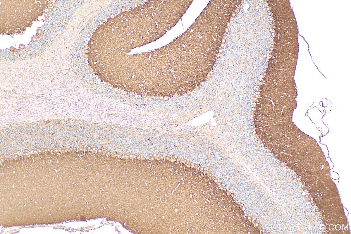
IHC staining of mouse brain using 31459-1-AP
Immunohistochemical analysis of paraffin-embedded mouse brain tissue slide using 31459-1-AP (DAB1 antibody) at dilution of 1:200 (under 10x lens). Heat mediated antigen retrieval with Tris-EDTA buffer (pH 9.0).
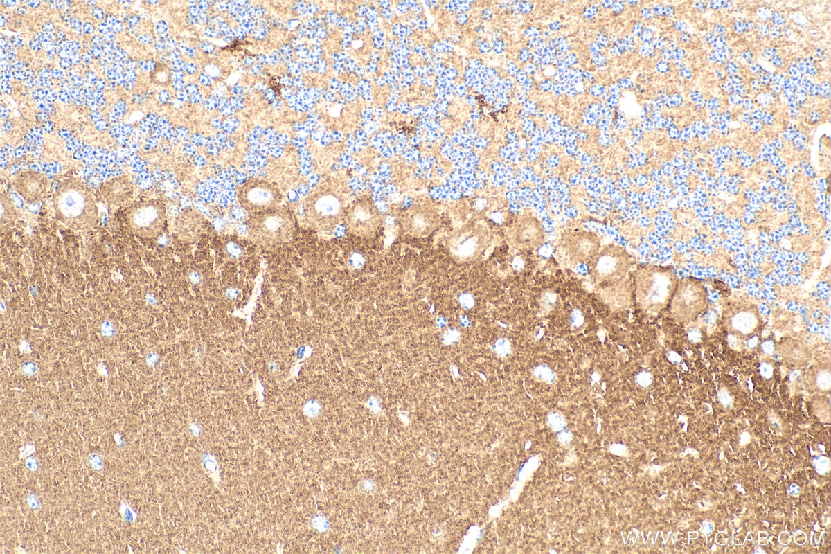
Immunohistochemical analysis of paraffin-embedded mouse cerebellum tissue slide using 31459-1-AP (DAB1 antibody) at dilution of 1:200 (under 40x lens). Heat mediated antigen retrieval with Tris-EDTA buffer (pH 9.0).
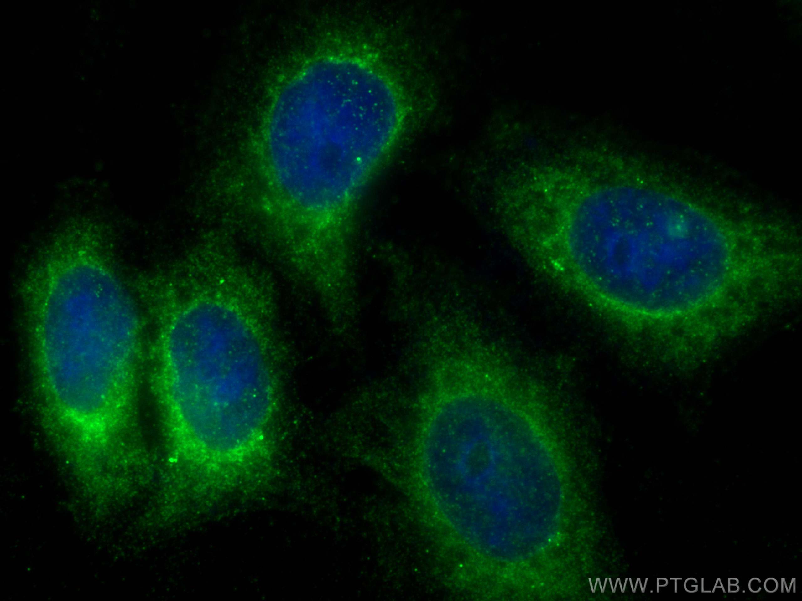
IF Staining of HepG2 using 31459-1-AP
Immunofluorescent analysis of (-20°C Ethanol) fixed HepG2 cells using DAB1 antibody (31459-1-AP) at dilution of 1:400 and CoraLite®488-Conjugated AffiniPure Goat Anti-Rabbit IgG(H+L) (SA00013-2).
Immunofluorescent analysis of (-20°C Ethanol) fixed HepG2 cells using DAB1 antibody (31459-1-AP) at dilution of 1:400 and CoraLite®488-Conjugated AffiniPure Goat Anti-Rabbit IgG(H+L) (SA00013-2).
The Proteintech guarantee covers Proteintech antibodies in any species and any application, including those not listed on the datasheet. If the antibody doesn’t perform, you can receive a hassle-free refund or credit note.
mouse cerebellum tissue, mouse brain tissue, rat brain tissue Note: suggested antigen retrieval with TE buffer pH 9.0; (*) Alternatively, antigen retrieval may be performed with citrate buffer pH 6.0
Positive IF/ICC detected in
HepG2 cells
Recommended dilution
Application
Dilution
Western Blot (WB)
WB : 1:1000-1:4000
Immunohistochemistry (IHC)
IHC : 1:50-1:500
Immunofluorescence (IF)/ICC
IF/ICC : 1:200-1:800
It is recommended that this reagent should be titrated in each testing system to obtain optimal results.
Sample-dependent, Check data in validation data gallery.
Product Information
31459-1-AP targets DAB1 in WB, IHC, IF/ICC, ELISA applications and shows reactivity with human, mouse, rat, pig samples.
PBS with 0.02% sodium azide and 50% glycerol, pH 7.3.
Storage Conditions
Store at -20°C. Stable for one year after shipment. Aliquoting is unnecessary for -20oC storage. 20ul sizes contain 0.1% BSA.
Background Information
Disabled-1 (Dab1) is an adaptor protein that is essential for neuronal migration and maturation in response to the extracellular protein Reelin. DAB1 protein docks to the intracellular part of the Reelin very low density lipoprotein receptor and apoE receptor type 2 and becomes tyrosine-phosphorylated following binding of Reelin to cortical neurons. The DAB1 protein contains a 180-amino acid N-terminal protein interaction/phosphotyrosine-binding (PTB) 1 domain that docks to the short cytoplasmic tail of the very low density lipoprotein receptor or apoE receptor type 2 at the level of NPXY motifs, with a preference for unphosphorylated motifs. Dab1 polypeptide bands ranging from 36 to 120 kDa have been identified in mouse embryonic brain, the 80-kDa Dab1 being the predominant form (PMID: 24313315).
Various lysates were subjected to SDS PAGE followed by western blot with 31459-1-AP (DAB1 antibody) at dilution of 1:2000 incubated at room temperature for 1.5 hours.
WB analysis using 31459-1-AP
Various lysates were subjected to SDS PAGE followed by western blot with 31459-1-AP (DAB1 antibody) at dilution of 1:1000 incubated at room temperature for 1.5 hours.
IHC Figures
IHC staining of rat brain using 31459-1-AP
Immunohistochemical analysis of paraffin-embedded rat brain tissue slide using 31459-1-AP (DAB1 antibody) at dilution of 1:200 (under 40x lens). Heat mediated antigen retrieval with Tris-EDTA buffer (pH 9.0).
IHC staining of mouse brain using 31459-1-AP
Immunohistochemical analysis of paraffin-embedded mouse brain tissue slide using 31459-1-AP (DAB1 antibody) at dilution of 1:200 (under 10x lens). Heat mediated antigen retrieval with Tris-EDTA buffer (pH 9.0).
IHC staining of mouse cerebellum using 31459-1-AP
Immunohistochemical analysis of paraffin-embedded mouse cerebellum tissue slide using 31459-1-AP (DAB1 antibody) at dilution of 1:200 (under 10x lens). Heat mediated antigen retrieval with Tris-EDTA buffer (pH 9.0).
IHC staining of mouse cerebellum using 31459-1-AP
Immunohistochemical analysis of paraffin-embedded mouse cerebellum tissue slide using 31459-1-AP (DAB1 antibody) at dilution of 1:200 (under 40x lens). Heat mediated antigen retrieval with Tris-EDTA buffer (pH 9.0).
IF/ICC Figures
IF Staining of HepG2 using 31459-1-AP
Immunofluorescent analysis of (-20°C Ethanol) fixed HepG2 cells using DAB1 antibody (31459-1-AP) at dilution of 1:400 and CoraLite®488-Conjugated AffiniPure Goat Anti-Rabbit IgG(H+L) (SA00013-2).
The species listed in Tested Reactivity are in-house verified and applicable species. For unlisted species, please refer to the homology analysis of the immunogen sequence and related species. For rabbit polyclonal antibodies, homology >70% is recommended. For mouse monoclonal antibodies and rabbit recombinant antibodies, homology >90% is recommended. Generally, the higher the homology, the greater the applicability. However, there will be certain differences in protein expression in different species, tissues or cells. Therefore, the homology analysis results are for reference only and do not serve as a guarantee.
At Proteintech, we pride ourselves on our antibody quality, customer service and transparency. As such, we are comparing our antibodies with other vendors, enabling easy identification and comparisons of key data to help you choose the suitable antibody for your needs.
We have selected the top cited antibodies from these vendors for you to compare.
 at dilution of 1:2000 incubated at room temperature for 1.5 hours.")
 at dilution of 1:1000 incubated at room temperature for 1.5 hours.")
 at dilution of 1:200 (under 40x lens). Heat mediated antigen retrieval with Tris-EDTA buffer (pH 9.0).")
 at dilution of 1:200 (under 10x lens). Heat mediated antigen retrieval with Tris-EDTA buffer (pH 9.0).")
 at dilution of 1:200 (under 10x lens). Heat mediated antigen retrieval with Tris-EDTA buffer (pH 9.0).")
 at dilution of 1:200 (under 40x lens). Heat mediated antigen retrieval with Tris-EDTA buffer (pH 9.0).")
 fixed HepG2 cells using DAB1 antibody (31459-1-AP) at dilution of 1:400 and CoraLite®488-Conjugated AffiniPure Goat Anti-Rabbit IgG(H+L) (SA00013-2).")