Tested Applications
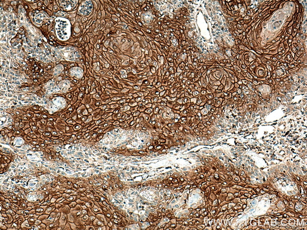
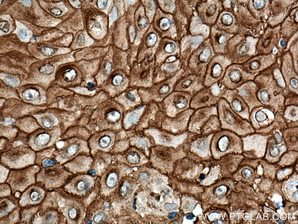
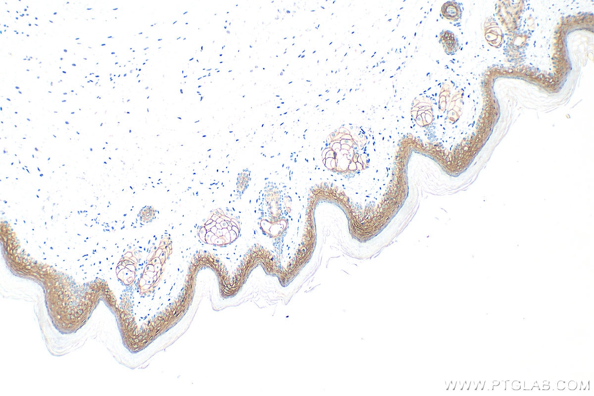
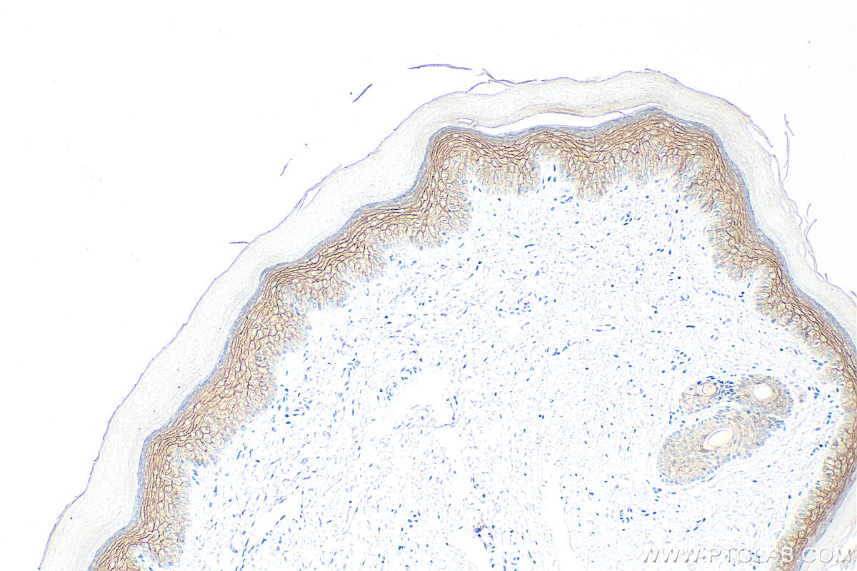
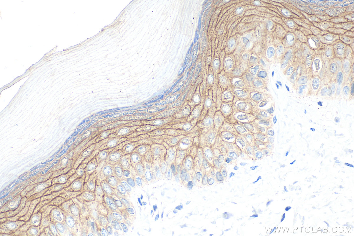
| Positive IHC detected in | mouse skin tissue, human skin cancer tissue, rat skin tissue Note: suggested antigen retrieval with TE buffer pH 9.0; (*) Alternatively, antigen retrieval may be performed with citrate buffer pH 6.0 |
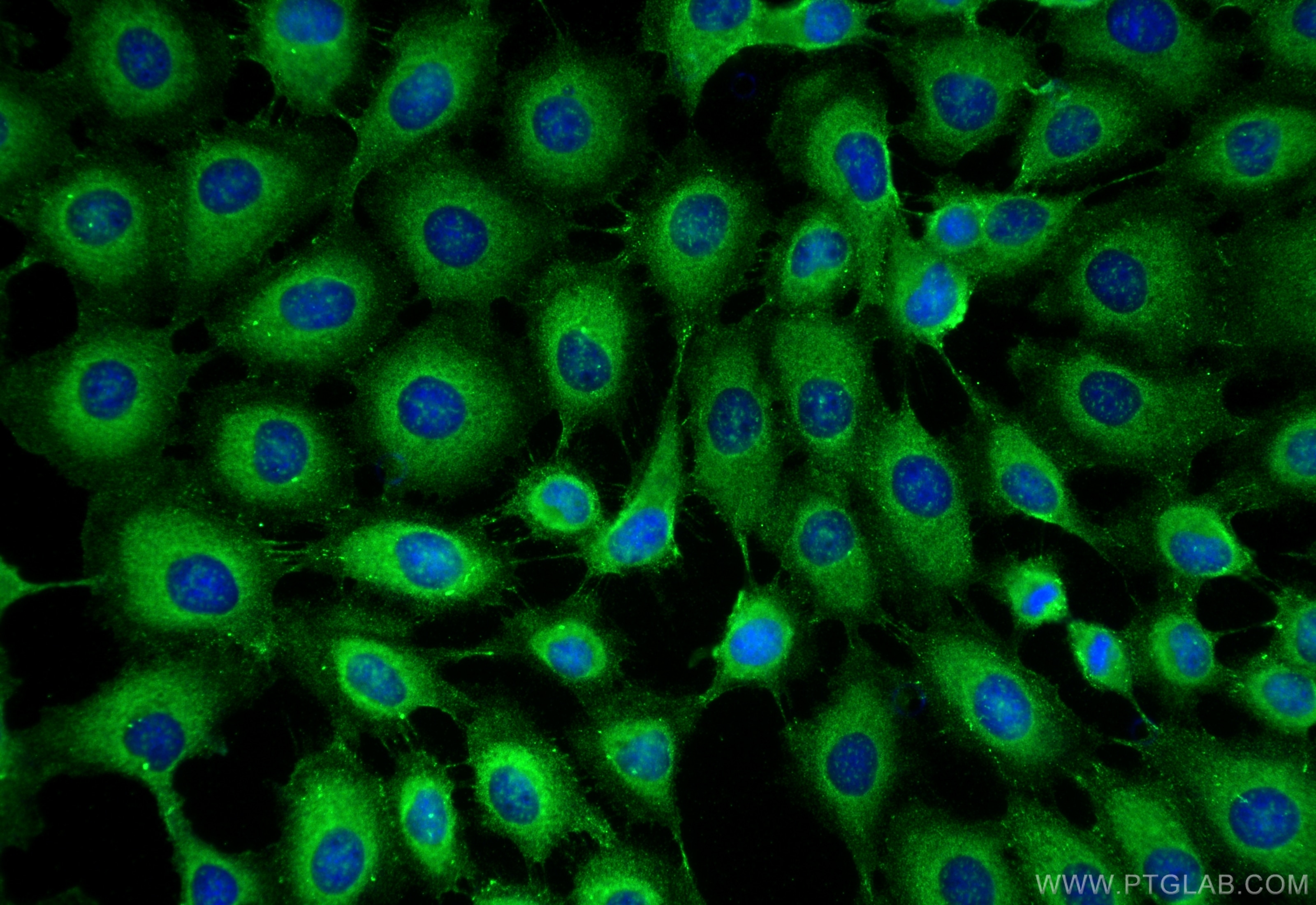
| Positive IF/ICC detected in | A431 cells |
Recommended dilution
| Application | Dilution |
|---|---|
| Immunohistochemistry (IHC) | IHC : 1:1000-1:4000 |
| Immunofluorescence (IF)/ICC | IF/ICC : 1:500-1:2000 |
| It is recommended that this reagent should be titrated in each testing system to obtain optimal results. | |
| Sample-dependent, Check data in validation data gallery. | |
Published Applications
| WB | See 4 publications below |
| IHC | See 2 publications below |
| IF | See 11 publications below |
Product Information
24587-1-AP targets DSG1 in WB, IHC, IF/ICC, ELISA applications and shows reactivity with human, mouse, rat samples.
| Tested Reactivity | human, mouse, rat |
| Cited Reactivity | human, mouse, rat |
| Host / Isotype | Rabbit / IgG |
| Class | Polyclonal |
| Type | Antibody |
| Immunogen |
CatNo: Ag20184 Product name: Recombinant human DSG1 protein Source: e coli.-derived, PGEX-4T Tag: GST Domain: 756-1049 aa of BC153001 Sequence: KKLADISLGKESYPDLDPSWPPQSTEPVCLPQETEPVVSGHPPISPHFGTTTVISESTYPSGPGVLHPKPILDPLGYGNVTVTESYTTSDTLKPSVHVHDNRPASNVVVTERVVGPISGADLHGMLEMPDLRDGSNVIVTERVIAPSSSLPTSLTIHHPRESSNVVVTERVIQPTSGMIGSLSMHPELANAHNVIVTERVVSGAGVTGISGTTGISGGIGSSGLVGTSMGAGSGALSGAGISGGGIGLSSLGGTASIGHMRSSSDHHFNQTIGSASPSTARSRITKYSTVQYSK Predict reactive species |
| Full Name | desmoglein 1 |
| Calculated Molecular Weight | 1049 aa, 114 kDa |
| GenBank Accession Number | BC153001 |
| Gene Symbol | DSG1 |
| Gene ID (NCBI) | 1828 |
| RRID | AB_2879624 |
| Conjugate | Unconjugated |
| Form | Liquid |
| Purification Method | Antigen affinity purification |
| UNIPROT ID | Q02413 |
| Storage Buffer | PBS with 0.02% sodium azide and 50% glycerol, pH 7.3. |
| Storage Conditions | Store at -20°C. Stable for one year after shipment. Aliquoting is unnecessary for -20oC storage. 20ul sizes contain 0.1% BSA. |
Background Information
Desmosomes are cell-cell junctions between epithelial, myocardial, and certain other cell types. Desmosomal cadherins, consisting of four desmogleins (DSG1-4) and three desmocollins (DSC1-3) in humans, mediate adhesion through calcium-dependent homophilic/heterophilic interactions. DSG1 is a single-pass transmembrane glycoprotein highly expressed in the epidermis and localized primarily within the suprabasal epithelial layers (PMID: 16286477; 24220297). DSG1 mediates intercellular adhesion and is crucial in maintaining epidermal integrity and barrier function (PMID: 23974871). It is also involved in epithelial cell differentiation (PMID: 23524961). Mutations in the DSG1 gene can cause the autosomal dominant disorder to striate palmoplantar keratoderma and a syndrome featuring severe dermatitis, multiple allergies, and metabolic wasting (SAM syndrome) (PMID: 29315490; 23974871).
Protocols
| Product Specific Protocols | |
|---|---|
| IF protocol for DSG1 antibody 24587-1-AP | Download protocol |
| IHC protocol for DSG1 antibody 24587-1-AP | Download protocol |
| Standard Protocols | |
|---|---|
| Click here to view our Standard Protocols |
Publications
| Species | Application | Title |
|---|---|---|
J Nanobiotechnology ZNPs reduce epidermal mechanical strain resistance by promoting desmosomal cadherin endocytosis via mTORC1-TFEB-BLOC1S3 axis | ||
EMBO Rep O-glycan initiation directs distinct biological pathways and controls epithelial differentiation. | ||
Stem Cell Res Ther Both Wnt signaling and epidermal stem cell-derived extracellular vesicles are involved in epidermal cell growth. | ||
J Cell Sci Novel stress granules-like structures are induced via a paracrine mechanism during viral infection. | ||
In Vitro Cell Dev Biol Anim S100A11 is involved in the progression of colorectal cancer through the desmosome-catenin-TCF signaling pathway | ||
Microscopy (Oxf) Inhibition of retinoid X receptor improved the morphology, localization of desmosomal proteins and paracellular permeability in three-dimensional cultures of mouse keratinocytes. |