Tested Applications
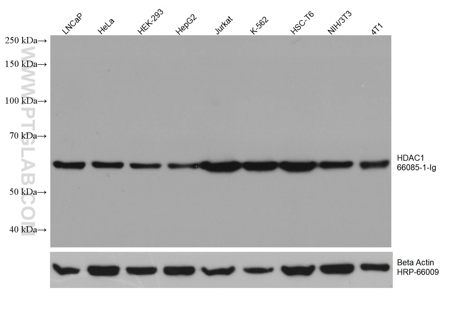
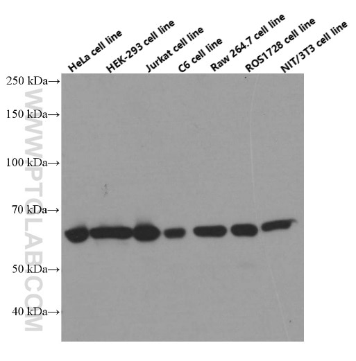
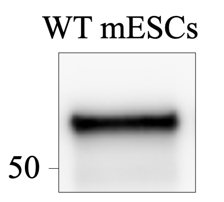
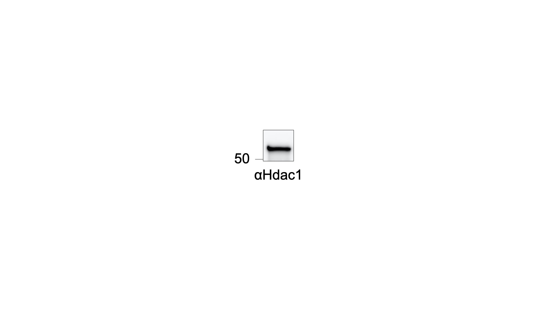
| Positive WB detected in | LNCaP cells, HeLa cells, HEK-293 cells, HepG2 cells, Jurkat cells, K-562 cells, HSC-T6 cells, NIH/3T3 cells, 4T1 cells |
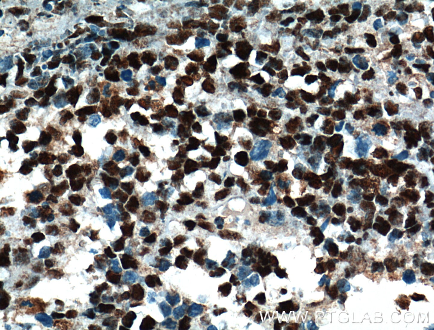
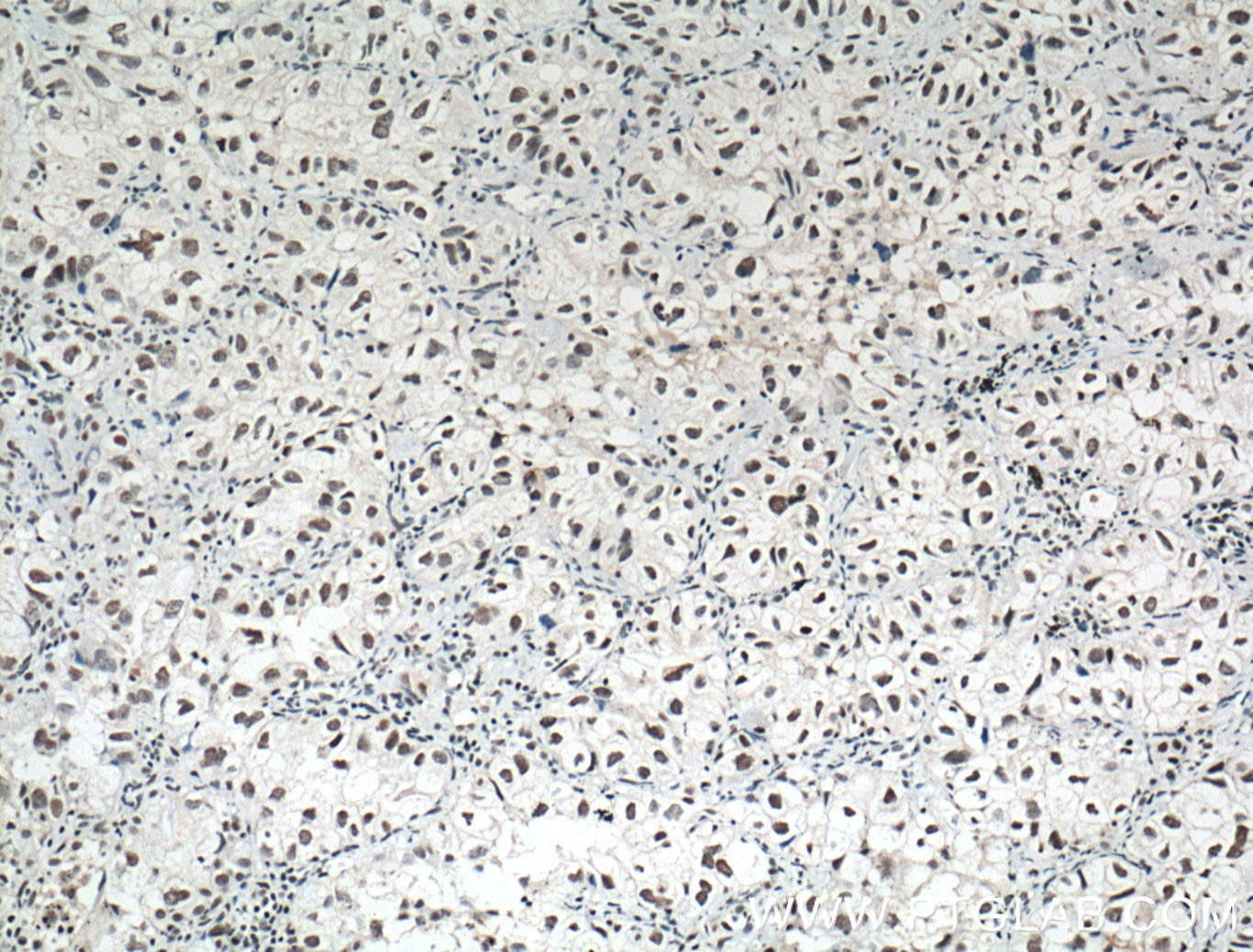
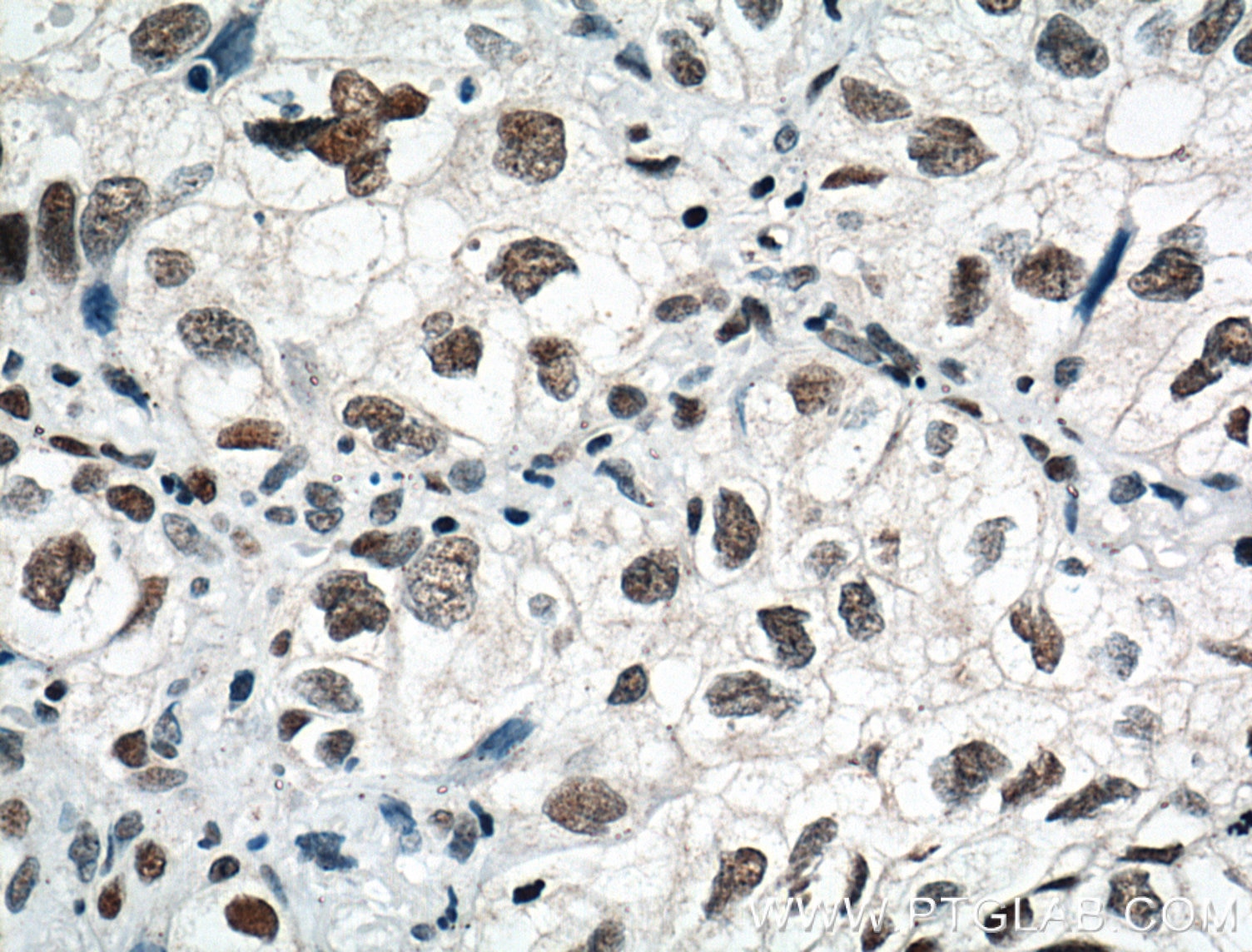
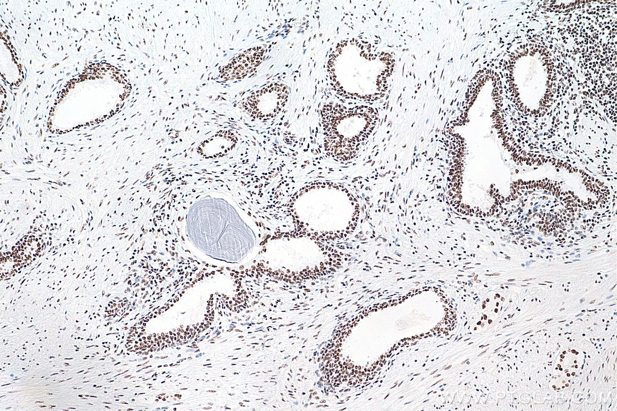
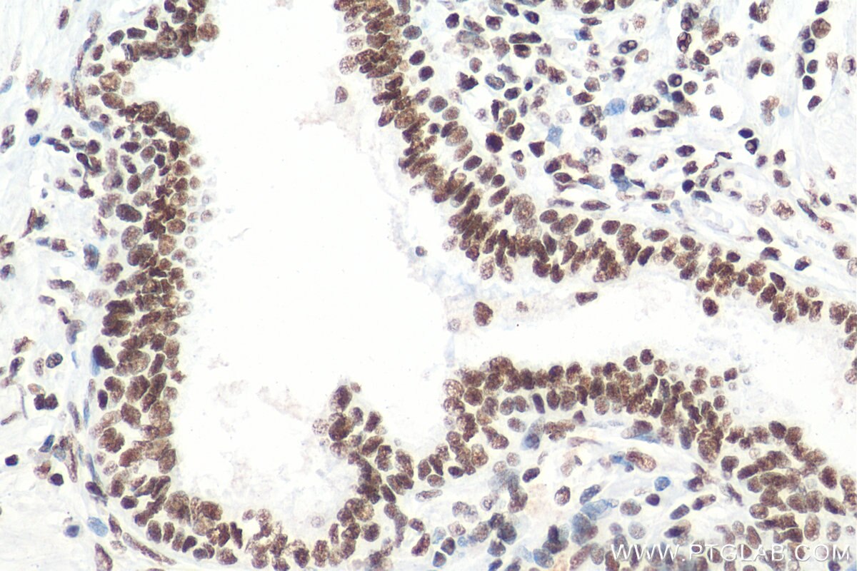
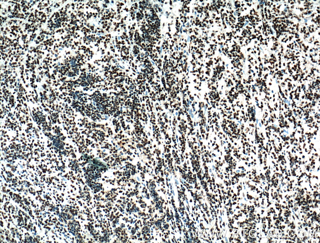
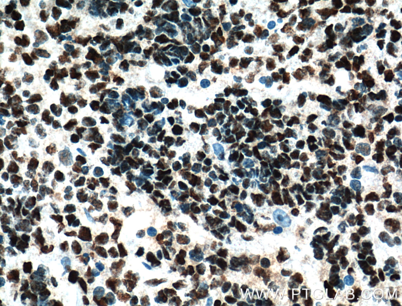
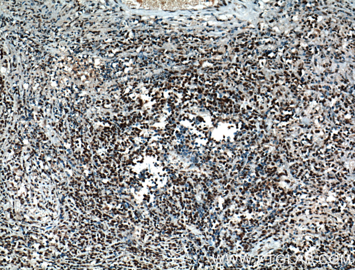
| Positive IHC detected in | human lymphoma tissue, human lung cancer tissue, human prostate cancer tissue Note: suggested antigen retrieval with TE buffer pH 9.0; (*) Alternatively, antigen retrieval may be performed with citrate buffer pH 6.0 |
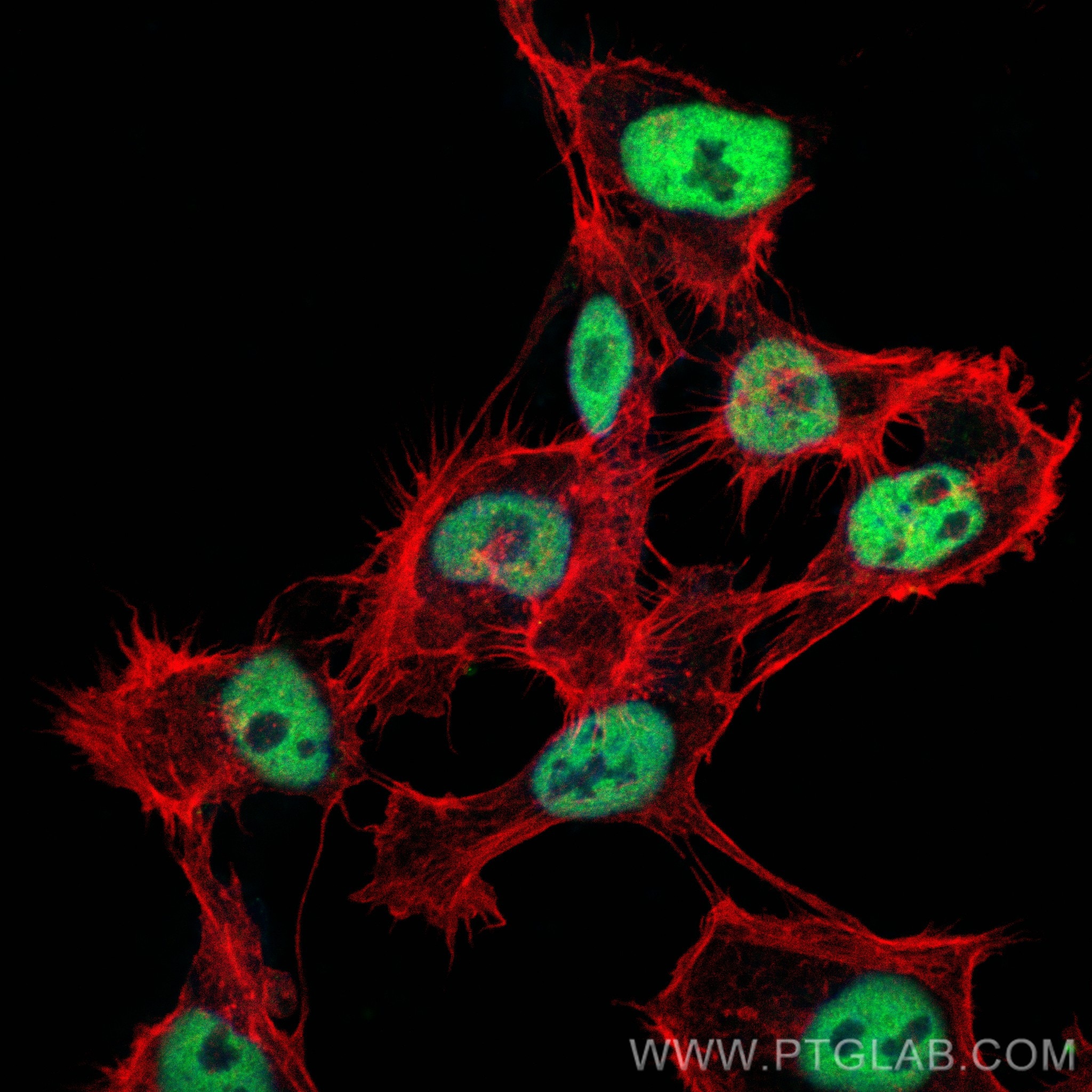
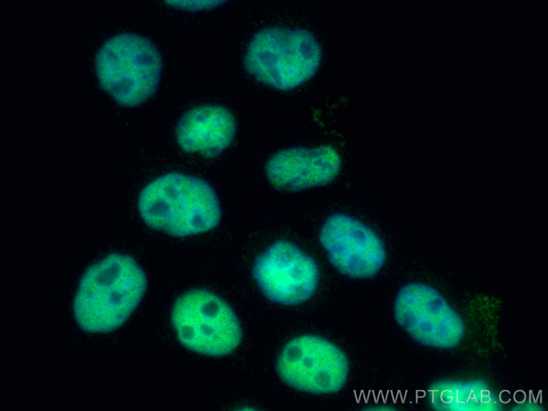
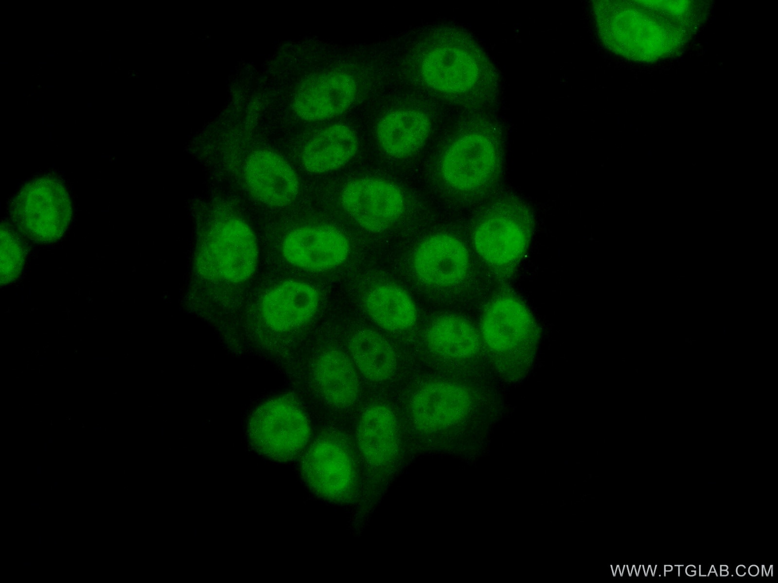
| Positive IF/ICC detected in | A431 cells, HeLa cells |
Recommended dilution
| Application | Dilution |
|---|---|
| Western Blot (WB) | WB : 1:5000-1:50000 |
| Immunohistochemistry (IHC) | IHC : 1:2000-1:10000 |
| Immunofluorescence (IF)/ICC | IF/ICC : 1:6000-1:24000 |
| It is recommended that this reagent should be titrated in each testing system to obtain optimal results. | |
| Sample-dependent, Check data in validation data gallery. | |
Published Applications
| KD/KO | See 1 publications below |
| WB | See 17 publications below |
| IHC | See 3 publications below |
| IF | See 3 publications below |
| IP | See 1 publications below |
| ChIP | See 1 publications below |
Product Information
66085-1-Ig targets HDAC1 in WB, IHC, IF/ICC, IP, ChIP, ELISA applications and shows reactivity with human, mouse, rat samples.
| Tested Reactivity | human, mouse, rat |
| Cited Reactivity | human, mouse, rat |
| Host / Isotype | Mouse / IgG1 |
| Class | Monoclonal |
| Type | Antibody |
| Immunogen |
CatNo: Ag19128 Product name: Recombinant human HDAC1 protein Source: e coli.-derived, PET28a Tag: 6*His Domain: 184-482 aa of BC000301 Sequence: EEAFYTTDRVMTVSFHKYGEYFPGTGDLRDIGAGKGKYYAVNYPLRDGIDDESYEAIFKPVMSKVMEMFQPSAVVLQCGSDSLSGDRLGCFNLTIKGHAKCVEFVKSFNLPMLMLGGGGYTIRNVARCWTYETAVALDTEIPNELPYNDYFEYFGPDFKLHISPSNMTNQNTNEYLEKIKQRLFENLRMLPHAPGVQMQAIPEDAIPEESGDEDEDDPDKRISICSSDKRIACEEEFSDSEEEGEGGRKNSSNFKKAKRVKTEDEKEKDPEEKKEVTEEEKTKEEKPEAKGVKEEVKLA Predict reactive species |
| Full Name | histone deacetylase 1 |
| Calculated Molecular Weight | 55 kDa |
| Observed Molecular Weight | 65 kDa |
| GenBank Accession Number | BC000301 |
| Gene Symbol | HDAC1 |
| Gene ID (NCBI) | 3065 |
| RRID | AB_11232033 |
| Conjugate | Unconjugated |
| Form | Liquid |
| Purification Method | Protein G purification |
| UNIPROT ID | Q13547 |
| Storage Buffer | PBS with 0.02% sodium azide and 50% glycerol, pH 7.3. |
| Storage Conditions | Store at -20°C. Stable for one year after shipment. Aliquoting is unnecessary for -20oC storage. 20ul sizes contain 0.1% BSA. |
Background Information
Histone deacetylases(HDAC) are a class of enzymes that remove the acetyl groups from the lysine residues leading to the formation of a condensed and transcriptionally silenced chromatin. The protein encoded by this gene belongs to the histone deacetylase/acuc/apha family and is a component of the histone deacetylase complex, which is responsible for gene expression silencing. It also plays an important role in the control of cell proliferation and differentiation by interacting with RB, p53 and other transcription factors. At least 4 classes of HDAC were identified. As a class I HDAC, HDAC 1 was primarily found in the nucleus. This antibody is raised against residues near the C terminus of human HDAC1. The calculated molecular weight of HDAC1 is 55 kDa, but modified HDAC1 is about 60-65 kDa .
Protocols
| Product Specific Protocols | |
|---|---|
| IF protocol for HDAC1 antibody 66085-1-Ig | Download protocol |
| IHC protocol for HDAC1 antibody 66085-1-Ig | Download protocol |
| WB protocol for HDAC1 antibody 66085-1-Ig | Download protocol |
| Standard Protocols | |
|---|---|
| Click here to view our Standard Protocols |
Publications
| Species | Application | Title |
|---|---|---|
Mol Cancer Exosomal circ_0006896 promotes AML progression via interaction with HDAC1 and restriction of antitumor immunity | ||
Immunity Histone Deacetylases 1 and 2 Regulate Microglia Function during Development, Homeostasis, and Neurodegeneration in a Context-Dependent Manner. | ||
Nat Commun YTHDF2 upregulation and subcellular localization dictate CD8 T cell polyfunctionality in anti-tumor immunity | ||
J Clin Invest The FOXN3-NEAT1-SIN3A repressor complex promotes progression of hormonally responsive breast cancer. | ||
Nat Commun ZNF516 suppresses EGFR by targeting the CtBP/LSD1/CoREST complex to chromatin. | ||
Br J Pharmacol Physalin B attenuates liver fibrosis via suppressing LAP2α-HDAC1-mediated deacetylation of the transcription factor GLI1 and hepatic stellate cell activation. |
Reviews
The reviews below have been submitted by verified Proteintech customers who received an incentive for providing their feedback.
FH Tsimafei (Verified Customer) (08-04-2024) | Antibody were incubated at 4C, ON
 |
FH Xiaoyu (Verified Customer) (06-28-2023) | Excellent antibody for WB
 |
FH Andrea (Verified Customer) (01-11-2023) | Excellent antibody
 |