Recombinant Human CXCL10/IP-10 protein (rFc Tag)(HPLC verified)
Species
Human
Purity
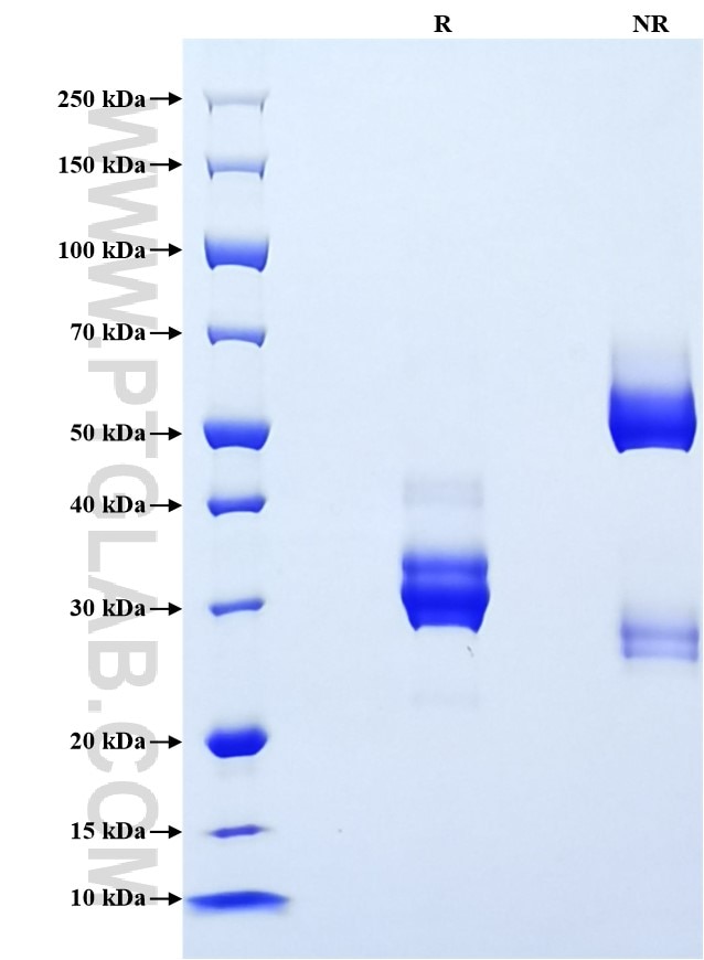
>90 %, SDS-PAGE
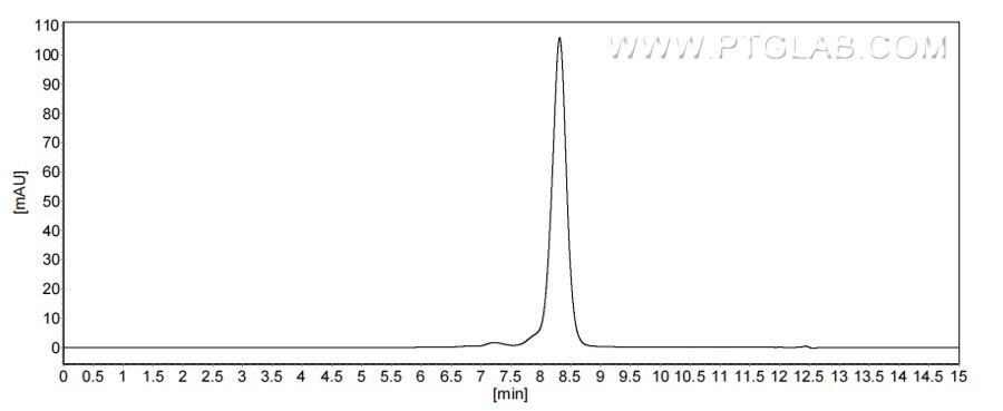
>90 %, SEC-HPLC
Tag
rFc Tag
Activity
not tested
Cat no : Eg2489
Validation Data Gallery
Product Information
| Purity | >90 %, SDS-PAGE >90 %, SEC-HPLC |
| Endotoxin | <0.1 EU/μg protein, LAL method |
| Activity |
Not tested |
| Expression | HEK293-derived Human CXCL10 protein Val22-Pro98 (Accession# P02778) with a rabbit IgG Fc tag at the C-terminus. |
| GeneID | 3627 |
| Accession | P02778 |
| PredictedSize | 34.9 kDa |
| SDS-PAGE | 28-35 kDa, reducing (R) conditions |
| Formulation | Lyophilized from 0.22 μm filtered solution in PBS, pH 7.4. Normally 5% trehalose and 5% mannitol are added as protectants before lyophilization. |
| Reconstitution | Briefly centrifuge the tube before opening. Reconstitute at 0.1-0.5 mg/mL in sterile water. |
| Storage Conditions |
It is recommended that the protein be aliquoted for optimal storage. Avoid repeated freeze-thaw cycles.
|
| Shipping | The product is shipped at ambient temperature. Upon receipt, store it immediately at the recommended temperature. |
Background
CXCL10 ( also known as IP-10) is a member of the CXC chemokine family which binds to the CXCR3 receptor to exert its biological effects. CXCL10 is a 12-kDa protein and constitutes two internal disulfide cross bridges. The predicted signal peptidase cleavage generates a 10-kDa secreted polypeptide with four conserved cysteine residues in the N-terminal. The CXCL10 gene localizes on chromosome 4 at band q21, a locus associated with an acute monocytic/B-lymphocyte lineage leukemia exhibiting translocation of t (4; 11) (q21; q23). CXCL10 mediates leukocyte trafficking, adaptive immunity, inflammation, haematopoiesis and angiogenesis. Under proinflammatory conditions CXCL10 is secreted from a variety of cells, such as leukocytes, activated neutrophils, eosinophils, monocytes, epithelial cells, endothelial cells, stromal cells (fibroblasts) and keratinocytes in response to IFN-γ.
References:
1. Liu M. et al. (2011). Oncol Lett. 2(4):583-589.
2. Walser TC. et al. (2006). Cancer Res. 66(15):7701-7.
3. Luster AD. et al. (1987). J Exp Med. 166(4):1084-97.
4. Lo BK. et al. (2010). Am J Pathol. 176(5):2435-46.
5. Groom JR. Et al. (2011). Immunol Cell Biol. 89(2):207-15.

