
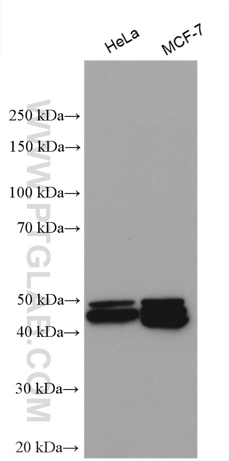
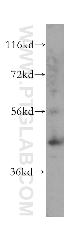
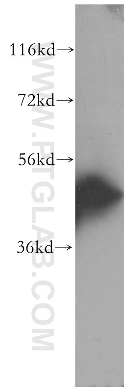
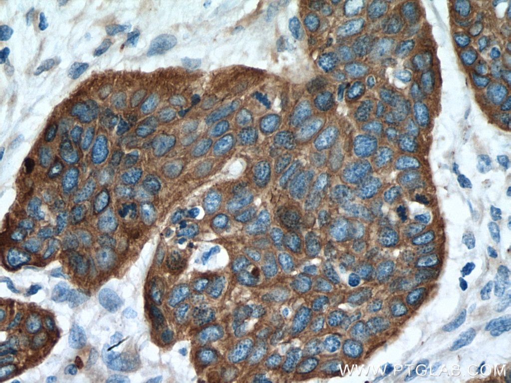
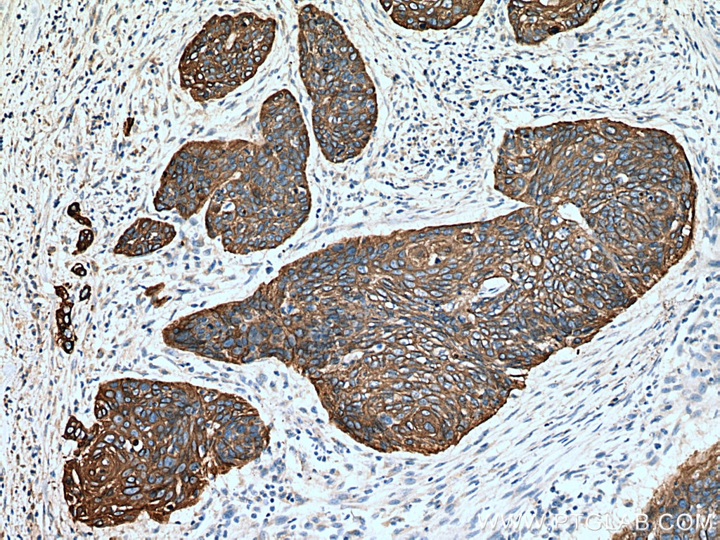
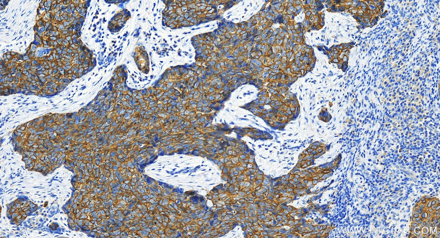
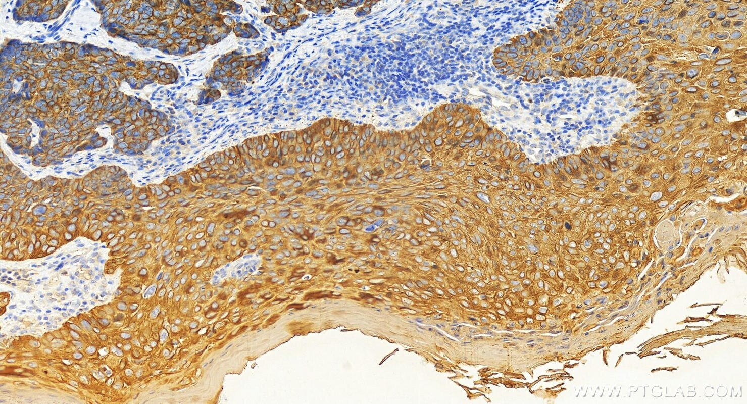
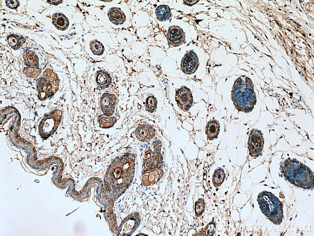

Tested Applications
| Positive WB detected in | HeLa cells, A375 cells, MCF-7 cells |
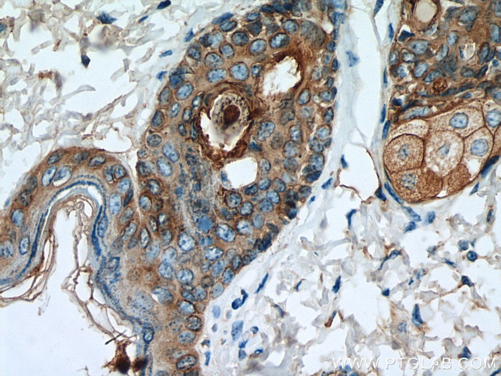
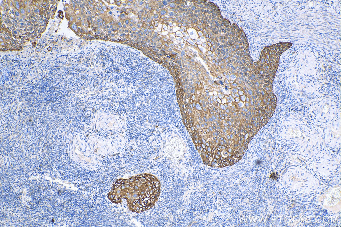
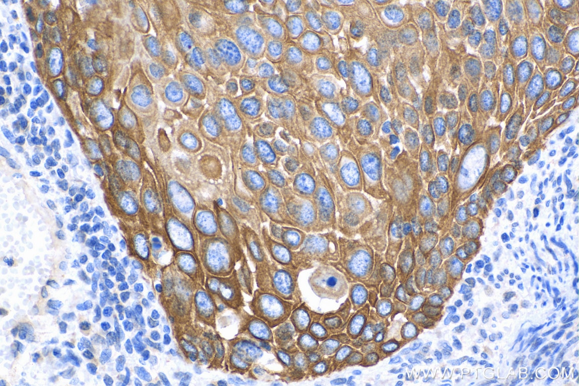
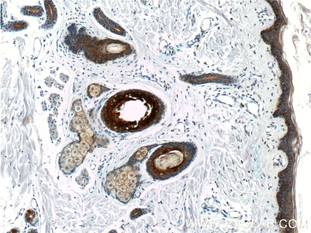
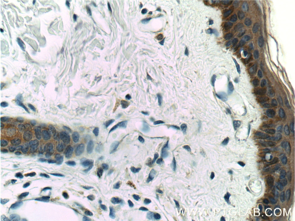
| Positive IHC detected in | human oesophagus cancer tissue, human bowen disease, human cervical cancer tissue, human skin tissue, mouse skin tissue Note: suggested antigen retrieval with TE buffer pH 9.0; (*) Alternatively, antigen retrieval may be performed with citrate buffer pH 6.0 |
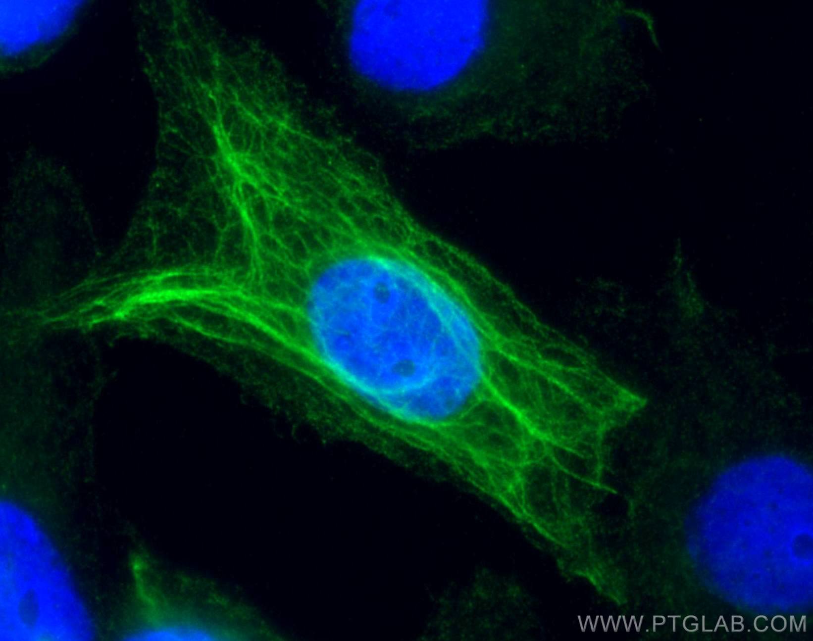
| Positive IF/ICC detected in | A431 cells |
Recommended dilution
| Application | Dilution |
|---|---|
| Western Blot (WB) | WB : 1:5000-1:50000 |
| Immunohistochemistry (IHC) | IHC : 1:500-1:2000 |
| Immunofluorescence (IF)/ICC | IF/ICC : 1:50-1:500 |
| It is recommended that this reagent should be titrated in each testing system to obtain optimal results. | |
| Sample-dependent, Check data in validation data gallery. | |
Product Information
17265-1-AP targets Cytokeratin 16 in WB, IHC, IF/ICC, ELISA applications and shows reactivity with human, mouse samples.
| Tested Reactivity | human, mouse |
| Cited Reactivity | human, mouse |
| Host / Isotype | Rabbit / IgG |
| Class | Polyclonal |
| Type | Antibody |
| Immunogen |
CatNo: Ag10999 Product name: Recombinant human KRT16 protein Source: e coli.-derived, PGEX-4T Tag: GST Domain: 121-473 aa of BC039169 Sequence: MQNLNDRLASYLDKVRALEEANADLEVKIRDWYQRQRPSEIKDYSPYFKTIEDLRNKIIAATIENAQPILQIDNARLAADDFRTKYEHELALRQTVEADVNGLRRVLDELTLARTDLEMQIEGLKEELAYLRKNHEEEMLALRGQTGGDVNVEMDAAPGVDLSRILNEMRDQYEQMAEKNRRDAETWFLSKTEELNKEVASNSELVQSSRSEVTELRRVLQGLEIELQSQLSMKASLENSLEETKGRYCMQLSQIQGLIGSVEEQLAQLRCEMEQQSQEYQILLDVKTRLEQEIATYRRLLEGEDAHLSSQQASGQSYSSREVFTSSSSSSSRQTRPILKEQSSSSFSQGQSS Predict reactive species |
| Full Name | keratin 16 |
| Calculated Molecular Weight | 473 aa, 51 kDa |
| Observed Molecular Weight | 48 kDa |
| GenBank Accession Number | BC039169 |
| Gene Symbol | Cytokeratin 16 |
| Gene ID (NCBI) | 3868 |
| RRID | AB_2132894 |
| Conjugate | Unconjugated |
| Form | Liquid |
| Purification Method | Antigen affinity purification |
| UNIPROT ID | P08779 |
| Storage Buffer | PBS with 0.02% sodium azide and 50% glycerol, pH 7.3. |
| Storage Conditions | Store at -20°C. Stable for one year after shipment. Aliquoting is unnecessary for -20oC storage. 20ul sizes contain 0.1% BSA. |
Background Information
Keratins are a large family of proteins that form the intermediate filament cytoskeleton of epithelial cells, which are classified into two major sequence types. Type I keratins are a group of acidic intermediate filament proteins, including K9-K23, and the hair keratins Ha1-Ha8. Type II keratins are the basic or neutral courterparts to the acidic type I keratins, including K1-K8, and the hair keratins, Hb1-Hb6. Keratin 16 is a type I cytokeratin. It is paired with keratin 6 in a number of epithelial tissues, including nail bed, esophagus, tongue, and hair follicles.
Protocols
| Product Specific Protocols | |
|---|---|
| IF protocol for Cytokeratin 16 antibody 17265-1-AP | Download protocol |
| IHC protocol for Cytokeratin 16 antibody 17265-1-AP | Download protocol |
| WB protocol for Cytokeratin 16 antibody 17265-1-AP | Download protocol |
| Standard Protocols | |
|---|---|
| Click here to view our Standard Protocols |
Publications
What published studies show
Cytokeratin 16 Polyclonal antibody (17265-1-AP) has 12 total citations. The publications appear in journals including Immunity, Cell Reports Medicine, Phytotherapy Research, Frontiers in Immunology, EMBO Reports, Communications Biology, Investigative Ophthalmology & Visual Science, Frontiers in Pharmacology, Journal of Biological Chemistry, Discover Oncology, and International Journal of Clinical and Experimental Pathology. The citations span research fields such as skin immunology and inflammation (including hidradenitis suppurativa, psoriasis, and prurigo nodularis), wound healing in diabetes, corneal pathology, squamous cell carcinoma and cancer stem cells, lung and colorectal cancer, and middle ear cholesteatoma. Applications used include IF, WB, and IHC across human and mouse samples.
| Species | Application | Title |
|---|---|---|
Immunity Skin immune-mesenchymal interplay within tertiarylymphoid structures promotes autoimmunepathogenesis in hidradenitis suppurativa | ||
Cell Rep Med Co-targeting BMI1 and MYC to eliminate cancer stem cells in squamous cell carcinoma | ||
Phytother Res Epigallocatechin-3-gallate promotes wound healing response in diabetic mice by activating keratinocytes and promoting re-epithelialization | ||
Front Immunol A murine model of prurigo nodularis-like skin lesions induced by persistent scratching under type 2 inflammatory conditions. | ||
EMBO Rep TMPRSS11B promotes an acidified microenvironment and immune suppression in squamous lung cancer. | ||
Commun Biol Macrophagic Ym1 orchestrates γδT cell-derived IL-17 production and keratinocyte functionality to mediate psoriasis-like skin inflammation. |















