
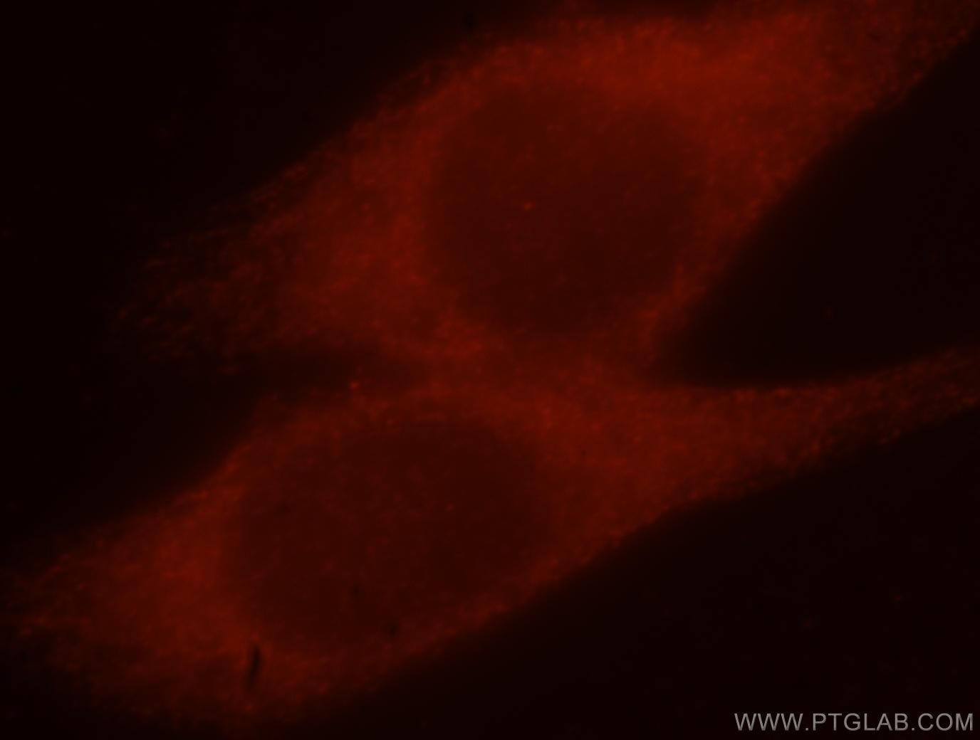

Tested Applications
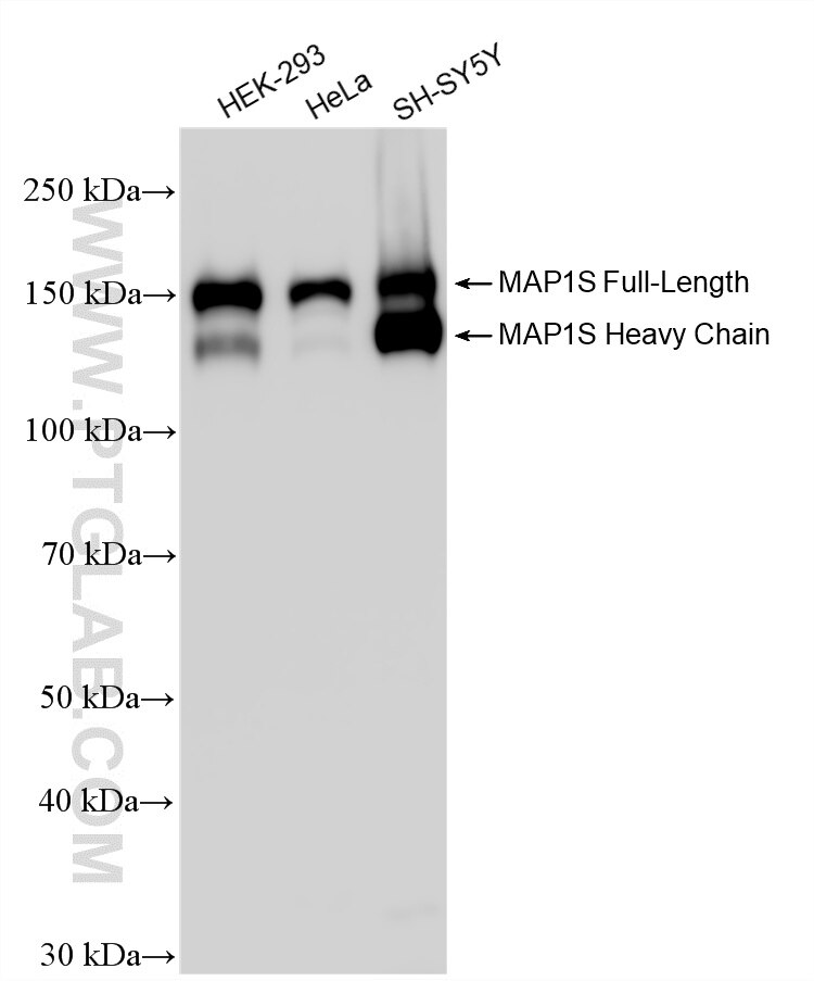
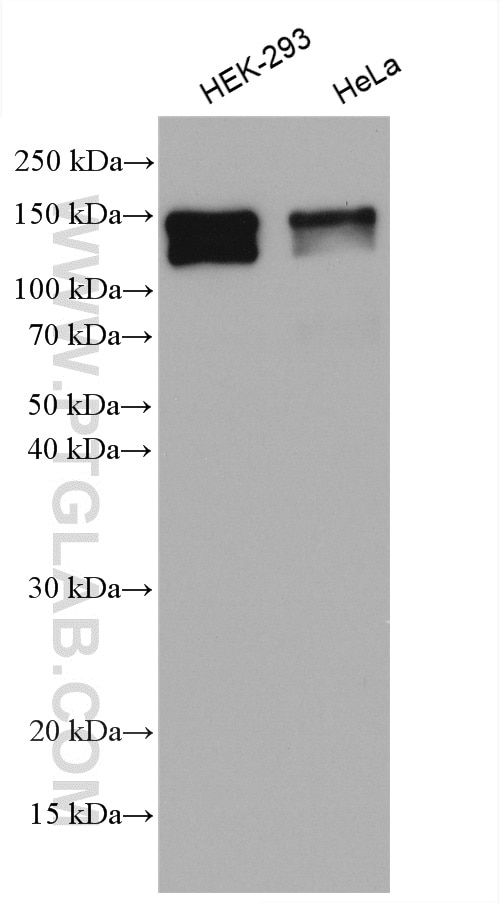
| Positive WB detected in | HEK-293 cells, HeLa cells, SH-SY5Y cells |
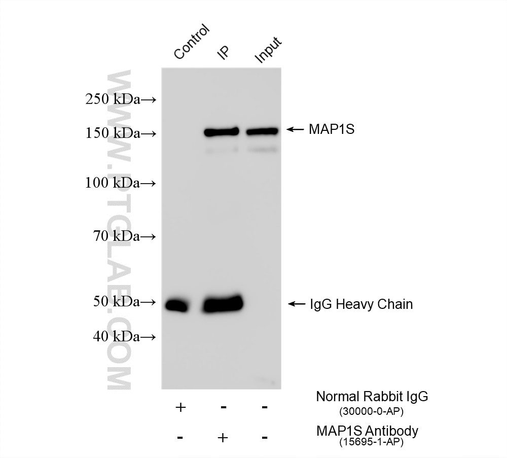
| Positive IP detected in | AC16 cells |
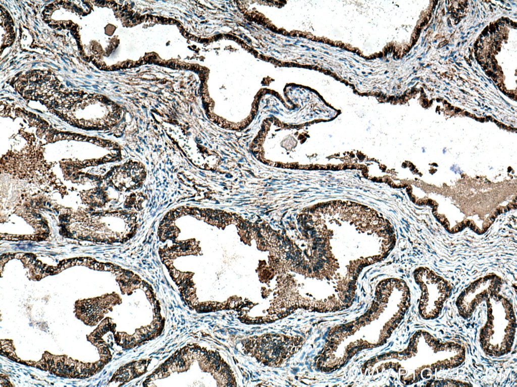
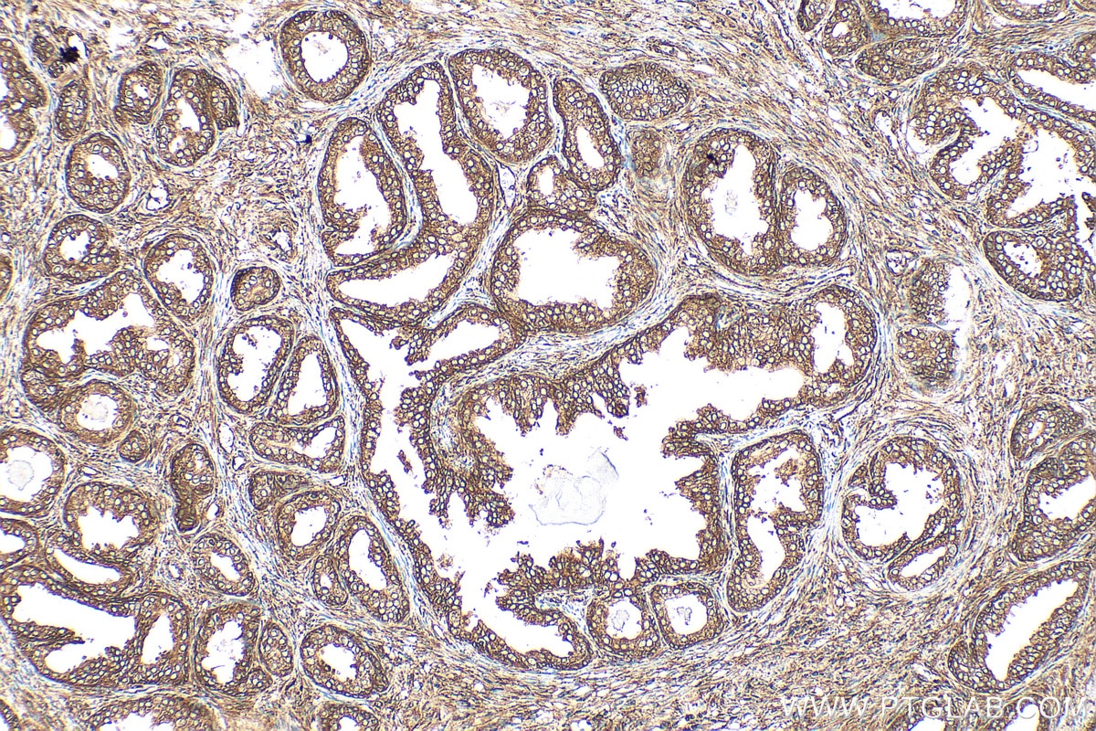
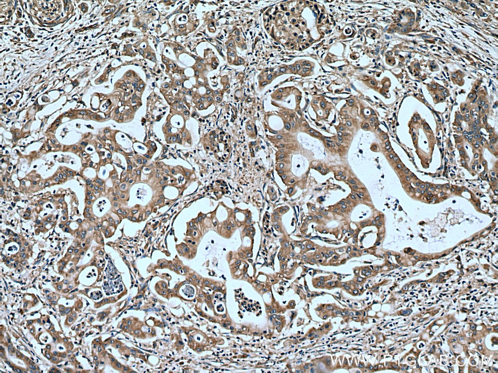
| Positive IHC detected in | human prostate cancer tissue, human pancreas cancer tissue Note: suggested antigen retrieval with TE buffer pH 9.0; (*) Alternatively, antigen retrieval may be performed with citrate buffer pH 6.0 |
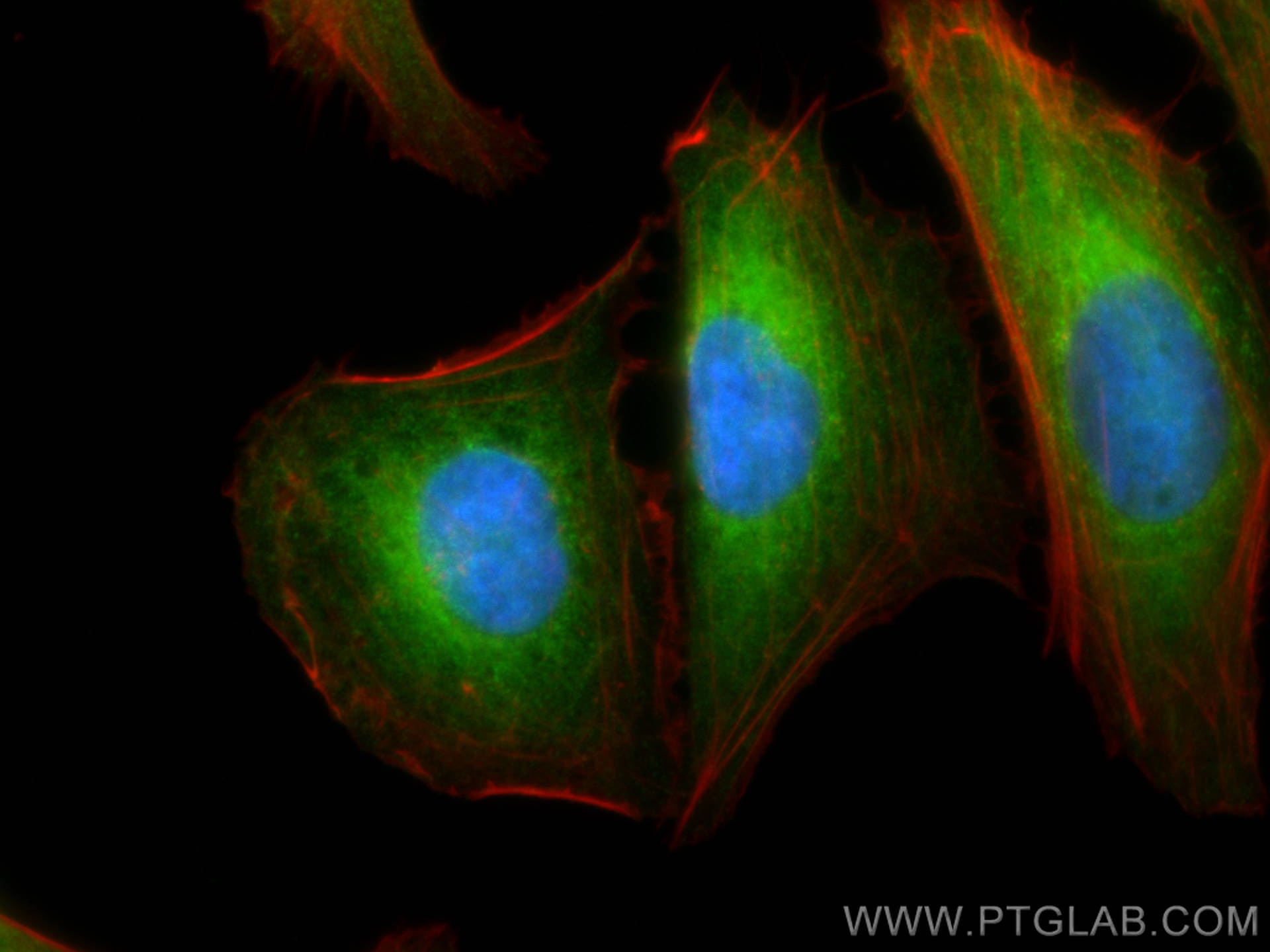
| Positive IF/ICC detected in | U2OS cells, HeLa cells |
Recommended dilution
| Application | Dilution |
|---|---|
| Western Blot (WB) | WB : 1:5000-1:50000 |
| Immunoprecipitation (IP) | IP : 0.5-4.0 ug for 1.0-3.0 mg of total protein lysate |
| Immunohistochemistry (IHC) | IHC : 1:200-1:800 |
| Immunofluorescence (IF)/ICC | IF/ICC : 1:200-1:800 |
| It is recommended that this reagent should be titrated in each testing system to obtain optimal results. | |
| Sample-dependent, Check data in validation data gallery. | |
Product Information
15695-1-AP targets MAP1S in WB, IHC, IF/ICC, IP, ELISA applications and shows reactivity with human samples.
| Tested Reactivity | human |
| Cited Reactivity | human, mouse |
| Host / Isotype | Rabbit / IgG |
| Class | Polyclonal |
| Type | Antibody |
| Immunogen |
CatNo: Ag8315 Product name: Recombinant human MAP1S protein Source: e coli.-derived, PET28a Tag: 6*His Domain: 457-806 aa of BC008806 Sequence: APTSEAGLSLPLRGPRARRSASPHDVDLCLVSPCEFEHRKAVPMAPAPASPGSSNDSSARSQERAGGLGAEETPPTSVSESLPTLSDSDPVPLAPGAADSDEDTEGFGVPRHDPLPDPLKVPPPLPDPSSICMVDPEMLPPKTARQTENVSRTRKPLARPNSRAAAPKATPVAAAKTKGLAGGDRASRPLSARSEPSEKGGRAPLSRKSSTPKTATRGPSGSASSRPGVSATPPKSPVYLDLAYLPSGSSAHLVDEEFFQRVRALCYVISGQDQRKEEGMRAVLDALLASKQHWDRDLQVTLIPTFDSVAMHTWYAETHARHQALGITVLGSNSMVSMQDDAFPACKVEF Predict reactive species |
| Full Name | microtubule-associated protein 1S |
| Calculated Molecular Weight | 806 aa, 85 kDa |
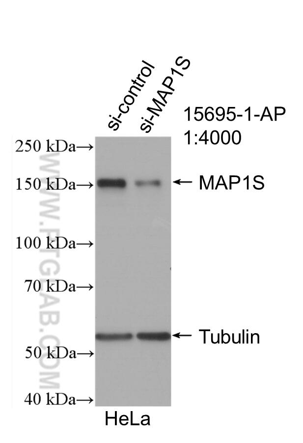
| Observed Molecular Weight | 130-150 kDa |
| GenBank Accession Number | BC008806 |
| Gene Symbol | MAP1S |
| Gene ID (NCBI) | 55201 |
| RRID | AB_2137868 |
| Conjugate | Unconjugated |
| Form | Liquid |
| Purification Method | Antigen affinity purification |
| UNIPROT ID | Q66K74 |
| Storage Buffer | PBS with 0.02% sodium azide and 50% glycerol, pH 7.3. |
| Storage Conditions | Store at -20°C. Stable for one year after shipment. Aliquoting is unnecessary for -20oC storage. 20ul sizes contain 0.1% BSA. |
Background Information
MAP1S (also known as C19ORF5 or VCY2IP1) is a novel member of the microtubule-associated protein 1 family and a homologue of the exclusively neuronal distributed microtubule-associated protein 1A and 1B (MAP1A/B). In contrast to MAP1A and MAP1B, MAP1S is expressed in a wide range of tissues in addition to neurons. MAP1S is synthesized as a precursor protein that is partially cleaved into heavy and light chains in a tissue-specific manner. In addition, a short chain isoform may be induced under prolonged mitotic arrest or inhibition of the 26S proteasome. Recently it has been reported that the short chain isoform associates with mitochondria in addition to microtubules and causes irreversible aggregation of dysfunctional mitochondria resulting in cell death. Western blot analysis using this antibody detected two main bands between 100-150 kDa corresponding to heavy chain and full-length of MAP1S.
Protocols
| Product Specific Protocols | |
|---|---|
| IF protocol for MAP1S antibody 15695-1-AP | Download protocol |
| IHC protocol for MAP1S antibody 15695-1-AP | Download protocol |
| IP protocol for MAP1S antibody 15695-1-AP | Download protocol |
| WB protocol for MAP1S antibody 15695-1-AP | Download protocol |
| Standard Protocols | |
|---|---|
| Click here to view our Standard Protocols |
Publications
What published studies show
The anti-MAP1S antibody (Cat# 15695-1-AP) has 10 citations spanning journals including Cell Research, J Cell Biol, EMBO Reports, Int J Biol Macromol, Front Synaptic Neurosci, Front Pharmacol, BBA Mol Cell Res, BMC Cancer, Cell Cycle, and bioRxiv. Research fields cover microtubule dynamics, hepatocellular carcinoma, gastric cancer, ovarian cancer, autophagy, dendritic filopodia, calpain splicing, tumor drug sensitivity, and neural development.
| Species | Application | Title |
|---|---|---|
Cell Res Regulators of tubulin polyglutamylation control nuclear shape and cilium disassembly by balancing microtubule and actin assembly. | ||
Int J Biol Macromol HBx-induced upregulation of MAP1S drives hepatocellular carcinoma proliferation and migration via MAP1S/Smad/TGF-β1 loop | ||
Front Synaptic Neurosci Proteomic Analysis of Dendritic Filopodia-Rich Fraction Isolated by Telencephalin and Vitronectin Interaction. | ||
EMBO Rep MAP1B-LC1 prevents autophagosome formation by linking syntaxin 17 to microtubules. | ||
Front Pharmacol Lipid droplets: a candidate new research field for epithelial ovarian cancer |
Reviews
What customers say
One reviewer reported that this antibody did not work in Western Blot using sheep atrial tissue lysates at a 1:200 dilution. No clear band was observed at the predicted molecular weight, resulting in a low rating.
The reviews below have been submitted by verified Proteintech customers who received an incentive for providing their feedback.
Alice (Verified Customer) (02-18-2026) | Didn't work in sheep atrial tissue lysates- no clear band at predicted MW.
|










