Tested Applications
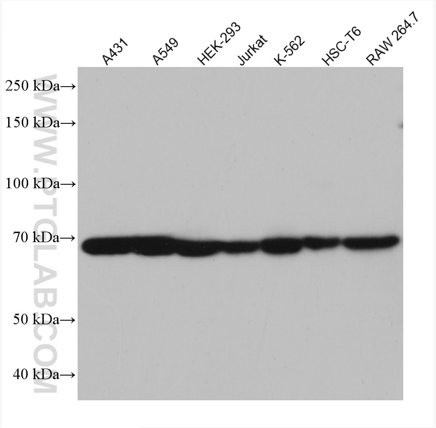
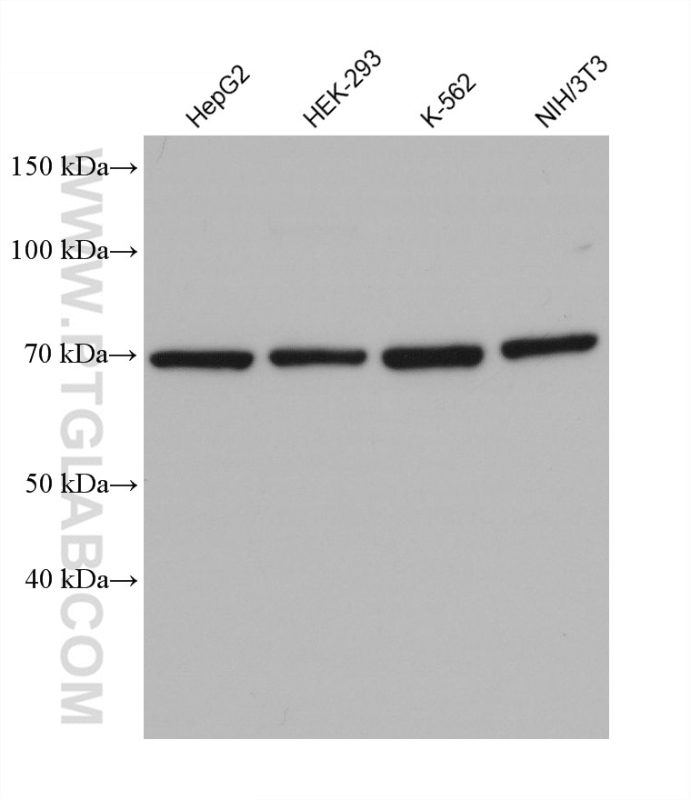
| Positive WB detected in | A431 cells, NIH/3T3 cells, HepG2 cells, A549 cells, HEK-293 cells, Jurkat cells, K-562 cells, HSC-T6 cells, RAW 264.7 cells |
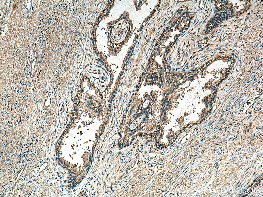
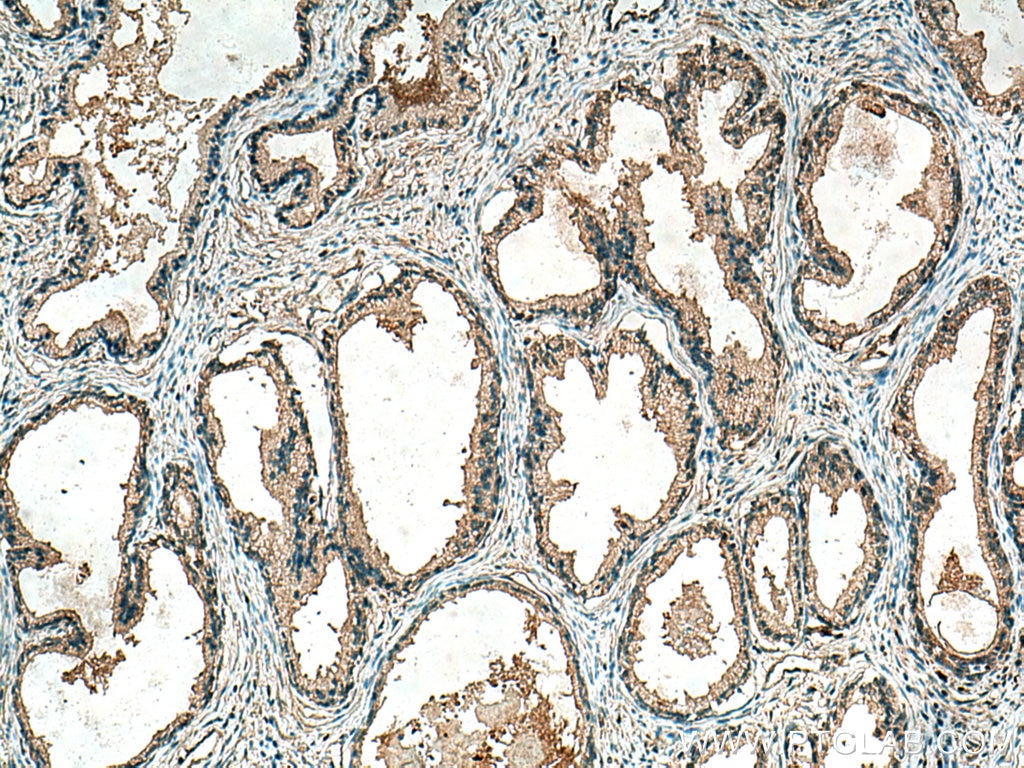
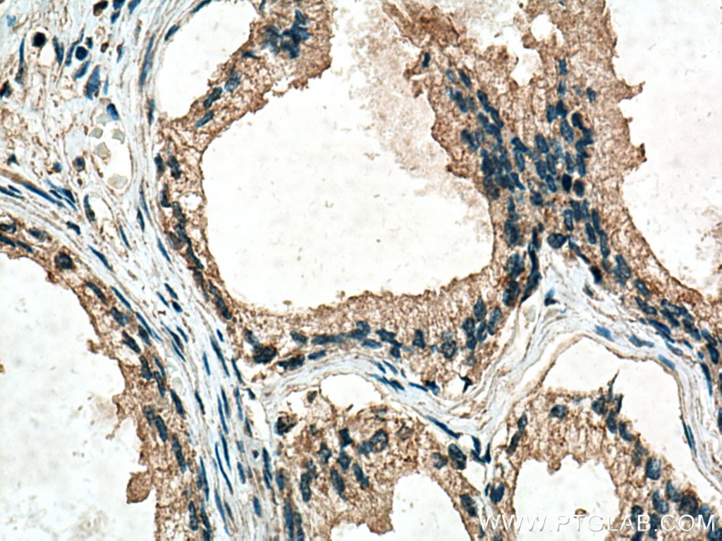
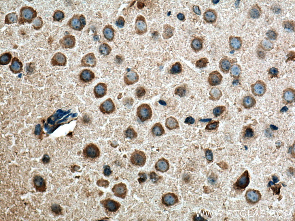
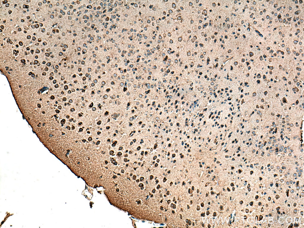
| Positive IHC detected in | human prostate cancer tissue, mouse brain tissue Note: suggested antigen retrieval with TE buffer pH 9.0; (*) Alternatively, antigen retrieval may be performed with citrate buffer pH 6.0 |
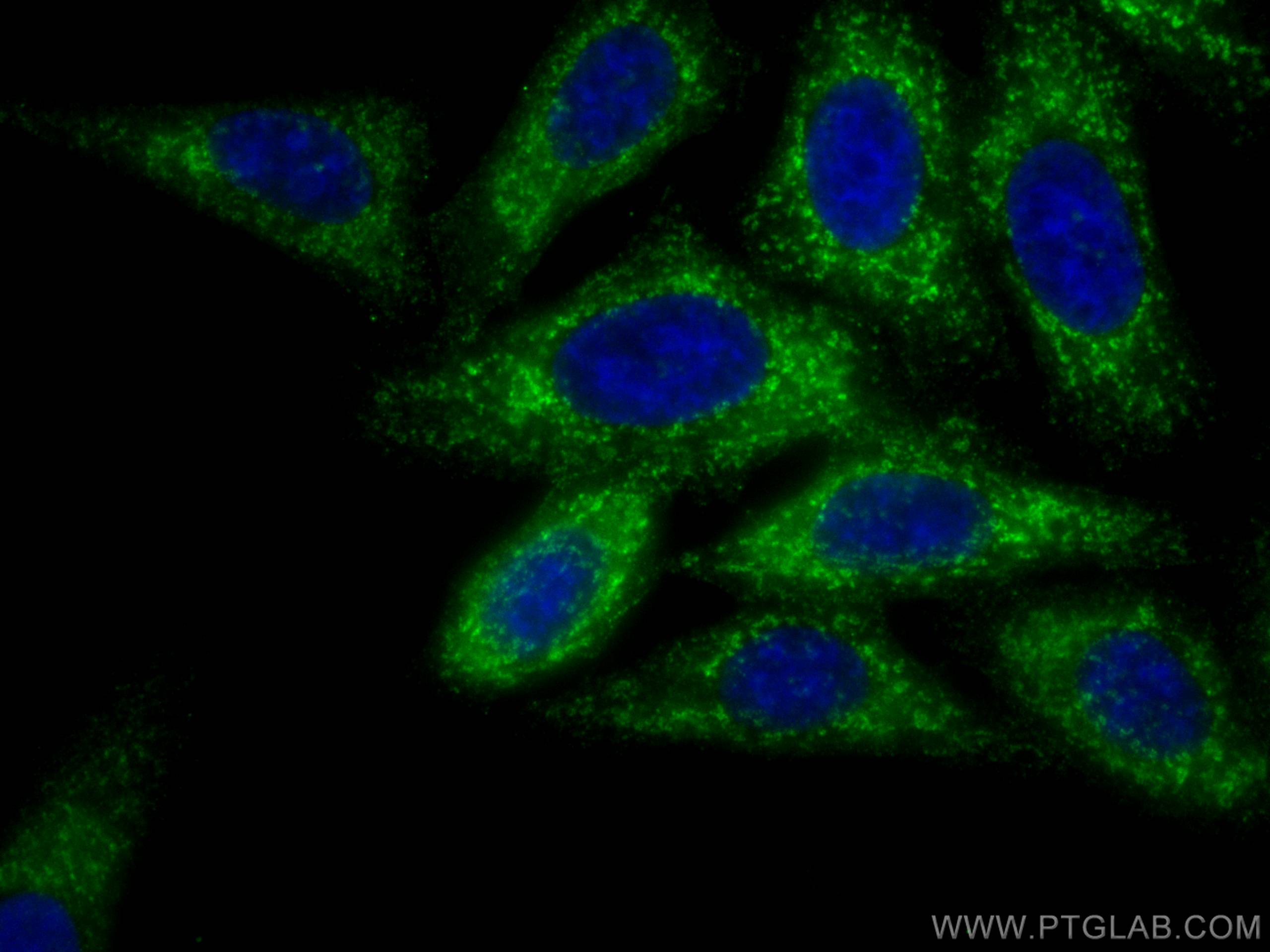
| Positive IF/ICC detected in | HepG2 cells |
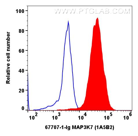
| Positive FC (Intra) detected in | K-562 cells |
Recommended dilution
| Application | Dilution |
|---|---|
| Western Blot (WB) | WB : 1:500-1:3000 |
| Immunohistochemistry (IHC) | IHC : 1:250-1:1000 |
| Immunofluorescence (IF)/ICC | IF/ICC : 1:50-1:500 |
| Flow Cytometry (FC) (INTRA) | FC (INTRA) : 0.20 ug per 10^6 cells in a 100 µl suspension |
| It is recommended that this reagent should be titrated in each testing system to obtain optimal results. | |
| Sample-dependent, Check data in validation data gallery. | |
Published Applications
| KD/KO | See 1 publications below |
| WB | See 8 publications below |
| IHC | See 2 publications below |
| IF | See 5 publications below |
| IP | See 1 publications below |
Product Information
67707-1-Ig targets TAK1 in WB, IHC, IF/ICC, FC (Intra), IP, ELISA applications and shows reactivity with human, mouse, rat samples.
| Tested Reactivity | human, mouse, rat |
| Cited Reactivity | human, mouse, rat |
| Host / Isotype | Mouse / IgG1 |
| Class | Monoclonal |
| Type | Antibody |
| Immunogen |
CatNo: Ag21286 Product name: Recombinant human MAP3K7 protein Source: e coli.-derived, PET28a Tag: 6*His Domain: 1-291 aa of BC017715 Sequence: MSTASAASSSSSSSAGEMIEAPSQVLNFEEIDYKEIEVEEVVGRGAFGVVCKAKWRAKDVAIKQIESESERKAFIVELRQLSRVNHPNIVKLYGACLNPVCLVMEYAEGGSLYNVLHGAEPLPYYTAAHAMSWCLQCSQGVAYLHSMQPKALIHRDLKPPNLLLVAGGTVLKICDFGTACDIQTHMTNNKGSAAWMAPEVFEGSNYSEKCDVFSWGIILWEVITRRKPFDEIGGPAFRIMWAVHNGTRPPLIKNLPKPIESLMTRCWSKDPSQRPSMEEIVKIMTHLMRYF Predict reactive species |
| Full Name | mitogen-activated protein kinase kinase kinase 7 |
| Calculated Molecular Weight | 579 aa, 64 kDa |
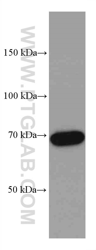
| Observed Molecular Weight | 67-70 kDa |
| GenBank Accession Number | BC017715 |
| Gene Symbol | TAK1 |
| Gene ID (NCBI) | 6885 |
| RRID | AB_2882898 |
| Conjugate | Unconjugated |
| Form | Liquid |
| Purification Method | Protein G purification |
| UNIPROT ID | O43318 |
| Storage Buffer | PBS with 0.02% sodium azide and 50% glycerol, pH 7.3. |
| Storage Conditions | Store at -20°C. Stable for one year after shipment. Aliquoting is unnecessary for -20oC storage. 20ul sizes contain 0.1% BSA. |
Background Information
MAP3K7(Mitogen-activated protein kinase kinase kinase 7) is also named as TAK1 and belongs to the MAP kinase kinase kinase subfamily. It plays an important role in the cascades of cellular responses evoked by changes in the environment. It has been linked to interleukin-1 receptor and tumor necrosis factor receptor signaling (PMID: 16186825). It has 4 isoforms (53-55 kDa, 64-70 kDa and 75-80 kDa) produced by alternative splicing.
Protocols
| Product Specific Protocols | |
|---|---|
| IF protocol for TAK1 antibody 67707-1-Ig | Download protocol |
| IHC protocol for TAK1 antibody 67707-1-Ig | Download protocol |
| WB protocol for TAK1 antibody 67707-1-Ig | Download protocol |
| Standard Protocols | |
|---|---|
| Click here to view our Standard Protocols |
Publications
| Species | Application | Title |
|---|---|---|
Cell Death Dis USP4 promotes the proliferation, migration, and invasion of esophageal squamous cell carcinoma by targeting TAK1 | ||
Inflammopharmacology Liensinine reduces acute lung injury brought on by lipopolysaccharide by inhibiting the activation of the NF-κB signaling pathway through modification of the Src/TRAF6/TAK1 axis | ||
Int Immunopharmacol Kongensin a attenuates intervertebral disc degeneration by inhibiting TAK1-mediated PANoptosis of nucleus pulposus cells | ||
Biochim Biophys Acta Mol Cell Res IL-17 promotes melanoma through TRAF2 as a scaffold protein recruiting PIAS2 and ELAVL1 to induce EPHA5 | ||
Exp Cell Res Deubiquitinase USP18 inhibits hepatic stellate cells activation and alleviates liver fibrosis via regulation of TAK1 activity | ||
Neurochem Res Exogenous TIPE2 Inhibit TAK1 to Improve Inflammation and Neuropathic Pain Induced by Sciatic Nerve Injury Through Inactivating NF-κB and JNK. |