Tested Applications
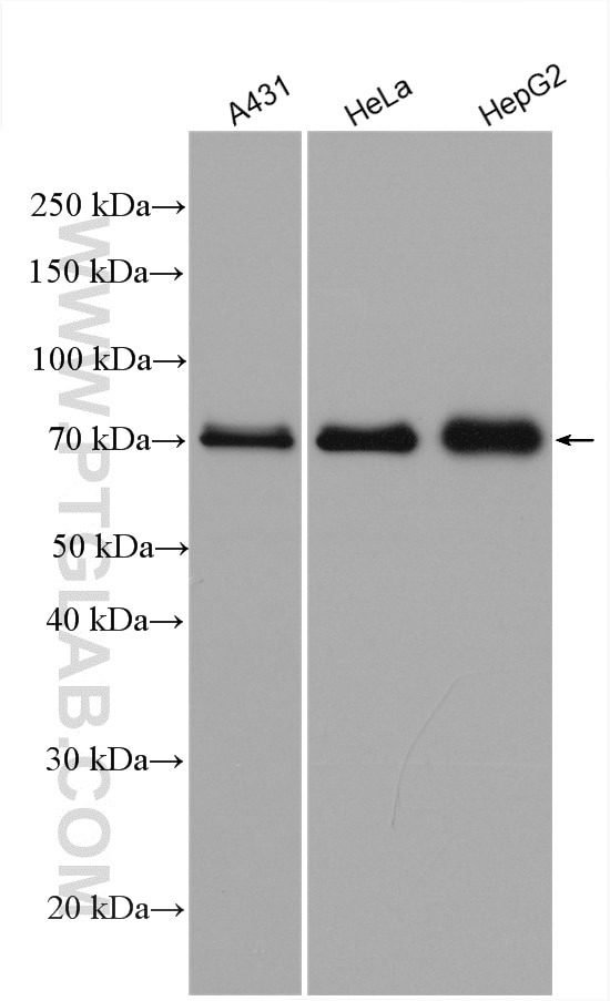
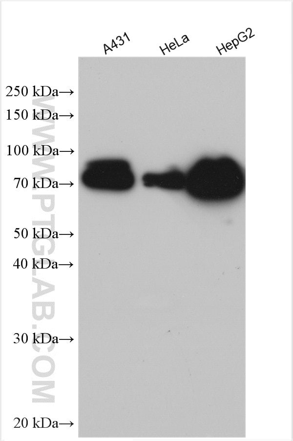
| Positive WB detected in | A431 cells, HeLa cells, HuH-7 cells, HepG2 cells |
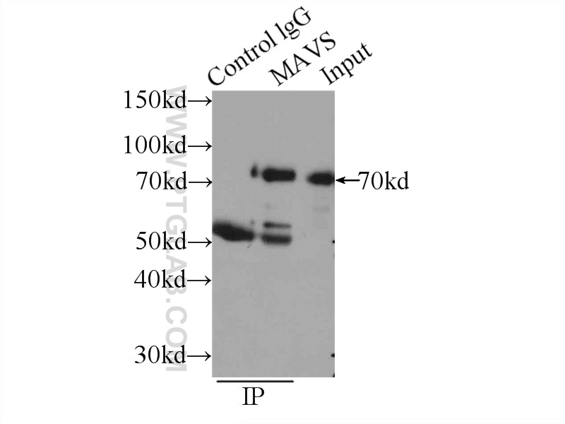
| Positive IP detected in | HEK-293 cells |
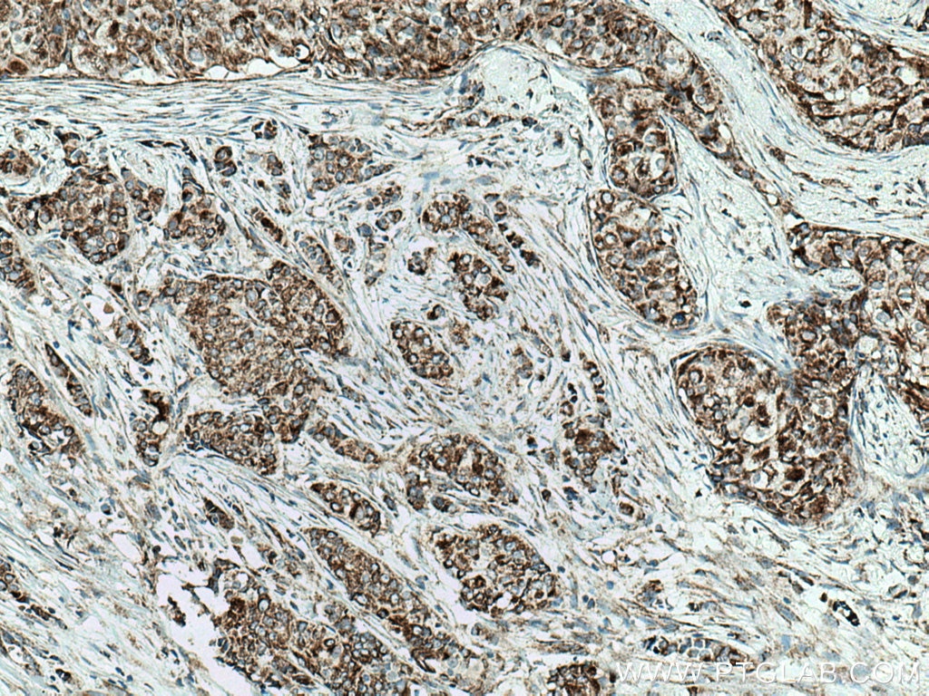
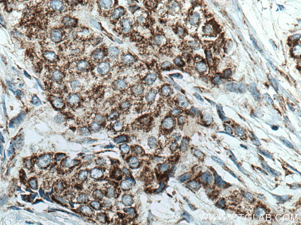
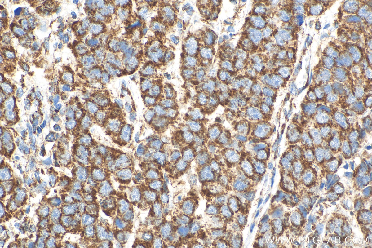
| Positive IHC detected in | human breast cancer tissue Note: suggested antigen retrieval with TE buffer pH 9.0; (*) Alternatively, antigen retrieval may be performed with citrate buffer pH 6.0 |
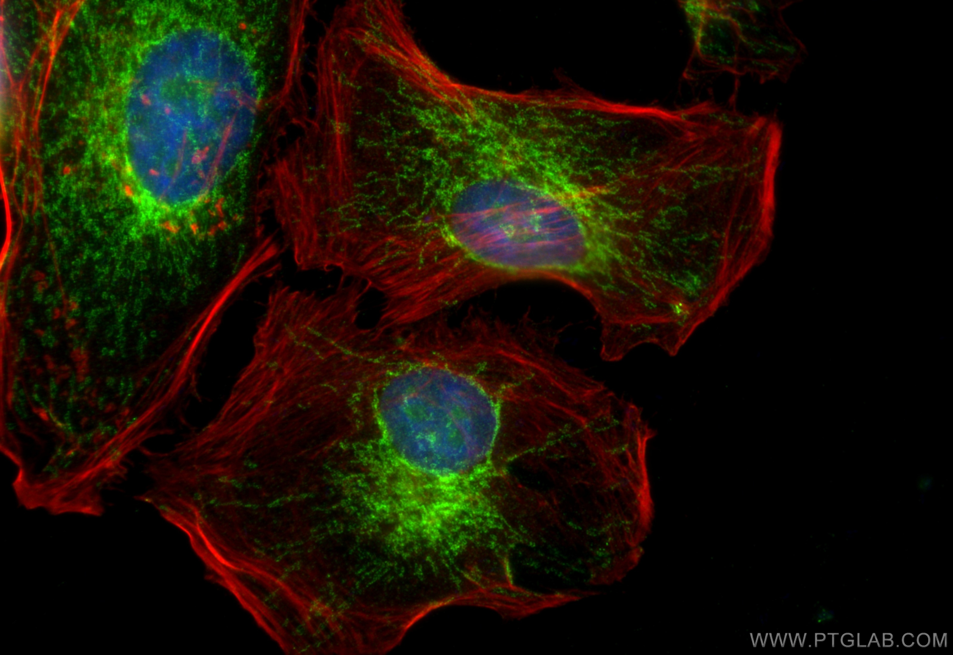
| Positive IF/ICC detected in | HeLa cells |
Recommended dilution
| Application | Dilution |
|---|---|
| Western Blot (WB) | WB : 1:2000-1:16000 |
| Immunoprecipitation (IP) | IP : 0.5-4.0 ug for 1.0-3.0 mg of total protein lysate |
| Immunohistochemistry (IHC) | IHC : 1:250-1:1000 |
| Immunofluorescence (IF)/ICC | IF/ICC : 1:50-1:500 |
| It is recommended that this reagent should be titrated in each testing system to obtain optimal results. | |
| Sample-dependent, Check data in validation data gallery. | |
Published Applications
| KD/KO | See 19 publications below |
| WB | See 97 publications below |
| IHC | See 2 publications below |
| IF | See 17 publications below |
| IP | See 9 publications below |
| CoIP | See 6 publications below |
Product Information
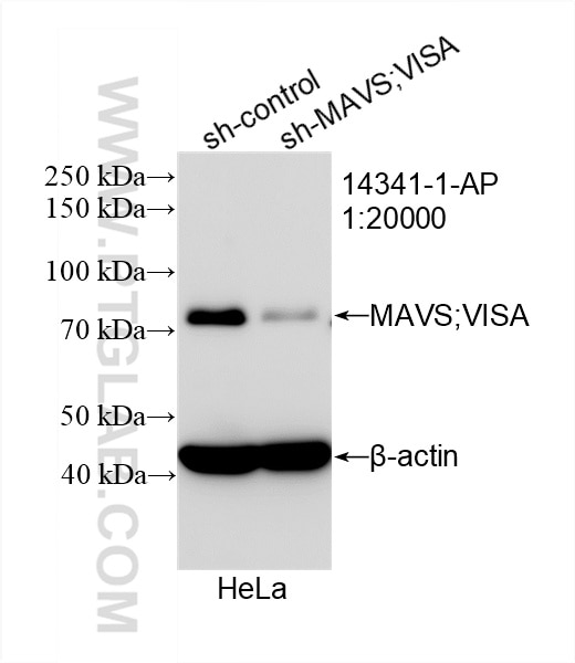
14341-1-AP targets MAVS; VISA in WB, IHC, IF/ICC, IP, CoIP, ELISA applications and shows reactivity with human samples.
| Tested Reactivity | human |
| Cited Reactivity | human, rat, pig, monkey, chicken, duck |
| Host / Isotype | Rabbit / IgG |
| Class | Polyclonal |
| Type | Antibody |
| Immunogen |
CatNo: Ag5655 Product name: Recombinant human MAVS; VISA protein Source: e coli.-derived, PGEX-4T Tag: GST Domain: 1-347 aa of BC044952 Sequence: MPFAEDKTYKYICRNFSNFCNVDVVEILPYLPCLTARDQDRLRATCTLSGNRDTLWHLFNTLQRRPGWVEYFIAALRGCELVDLADEVASVYESYQPRTSDRPPDPLEPPSLPAERPGPPTPAAAHSIPYNSCREKEPSYPMPVQETQAPESPGENSEQALQTLSPRAIPRNPDGGPLESSSDLAALSPLTSSGHQEKDTELGSTHTAGATSSLTPSRGPVSPSVSFQPLARSTPRASRLPGPTGSVVSTGTSFSSSSPGLASAGAAEGKQGAESDQAEPIICSSGAEAPANSLPSKVPTTLMPVNTVALKVPANPASVSTVPSKLPTSSKPPGAVPSNALTNPAPS Predict reactive species |
| Full Name | mitochondrial antiviral signaling protein |
| Calculated Molecular Weight | 57 kDa |
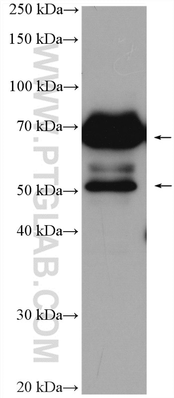
| Observed Molecular Weight | 50-55 kDa, 70-75 kDa |
| GenBank Accession Number | BC044952 |
| Gene Symbol | MAVS |
| Gene ID (NCBI) | 57506 |
| RRID | AB_10548408 |
| Conjugate | Unconjugated |
| Form | Liquid |
| Purification Method | Antigen affinity purification |
| UNIPROT ID | Q7Z434 |
| Storage Buffer | PBS with 0.02% sodium azide and 50% glycerol, pH 7.3. |
| Storage Conditions | Store at -20°C. Stable for one year after shipment. Aliquoting is unnecessary for -20oC storage. 20ul sizes contain 0.1% BSA. |
Background Information
Mitochondrial antiviral-signaling protein (MAVS) is also known as virus-induced-signaling adapter (VISA) or IFN-β promoter stimulator protein 1 (IPS-1), it is widely involved and required for innate immune defense against viruses. MAVS, present in T cells, monocytes, epithelial cells and hepatocytes, contains CARD and transmembrane domains which are essential for antiviral functions. MAVS is able to interact with various cellular proteins including DDX58/RIG-I, IFIH1/MDA5, TRAF2, TRAF6, TMEM173/MITA, IFIT3 and etc. It can undergoe phosphorylation on multiple sites and ubiquitination, which may together cause the molecular weight migrate to about 70 kDa despite the predicated 57 kDa.
Protocols
| Product Specific Protocols | |
|---|---|
| IF protocol for MAVS; VISA antibody 14341-1-AP | Download protocol |
| IHC protocol for MAVS; VISA antibody 14341-1-AP | Download protocol |
| IP protocol for MAVS; VISA antibody 14341-1-AP | Download protocol |
| WB protocol for MAVS; VISA antibody 14341-1-AP | Download protocol |
| Standard Protocols | |
|---|---|
| Click here to view our Standard Protocols |
Publications
| Species | Application | Title |
|---|---|---|
Signal Transduct Target Ther TRAF3 activates STING-mediated suppression of EV-A71 and target of viral evasion | ||
Nat Immunol ALOX15 orchestrates mitochondrial antiviral immunity and serves as a host target for anti-influenza therapy | ||
Immunity Decreased Expression of the Host Long-Noncoding RNA-GM Facilitates Viral Escape by Inhibiting the Kinase activity TBK1 via S-glutathionylation. | ||
Mol Cell An Epstein-Barr virus protein interaction map reveals NLRP3 inflammasome evasion via MAVS UFMylation | ||
Nat Commun MLL5 suppresses antiviral innate immune response by facilitating STUB1-mediated RIG-I degradation. | ||
EMBO J CircPVT1 promotes ER-positive breast tumorigenesis and drug resistance by targeting ESR1 and MAVS
|
Reviews
The reviews below have been submitted by verified Proteintech customers who received an incentive for providing their feedback.
FH Damien (Verified Customer) (09-03-2019) | No convince by this anti-MAVS antibody.In fact, in human cells MAVS protein is expressed in two differents forms : short-MAVS (55kDa) and full-length MAVS (72 kDa). So I have to observe two bands by WB. However, after using different conditions with this antibody according to the manufacturer's protocol I always observe a smear of protein, and one band at 72 kDa. The expected on around 55 kDa never appear. Note that my cells lysates were not degraded since the others proteins that I want to reveal were great.
|