Recombinant Mouse CD36 protein (rFc Tag)
Species
Mouse
Purity
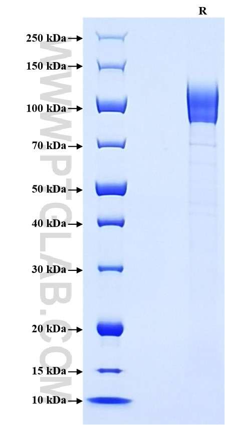
>90 %, SDS-PAGE
Tag
rFc Tag
Activity
not tested
Cat no : Eg3464
Validation Data Gallery
Product Information
| Purity | >90 %, SDS-PAGE |
| Endotoxin | <0.1 EU/μg protein, LAL method |
| Activity |
Not tested |
| Expression | HEK293-derived Mouse CD36 protein Gly30-Lys439 (Accession# Q08857) with a rabbit IgG Fc tag at the C-terminus. |
| GeneID | 12491 |
| Accession | Q08857 |
| PredictedSize | 72.7 kDa |
| SDS-PAGE | 85-120 kDa, reducing (R) conditions |
| Formulation | Lyophilized from 0.22 μm filtered solution in PBS, pH 7.4. Normally 5% trehalose and 5% mannitol are added as protectants before lyophilization. |
| Reconstitution | Briefly centrifuge the tube before opening. Reconstitute at 0.1-0.5 mg/mL in sterile water. |
| Storage Conditions |
It is recommended that the protein be aliquoted for optimal storage. Avoid repeated freeze-thaw cycles.
|
| Shipping | The product is shipped at ambient temperature. Upon receipt, store it immediately at the recommended temperature. |
Background
CD36 is also named as GP3B, GP4, Glycoprotein IIIb (GPIIIB), PAS-4 and Fatty acid translocase (FAT). CD36, also known as the scavenger receptor B2, is a multifunctional receptor widely expressed in various organs. CD36 is a vital molecule in lipid metabolism. In heart and muscle, CD36 is the main (long-chain) fatty acid transporter, regulating myocellular fatty acid uptake via its vesicle-mediated reversible trafficking (recycling) between intracellular membrane compartments and the cell surface. CD36 is subject to various types of post-translational modification. CD36 is expressed in various tissues, including endothelial cells, cardiac muscle cells, renal tubular epithelial cells, liver cells, adipocytes, platelets, and macrophages, and is involved in many pathophysiological processes, including immune regulation and metabolic regulation. CD36 mediates a mitochondrial metabolic switch from oxidative phosphorylation to superoxide production in response to its ligand, oxidized LDL.
References:
1.Shu H, et al. (2022) Cardiovasc Res. 118(1):115-129.
2.Kim TT, Dyck JR. (2016) Biochim Biophys Acta. 1861(10):1450-60.
3.Luiken JJ, et al. (2016) Biochim Biophys Acta. 1862(12):2253-2258.
4.Campbell SE, et al. (2004) J Biol Chem. 279(35):36235-41.
5.Bezaire V, et al. (2006) Am J Physiol Endocrinol Metab. 290(3):E509-15.
