Recombinant Mouse Tie-2/CD202b protein (rFc Tag)
Species
Mouse
Purity
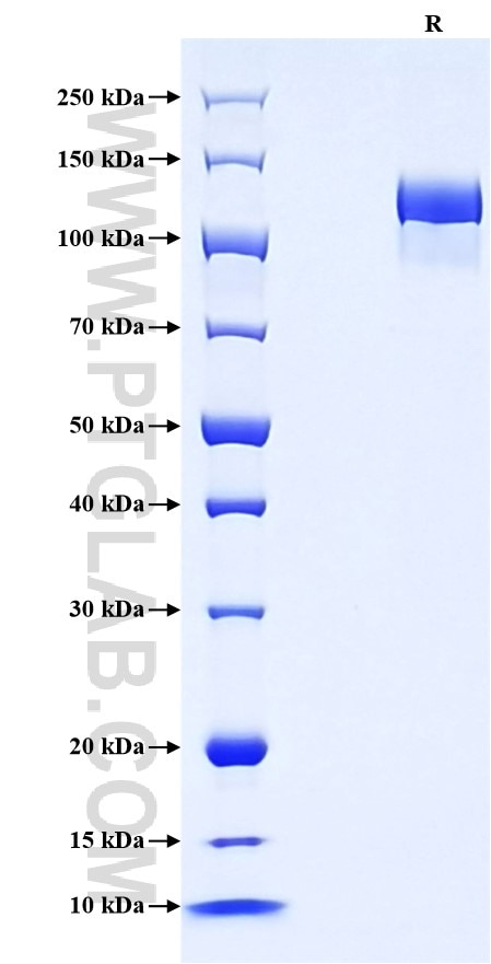
>90 %, SDS-PAGE
Tag
rFc Tag
Activity
not tested
Cat no : Eg1514
Validation Data Gallery
Product Information
| Purity | >90 %, SDS-PAGE |
| Endotoxin | <0.1 EU/μg protein, LAL method |
| Activity |
Not tested |
| Expression | HEK293-derived Mouse Tie-2 protein Ala23-Lys744 (Accession# Q02858) with a rabbit IgG Fc tag at the C-terminus. |
| GeneID | 21687 |
| Accession | Q02858 |
| PredictedSize | 106.9 kDa |
| SDS-PAGE | 120-140 kDa, reducing (R) conditions |
| Formulation | Lyophilized from 0.22 μm filtered solution in PBS, pH 7.4. Normally 5% trehalose and 5% mannitol are added as protectants before lyophilization. |
| Reconstitution | Briefly centrifuge the tube before opening. Reconstitute at 0.1-0.5 mg/mL in sterile water. |
| Storage Conditions |
It is recommended that the protein be aliquoted for optimal storage. Avoid repeated freeze-thaw cycles.
|
| Shipping | The product is shipped at ambient temperature. Upon receipt, store it immediately at the recommended temperature. |
Background
Tie-2, also known as CD202b or TEK, is a receptor tyrosine kinase (RTK) specifically expressed in endothelial cells. It plays a crucial role in angiogenesis, forming new blood vessels, and maintaining vascular stability. TIE-2 is a tyrosine kinase receptor expressed mainly by endothelial cells, cancer cells, and some immune cells like monocytes. Tie-2 is a member of the Tie receptor family, characterized by the presence of immunoglobulin and epidermal growth factor (EGF) homology domains. Tie-2 is selectively expressed in endothelial cells and gene deficiency of Tie-2 leads to embryonic lethality due to abnormal vascular development. Ang-1, which is secreted from perivascular cells, has been identified as the major ligand for Tie-2. Binding of Ang-1 to Tie-2 elicits different downstream signaling pathways mainly regulated by Akt, which induces pro-angiogenic responses including the promotion of endothelial cell migration, tube formation and survival.
References:
1. Dumont DJ. et al. (1992) Oncogene. 7(8):1471-80.
2. Duran CL. et al. (2021) Cancers (Basel). 13(22):5730.
3. Sato TN. et al. (1995) Nature. 376(6535):70-4.
4. Isidori AM. et al. (2016) J Endocrinol Invest. 39(11):1235-1246.
5. Fukuhara S. et al. (2010) Histol Histopathol. 25(3):387-96.
