Tested Applications
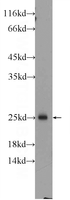
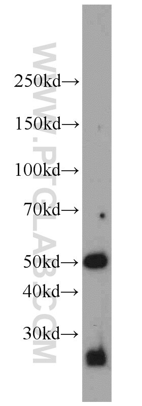
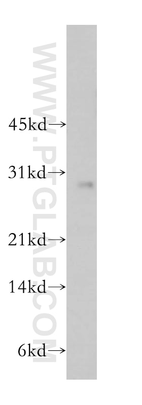
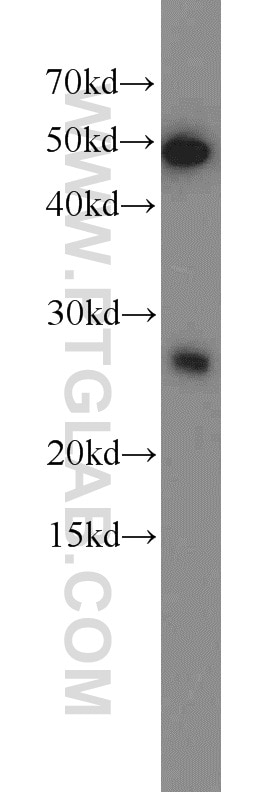
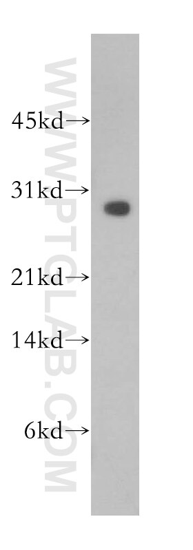
| Positive WB detected in | rat spleen tissue, A375 cells, DU 145 cells, MCF-7 cells, PC-3 cells |
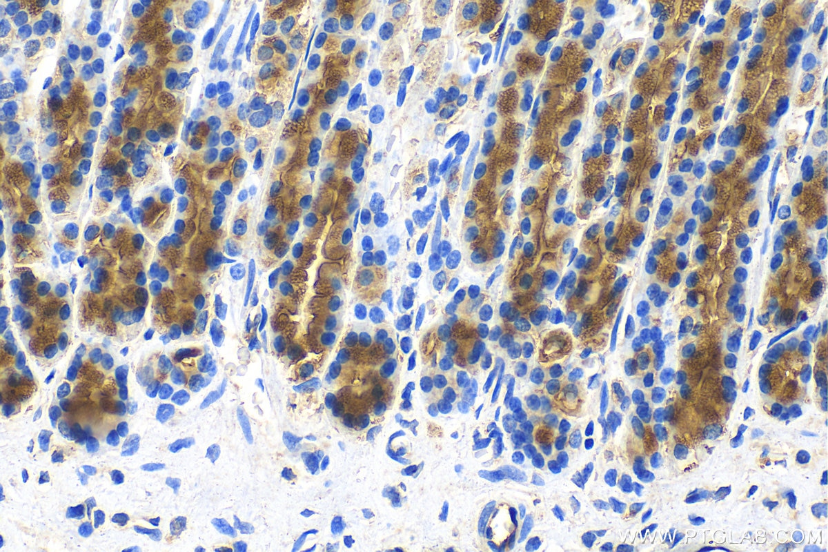
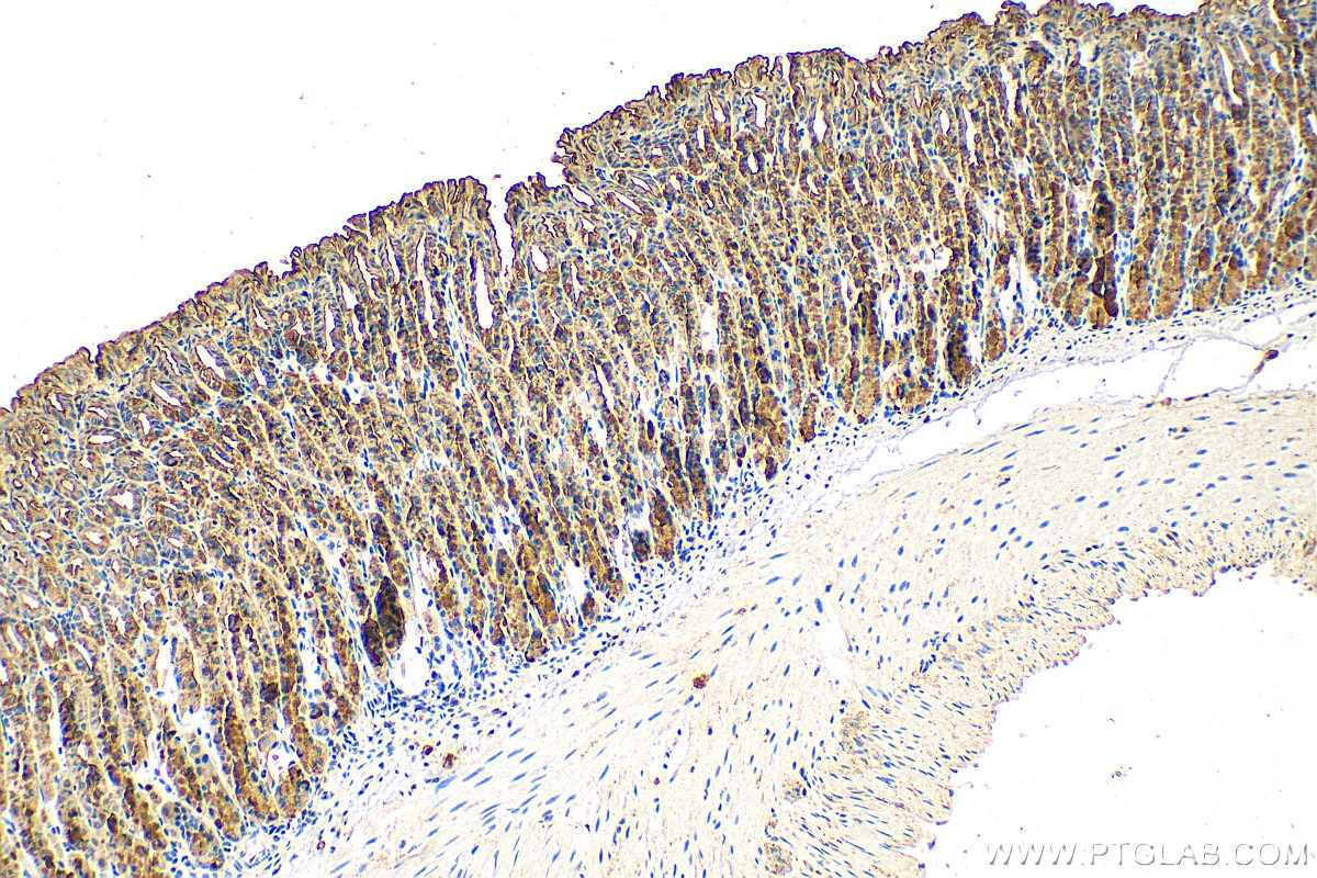
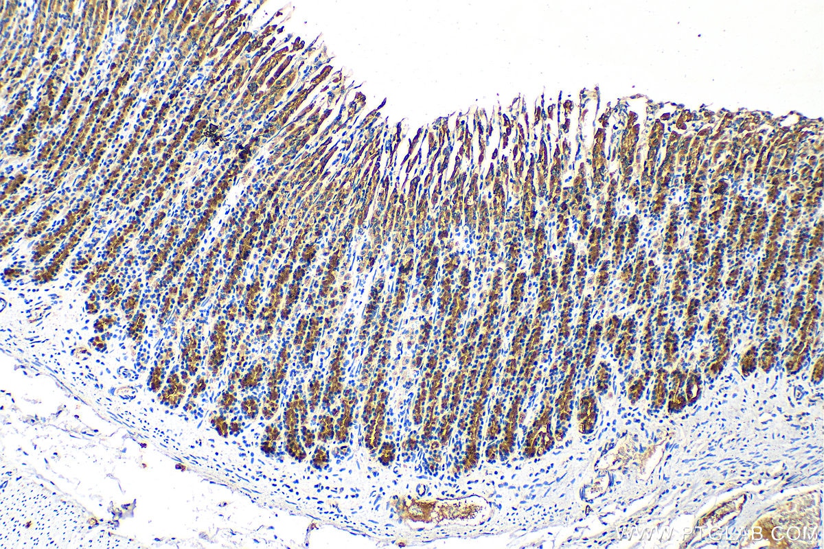
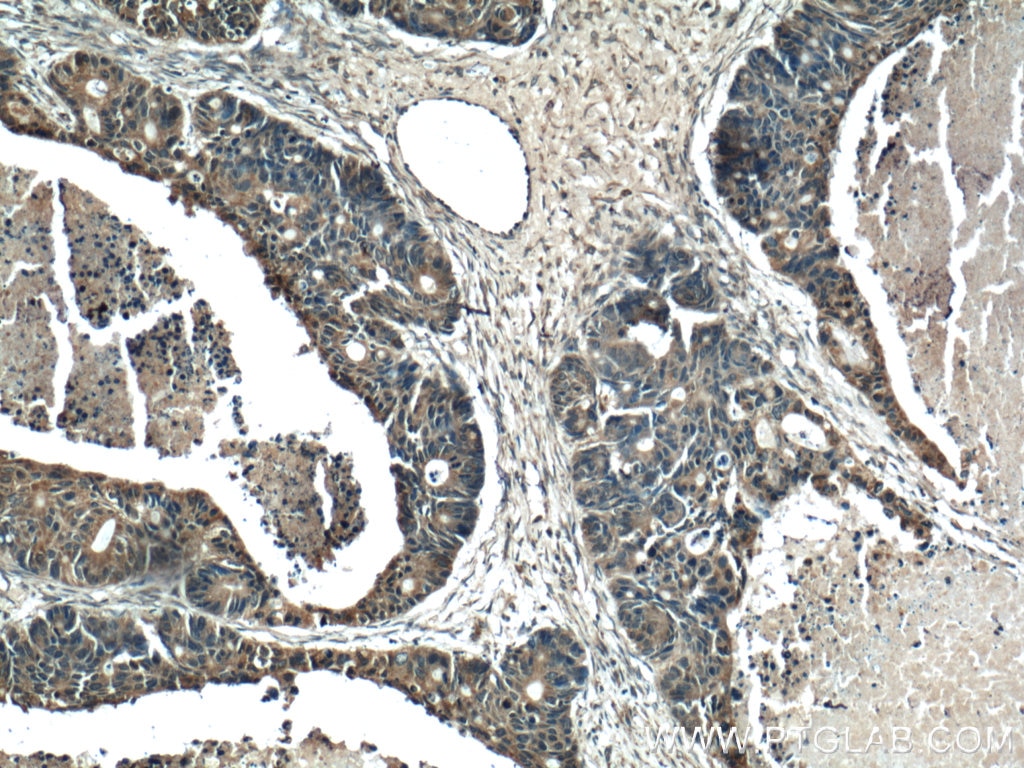
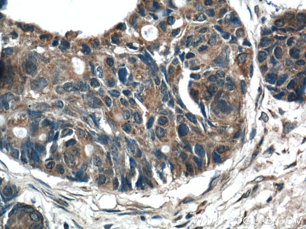
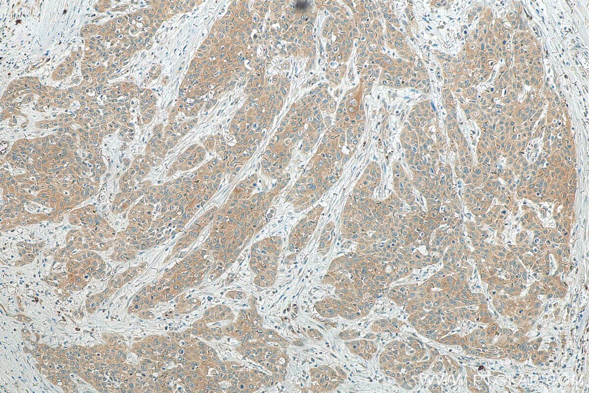
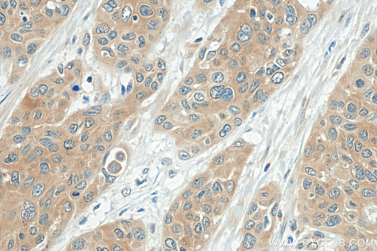
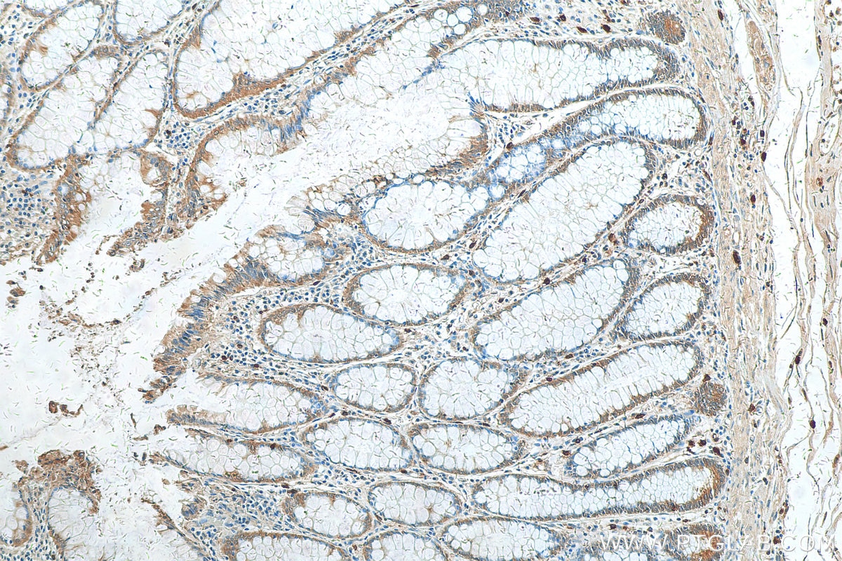
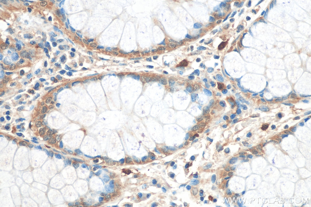
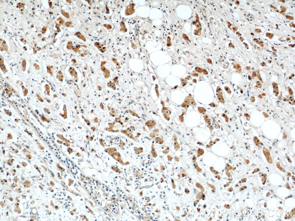
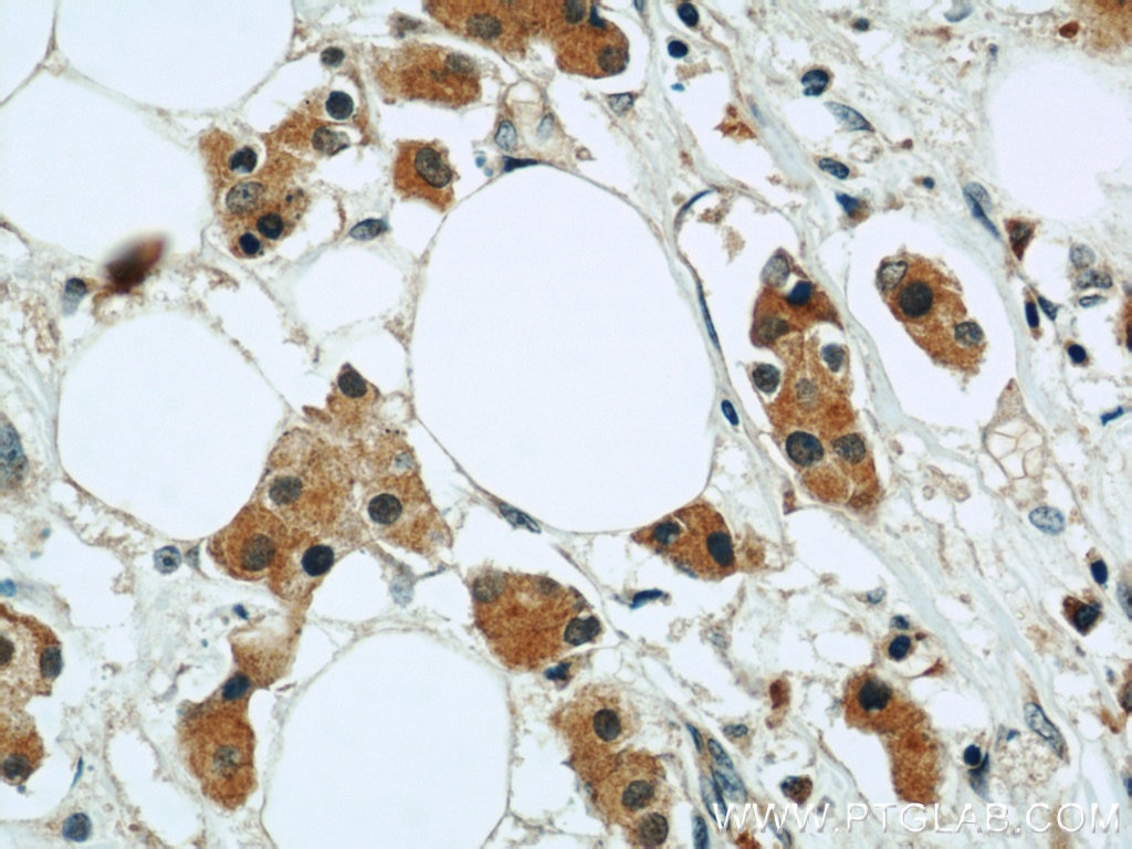
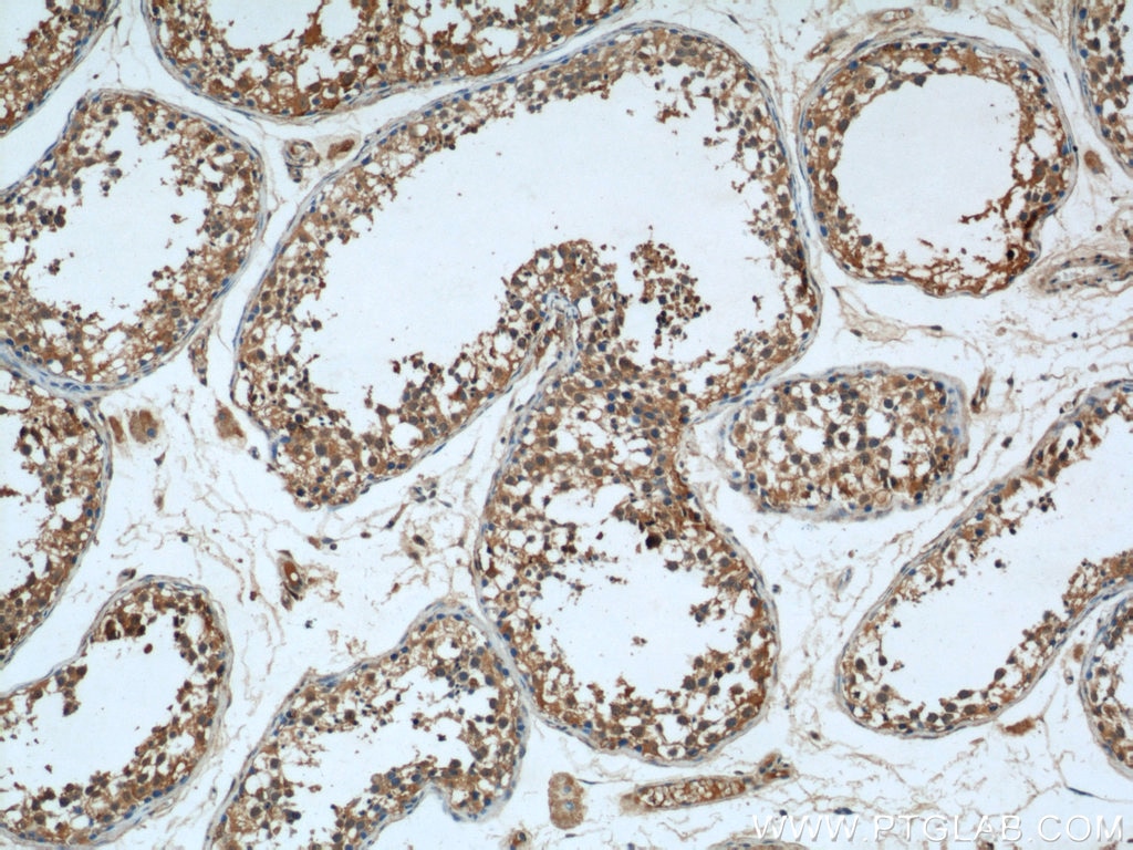
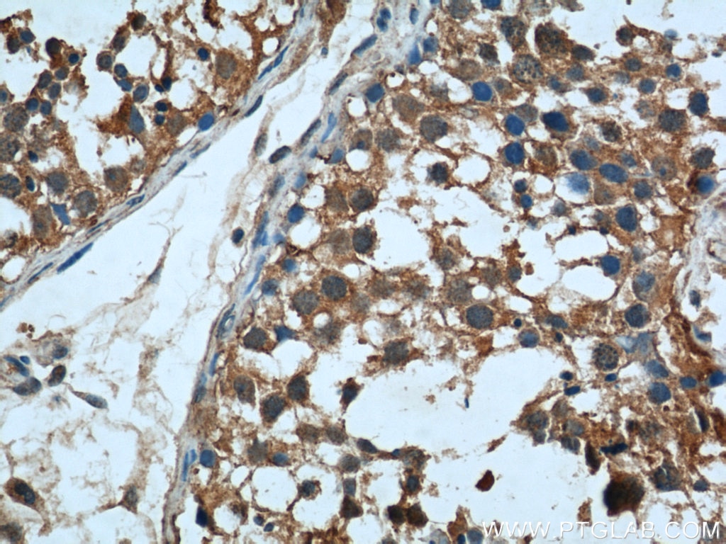
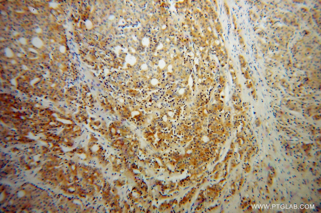
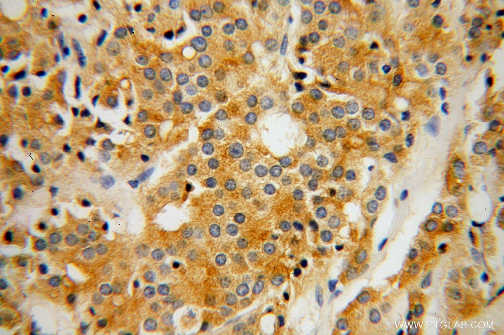
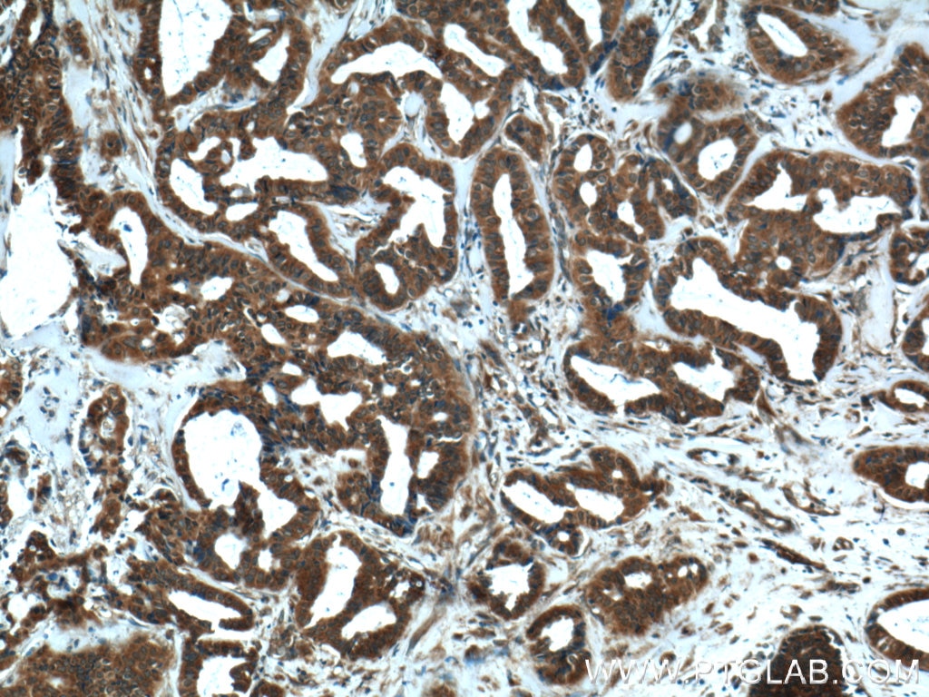
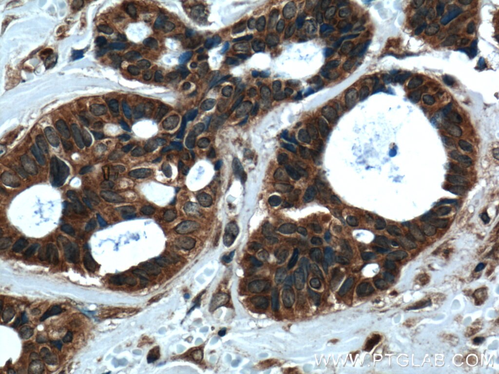
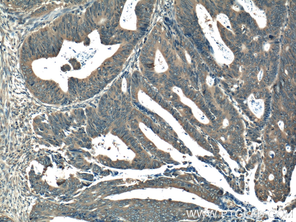
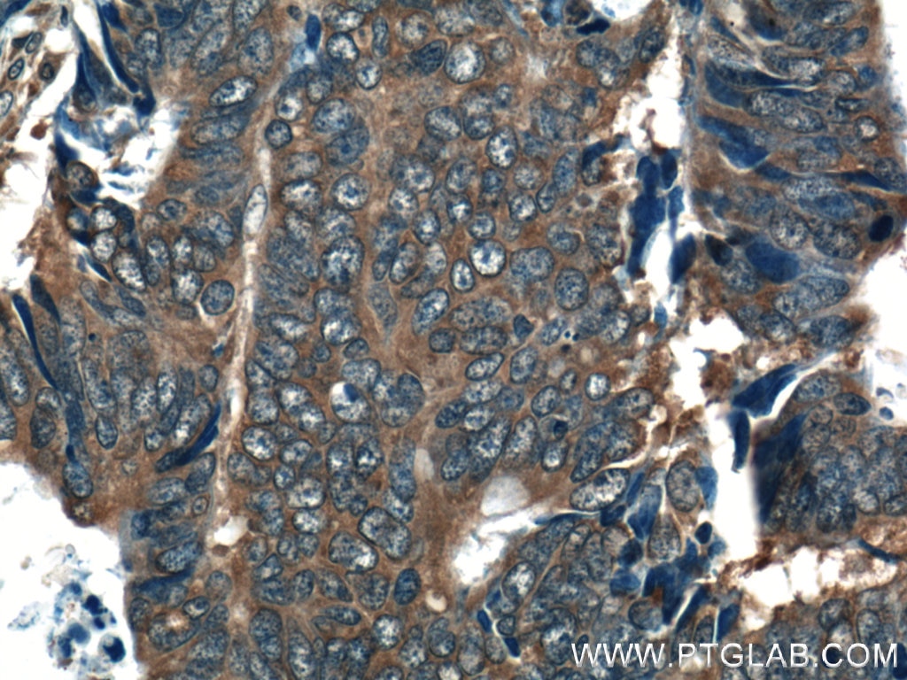
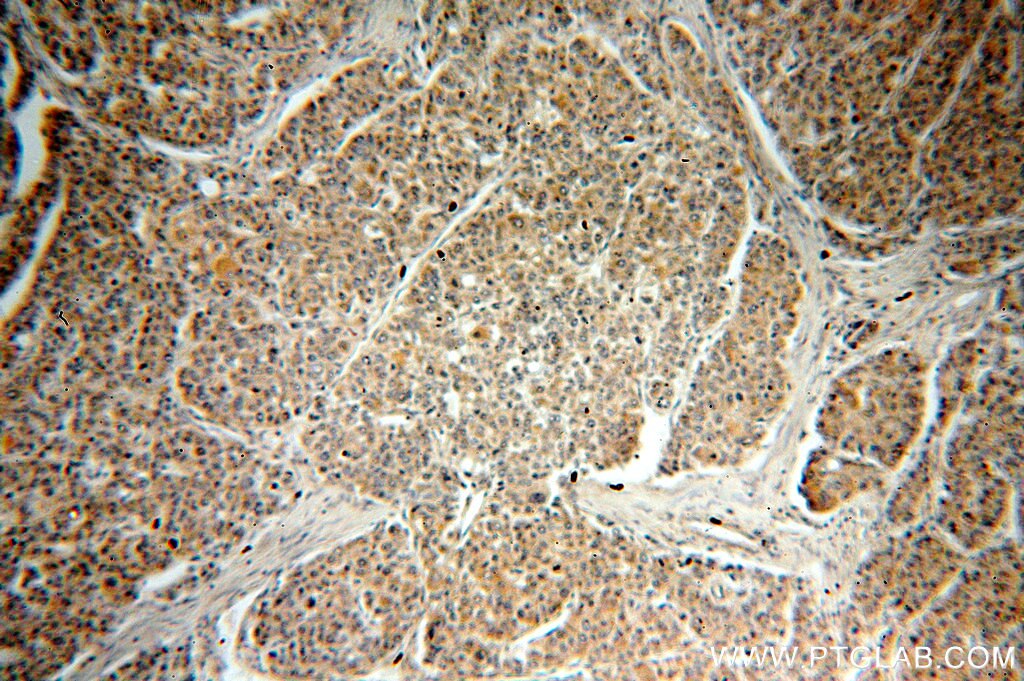
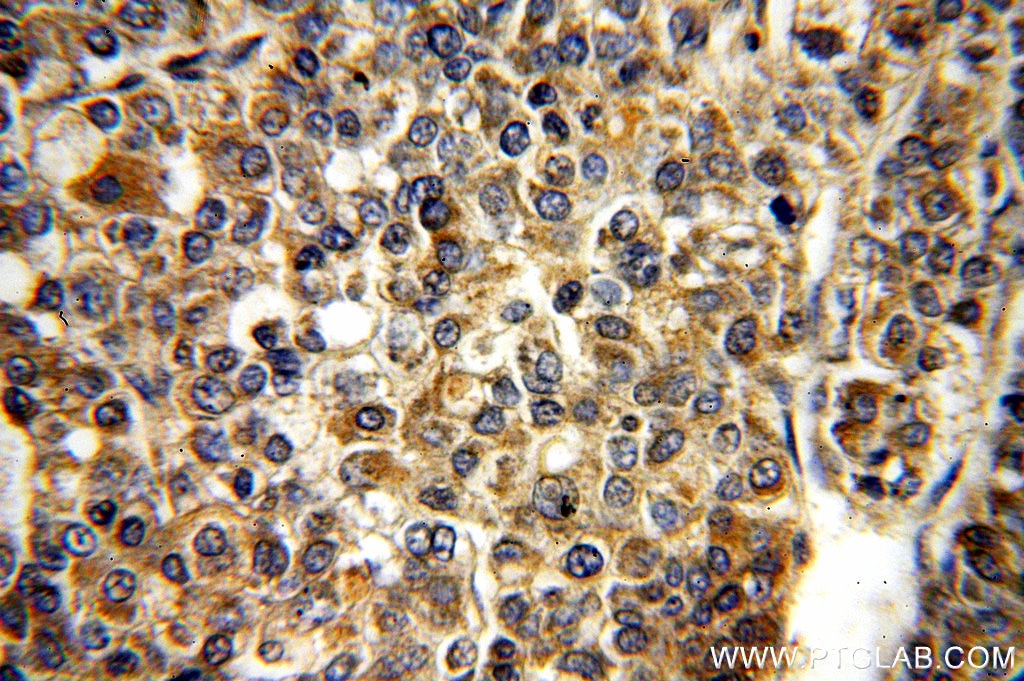
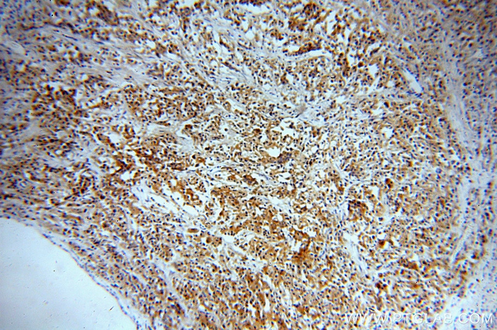
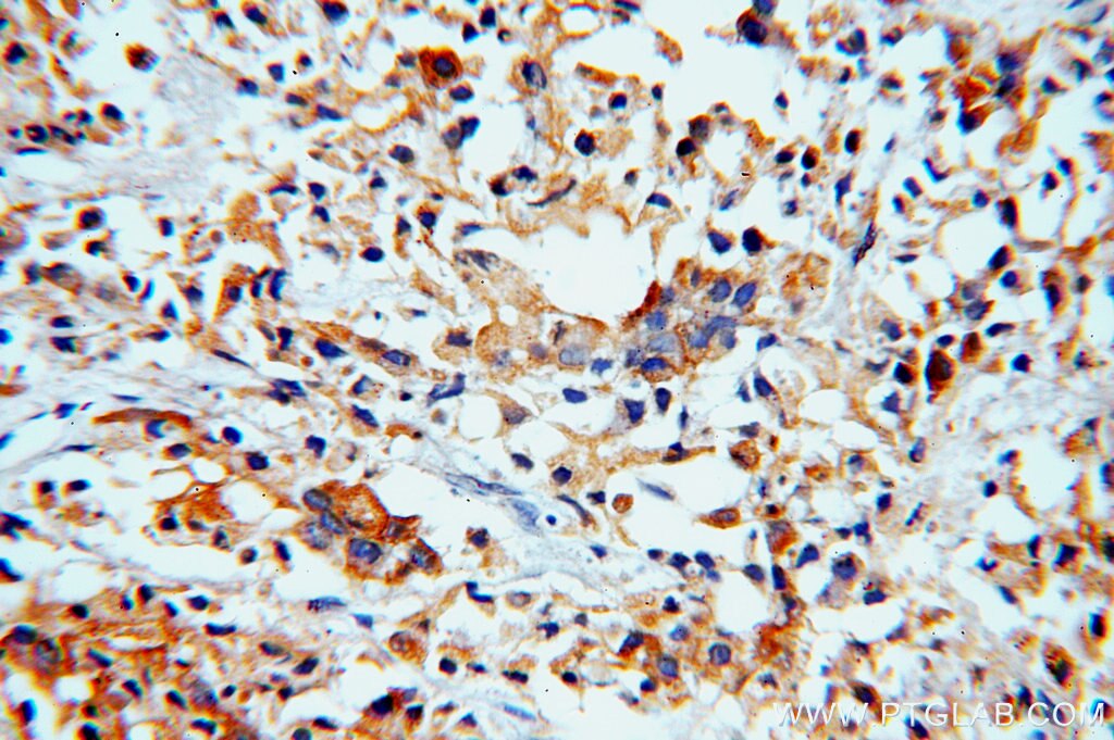
| Positive IHC detected in | rat stomach tissue, human breast cancer tissue, human colon cancer tissue, human liver cancer tissue, human oesophagus cancer tissue, human pancreas cancer tissue, human prostate cancer tissue, human testis tissue, mouse stomach tissue Note: suggested antigen retrieval with TE buffer pH 9.0; (*) Alternatively, antigen retrieval may be performed with citrate buffer pH 6.0 |
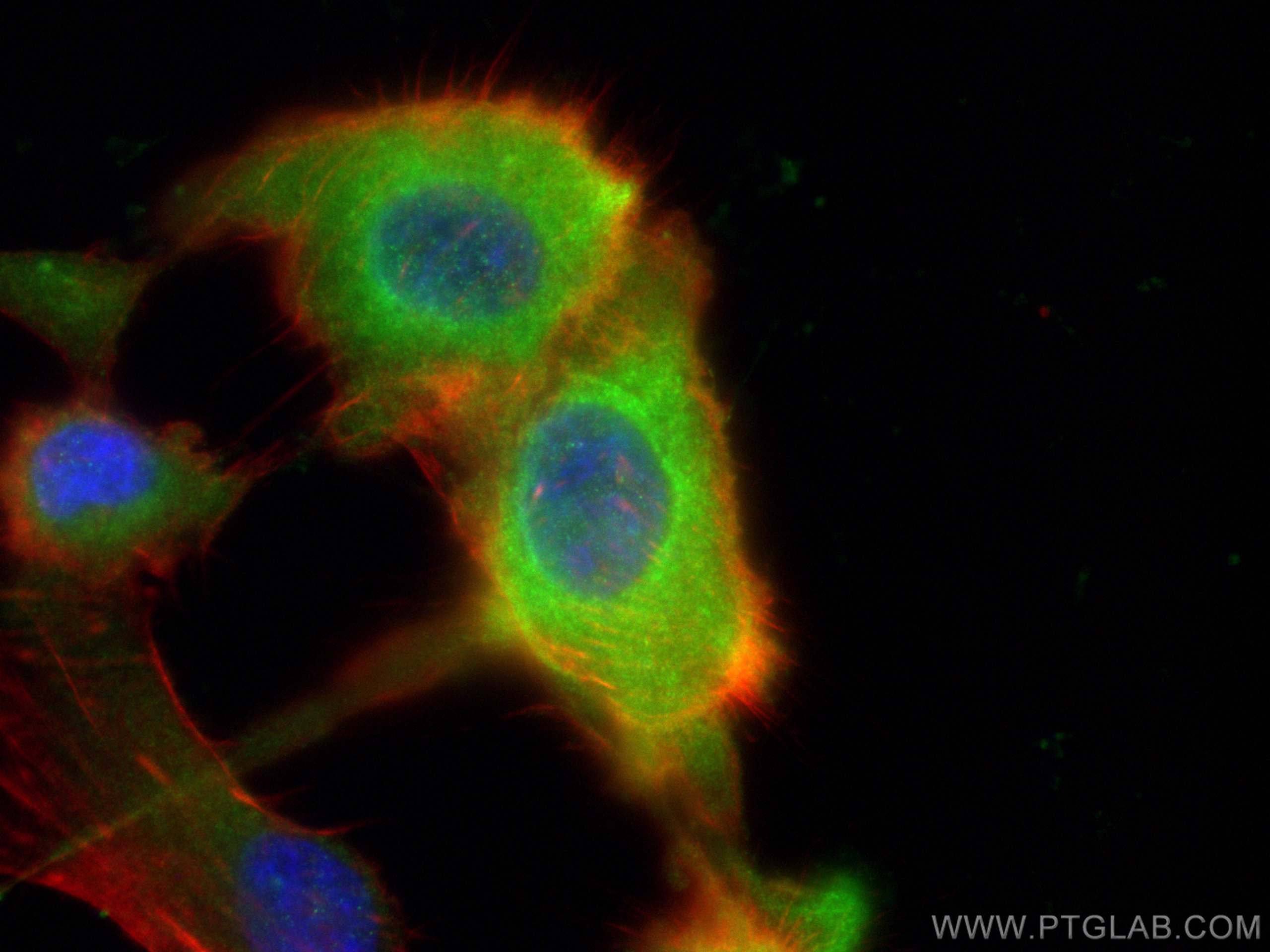
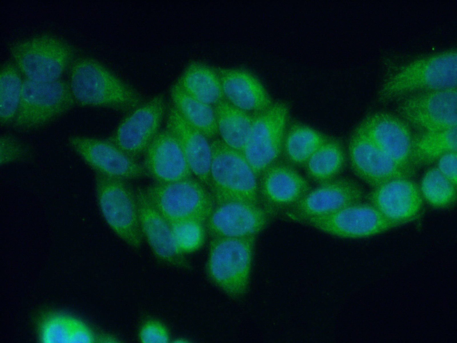
| Positive IF/ICC detected in | U-87 MG cells, HeLa cells |
Recommended dilution
| Application | Dilution |
|---|---|
| Western Blot (WB) | WB : 1:500-1:2000 |
| Immunohistochemistry (IHC) | IHC : 1:250-1:1000 |
| Immunofluorescence (IF)/ICC | IF/ICC : 1:50-1:500 |
| It is recommended that this reagent should be titrated in each testing system to obtain optimal results. | |
| Sample-dependent, Check data in validation data gallery. | |
Published Applications
| KD/KO | See 13 publications below |
| WB | See 43 publications below |
| IHC | See 16 publications below |
| IF | See 13 publications below |
| CoIP | See 1 publications below |
Product Information
13412-1-AP targets RAB27B in WB, IHC, IF/ICC, CoIP, ELISA applications and shows reactivity with human, mouse, rat samples.
| Tested Reactivity | human, mouse, rat |
| Cited Reactivity | human, mouse, rat, canine |
| Host / Isotype | Rabbit / IgG |
| Class | Polyclonal |
| Type | Antibody |
| Immunogen |
CatNo: Ag4064 Product name: Recombinant human RAB27B protein Source: e coli.-derived, PGEX-4T Tag: GST Domain: 1-218 aa of BC027474 Sequence: MTDGDYDYLIKLLALGDSGVGKTTFLYRYTDNKFNPKFITTVGIDFREKRVVYNAQGPNGSSGKAFKVHLQLWDTAGQERFRSLTTAFFRDAMGFLLMFDLTSQQSFLNVRNWMSQLQANAYCENPDIVLIGNKADLPDQREVNERQARELADKYGIPYFETSAATGQNVEKAVETLLDLIMKRMEQCVEKTQIPDTVNGGNSGNLDGEKPPEKKCIC Predict reactive species |
| Full Name | RAB27B, member RAS oncogene family |
| Calculated Molecular Weight | 218 aa, 25 kDa |
| Observed Molecular Weight | 25-30 kDa |
| GenBank Accession Number | BC027474 |
| Gene Symbol | RAB27B |
| Gene ID (NCBI) | 5874 |
| RRID | AB_2176732 |
| Conjugate | Unconjugated |
| Form | Liquid |
| Purification Method | Antigen affinity purification |
| UNIPROT ID | O00194 |
| Storage Buffer | PBS with 0.02% sodium azide and 50% glycerol, pH 7.3. |
| Storage Conditions | Store at -20°C. Stable for one year after shipment. Aliquoting is unnecessary for -20oC storage. 20ul sizes contain 0.1% BSA. |
Background Information
The Rab27 GTPase subfamily consists of two closely related homologs, Rab27a and Rab27b. Northern blot analysis revealed that Rab27b is expressed significantly only in testis, in which the predominant transcript was 1.4 kb in size. An 8-kb mRNA of unknown origin was detected in many tissues. Rab27b is associated with tubulovesicle membranes in the parietal cell and Rab27b may play a role in stimulation-associated membrane recruitment and gastric acid secretion. Rab27b is recently reported as a marker for breast cancer progression. It regulates invasive growth and metastasis in ER-positive breast cancer cell lines, and increased expression is associated with poor prognosis in humans.
Protocols
| Product Specific Protocols | |
|---|---|
| IF protocol for RAB27B antibody 13412-1-AP | Download protocol |
| IHC protocol for RAB27B antibody 13412-1-AP | Download protocol |
| WB protocol for RAB27B antibody 13412-1-AP | Download protocol |
| Standard Protocols | |
|---|---|
| Click here to view our Standard Protocols |
Publications
| Species | Application | Title |
|---|---|---|
Nature Microenvironment-induced PTEN loss by exosomal microRNA primes brain metastasis outgrowth. | ||
Cell Exosome transfer from stromal to breast cancer cells regulates therapy resistance pathways. | ||
Immunity MLKL, the Protein that Mediates Necroptosis, Also Regulates Endosomal Trafficking and Extracellular Vesicle Generation. | ||
Nat Commun Identification of genes associated with cortical malformation using a transposon-mediated somatic mutagenesis screen in mice. | ||
Reviews
The reviews below have been submitted by verified Proteintech customers who received an incentive for providing their feedback.
FH Kacie (Verified Customer) (09-12-2022) | Antibody works well for both ICC and WB.
|