
Validation Data Gallery
![Dilution series of mCherry in HEK 293T
cell lysate
Primary antibody: [6G6] 1:1,000
Secondary antibody: goat anti-mouse
Alexa Fluor® 647 1:1,000](https://www.ptglab.com/products/pictures/Result-6g6.png "Dilution series of mCherry in HEK 293T
cell lysate
Primary antibody: [6G6] 1:1,000
Secondary antibody: goat anti-mouse
Alexa Fluor® 647 1:1,000")
Product Information
Mouse monoclonal antibody [6G6] to Red Fluorescent Proteins
| Description | RFP mouse monoclonal [6G6] for WB |
| Applications | Western blotting |
| Specificity/Target | Tested on mOrange, OFP, DsRed, mCherry, mRFP, mRFPRuby, mScarlet, mScarlet-I, tdTomato, mPlum. For the complete list, please click here: Fluorescent protein specificity table antibodies |
| Recommended Dilution | Western Blot: 1:2,000 |
| Host/IsoType | Mouse/IgG2c |
| Class | Monoclonal |
| Type | Primary Antibody |
| Immunogen | Red fluorescent protein (RFP) |
| UniProtKB | Q9U6Y8 |
| Clone No. | 6G6 |
| Conjugate | Unconjugated |
| Form | Liquid |
| Purification Method | Protein A or G purified |
| RRID | AB_2631395 |
| Storage Buffer | PBS, 50% glycerol, preservative: 0.09% sodium azide. |
| Storage Condition | Shipped on ice. Store at -20°C/-4°F. Stable for 1 year after shipment. |
| Size | 20 μL; 150 μL |
| Background | Red fluorescent proteins (RFPs) is a collective term referring to a heterogenous group of red chromophore-carrying proteins, originating from various species and forming different protein lineages. The original RFP (dsRed) is a 225 amino acid fluorescent protein (25.9 kDa) derived from Discosoma sp.. It emits red light with a peak wavelength of 593 nm upon excitation by green light (excitation peak at 558 nm). When fused with other proteins, RFP serves as a versatile reporter protein e.g. for quantifying expression levels or facilitates visualization of subcellular localization through fluorescence microscopy. This antibody is a mouse (IgG2c) monoclonal antibody raised against RFP, reactive to a variety of RFP derivates, including mOrange, OFP, DsRed, mCherry, mRFP, mRFPRuby, mScarlet, mScarlet-I, tdTomato, and mPlum. |
Publications
| Application | Title |
|---|---|
Nature POLAR-guided signalling complex assembly and localization drive asymmetric cell division. | |
Cell Deficient Endoplasmic Reticulum-Mitochondrial Phosphatidylserine Transfer Causes Liver Disease. | |
Cell Formation of NPR1 Condensates Promotes Cell Survival during the Plant Immune Response. |
Reviews
The reviews below have been submitted by verified Proteintech customers who received an incentive for providing their feedback.
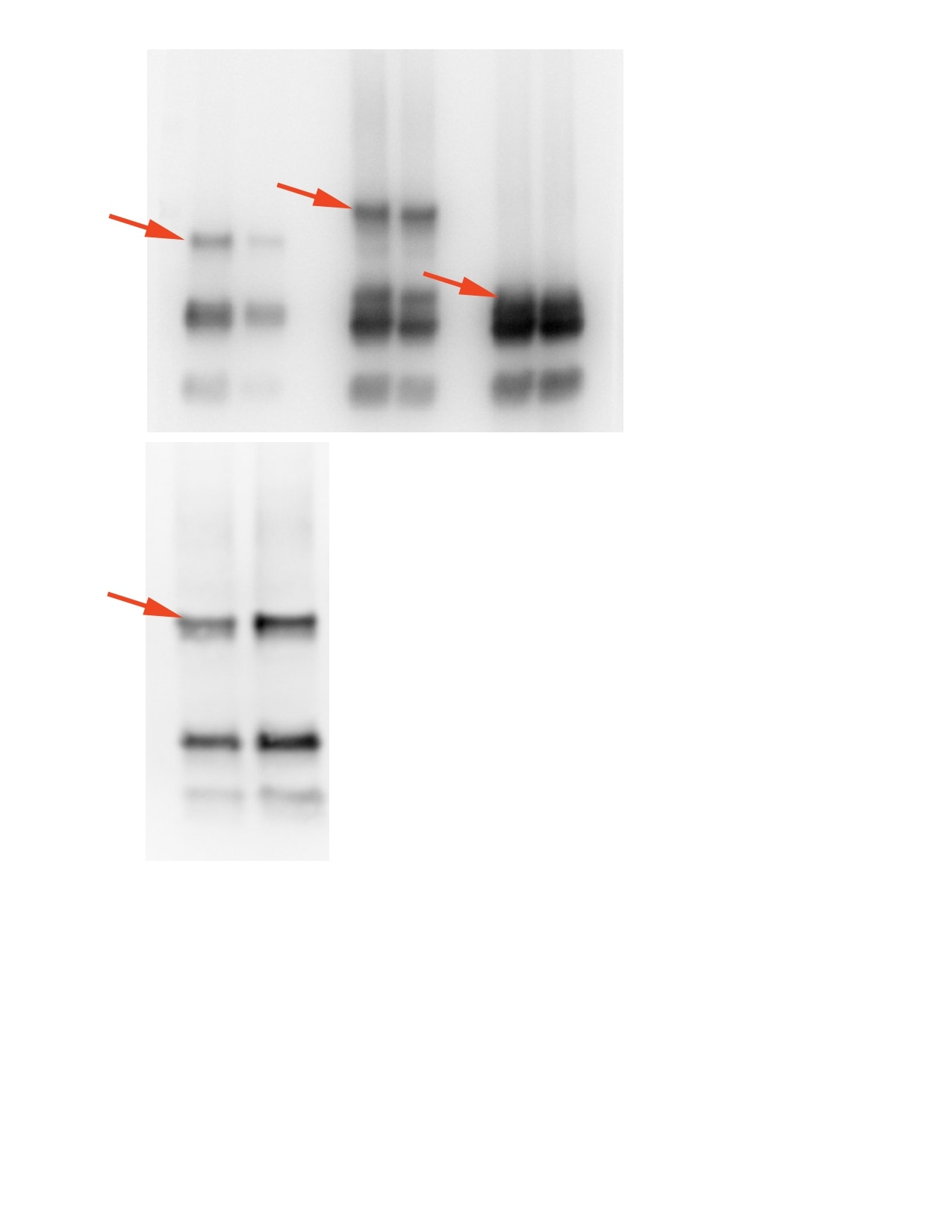
FH Iga (Verified Customer) (08-17-2022) | The picture shows samples of four different mCherry tagged proteins produced by Phytophthora capsici (two independent transgenic lines each time). Each sample was immunoprecipitated from plant tissue infected by transgenic Phytophthora. The correct size of the proteins of interest is indicated by the red arrow. The explanation of the presence of additional bands on western blot: 1) could be that it's the matter of mCherry cleavage that occurs inside Phytophthora mycelium/ 2) modification of protein size that is the consequence of protein maturation inside mycelium/ 3) these are proteins that are secreted by Phytophthora during infection to manipulate plant immunity- it's possible that the host plant, in order to defend from the pathogen, has proteases that cleaves Phytophthora proteins. From my experience I wouldn't interpret these additional bands as the consequence of the revalation with antibody (confocal microscopy pictures showed that the proteins of interest are localised in additional compartments than only extrahaustorial space).
 |