Tested Applications
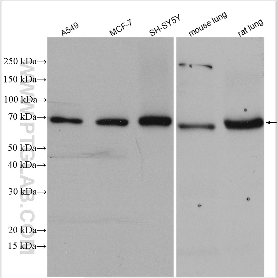
| Positive WB detected in | A549 cells, MCF-7 cells, rat kidney tissue, Jurkat cells, mouse kidney tissue, L02 cells, SH-SY5Y cells, mouse lung tissue, rat lung tissue |
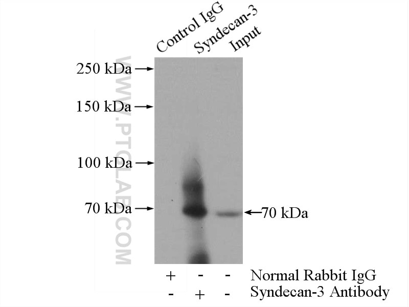
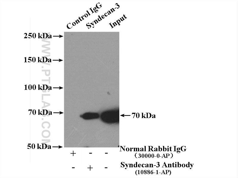
| Positive IP detected in | mouse lung tissue, A549 cells |
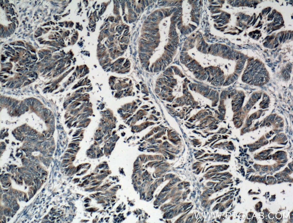
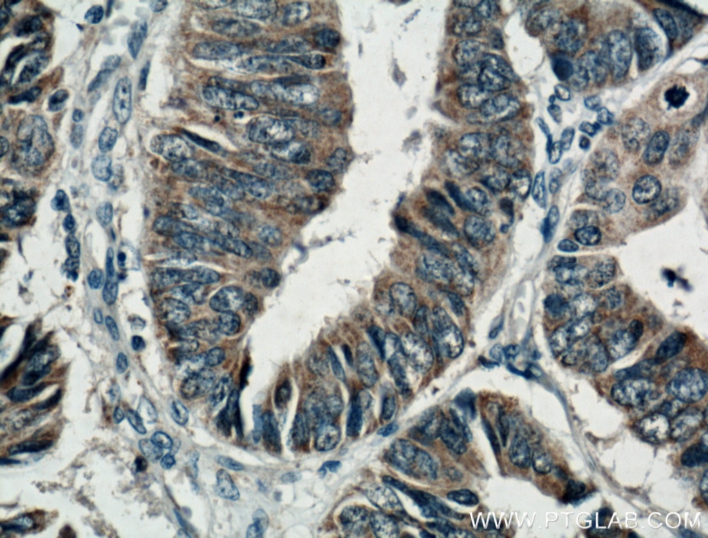
| Positive IHC detected in | human colon cancer tissue Note: suggested antigen retrieval with TE buffer pH 9.0; (*) Alternatively, antigen retrieval may be performed with citrate buffer pH 6.0 |
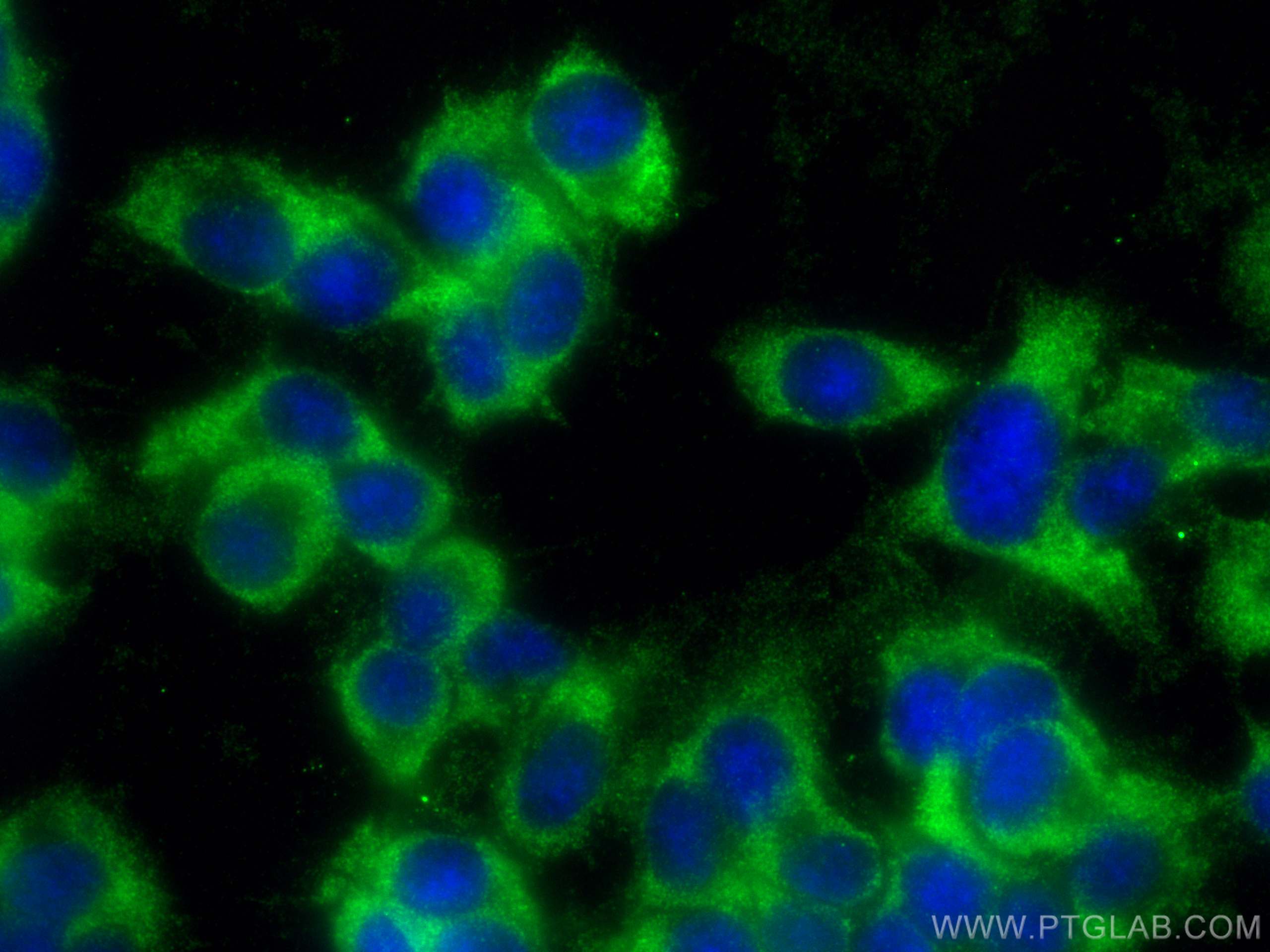
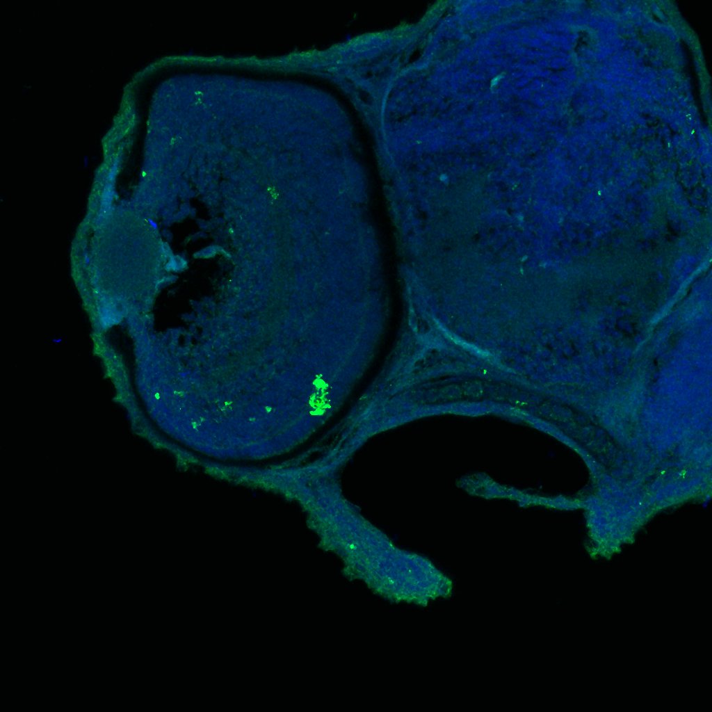
| Positive IF/ICC detected in | PC-12 cells, MCF-7 cells |
Recommended dilution
| Application | Dilution |
|---|---|
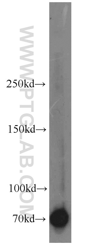
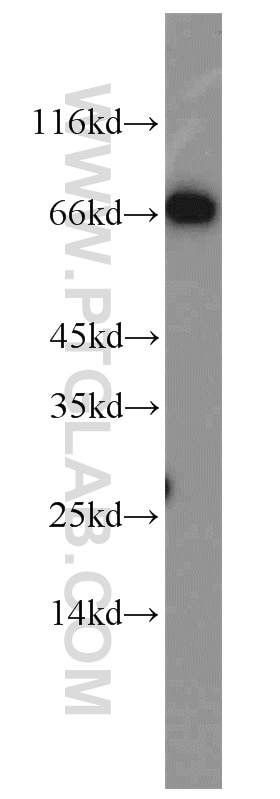
| Western Blot (WB) | WB : 1:500-1:2000 |
| Immunoprecipitation (IP) | IP : 0.5-4.0 ug for 1.0-3.0 mg of total protein lysate |
| Immunohistochemistry (IHC) | IHC : 1:50-1:500 |
| Immunofluorescence (IF)/ICC | IF/ICC : 1:50-1:500 |
| It is recommended that this reagent should be titrated in each testing system to obtain optimal results. | |
| Sample-dependent, Check data in validation data gallery. | |
Published Applications
| WB | See 2 publications below |
| IHC | See 4 publications below |
| IF | See 2 publications below |
Product Information
10886-1-AP targets Syndecan-3 in WB, IHC, IF/ICC, IP, ELISA applications and shows reactivity with human, mouse, rat samples.
| Tested Reactivity | human, mouse, rat |
| Cited Reactivity | human, mouse |
| Host / Isotype | Rabbit / IgG |
| Class | Polyclonal |
| Type | Antibody |
| Immunogen |
CatNo: Ag1317 Product name: Recombinant human SDC3 protein Source: e coli.-derived, PGEX-4T Tag: GST Domain: 1-370 aa of BC013974 Sequence: MAIAYLGSSCPSQPPSSLALSLSPTPSDFEQESGIETAMRFSPDVALAVSTTPFEELPSERPTLEPATSPLVVTEVPEEPSQRATTVSTTMATTAATSTGDPTVATVPATVATATPSTPAAPPFTATTAVIRTTGVRRLLPLPLTTVATARATTPEAPSPPTTAAVLDTEAPTPRLVSTATSRPRALPRPATTQEPDIPERSTLPLGTTAPGPTEVAQTPTPETFLTTIRDEPEVPVSGGPSGDFELPEEETTQPDTANEVVAVGGAAAKASSPPGTLPKGARPGPGLLDNAIDSGSSAAQLPQKSILERKEVLVAVIVGGVVGALFAAFLVTLLIYRMKKKDEGSYTLEEPKQASVTYQKPDKQEEFYA Predict reactive species |
| Full Name | syndecan 3 |
| Calculated Molecular Weight | 38 kDa |
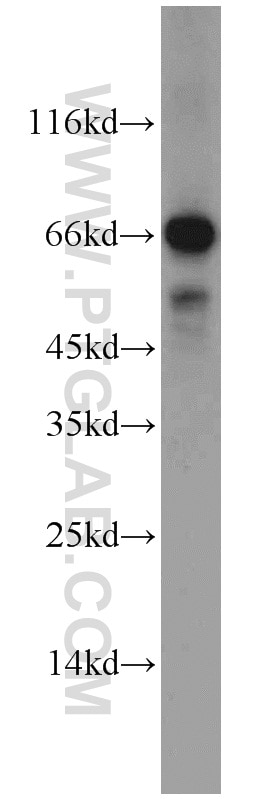
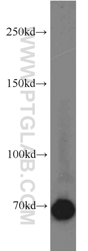
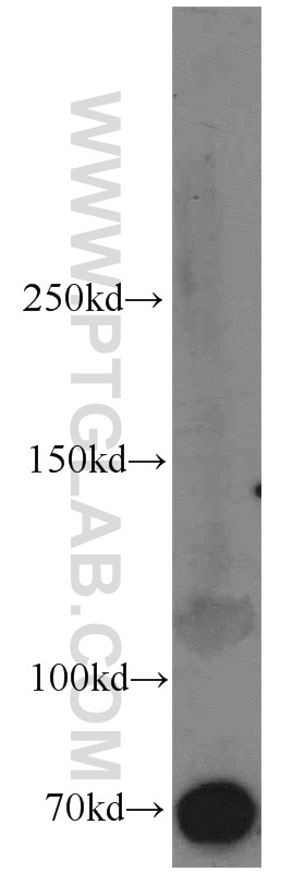
| Observed Molecular Weight | 60-70 kDa |
| GenBank Accession Number | BC013974 |
| Gene Symbol | Syndecan-3 |
| Gene ID (NCBI) | 9672 |
| RRID | AB_2182999 |
| Conjugate | Unconjugated |
| Form | Liquid |
| Purification Method | Antigen affinity purification |
| UNIPROT ID | O75056 |
| Storage Buffer | PBS with 0.02% sodium azide and 50% glycerol, pH 7.3. |
| Storage Conditions | Store at -20°C. Stable for one year after shipment. Aliquoting is unnecessary for -20oC storage. 20ul sizes contain 0.1% BSA. |
Background Information
Syndecan-3 is a member o f the Syndecan proteoglycan family. It plays a role in the organization of cell shape by affecting the actin cytoskeleton, possibly by transferring signals from the cell surface in a sugar-dependent mechanism.
Protocols
| Product Specific Protocols | |
|---|---|
| IF protocol for Syndecan-3 antibody 10886-1-AP | Download protocol |
| IHC protocol for Syndecan-3 antibody 10886-1-AP | Download protocol |
| IP protocol for Syndecan-3 antibody 10886-1-AP | Download protocol |
| WB protocol for Syndecan-3 antibody 10886-1-AP | Download protocol |
| Standard Protocols | |
|---|---|
| Click here to view our Standard Protocols |
Publications
| Species | Application | Title |
|---|---|---|
Nucleic Acids Res Metabolic and chemical regulation of tRNA modification associated with taurine deficiency and human disease. | ||
Am J Pathol No haploinsufficiency but loss of heterozygosity for EXT in multiple osteochondromas. | ||
Am J Pathol Screening for potential targets for therapy in mesenchymal, clear cell, and dedifferentiated chondrosarcoma reveals Bcl-2 family members and TGFβ as potential targets. | ||
J Cell Sci Electrophoresis of cell membrane heparan sulfate regulates galvanotaxis in glial cells. | ||
BMC Cancer Prognostic significance of the expression of GFRα1, GFRα3 and syndecan-3, proteins binding ARTEMIN, in mammary carcinoma. | ||
MedComm (2020) Long-Term High-Fat Diet Affected Bone Marrow Microenvironment During Aging at Single-Cell Resolution. |
Reviews
The reviews below have been submitted by verified Proteintech customers who received an incentive for providing their feedback.
FH Kamal (Verified Customer) (02-15-2024) | Mouse liver lysates were subjected to SDS PAGE followed by western blot with 10886-1-AP (Syndecan-3 antibody) at dilution of 1:2000 in 1X TBST incubated overnight at 4 degree C. Syndecan-3 appeared at 65 kDa.
|
FH Ryan (Verified Customer) (02-28-2019) | Tissue was fixed in PFA with tris-HCl antigen retrieval ph=6. Co-localisation with microglia based on known markers (not shown).
 |