

Tested Applications
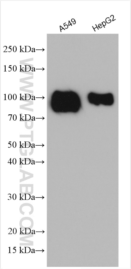
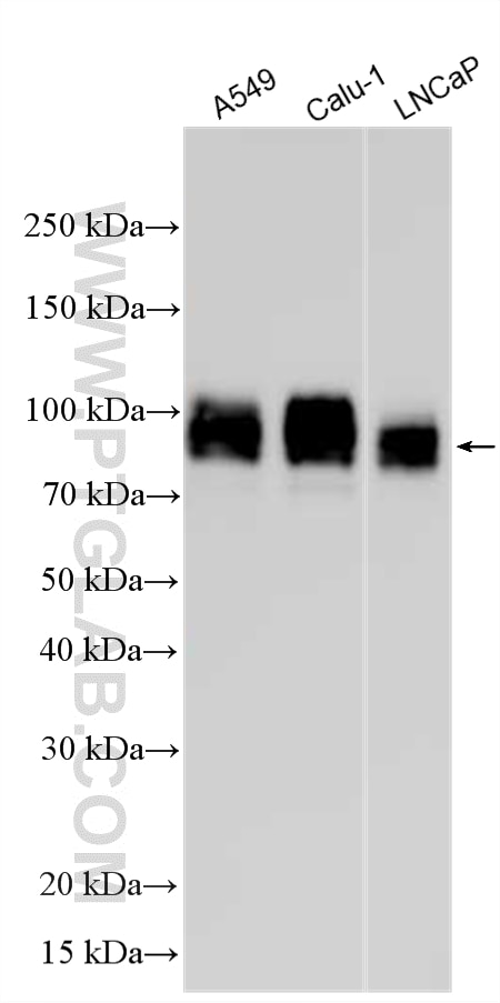
| Positive WB detected in | A549 cells, HepG2 cells, Calu-1 cells, LNCaP cells |
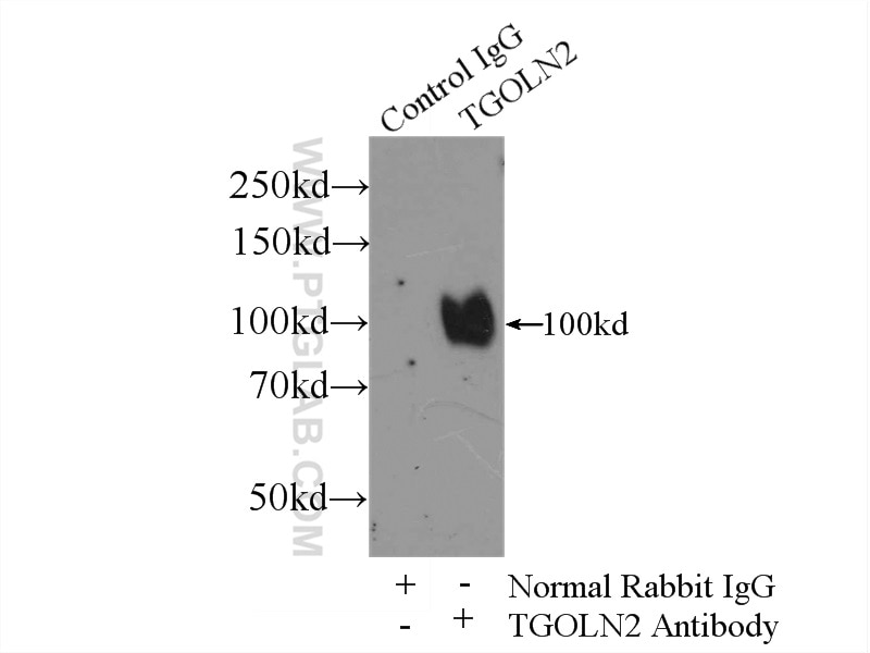
| Positive IP detected in | HeLa cells |
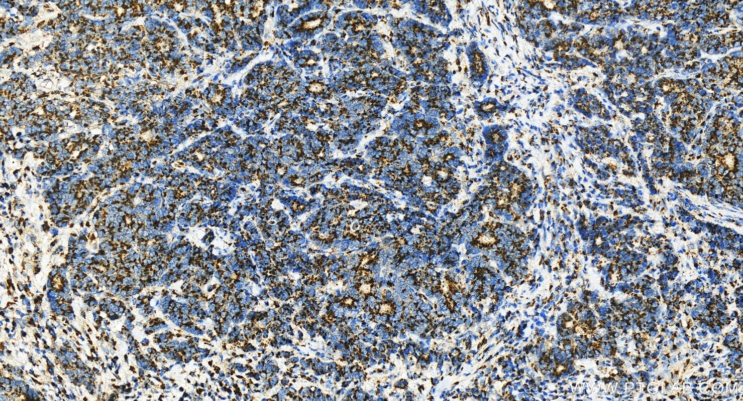
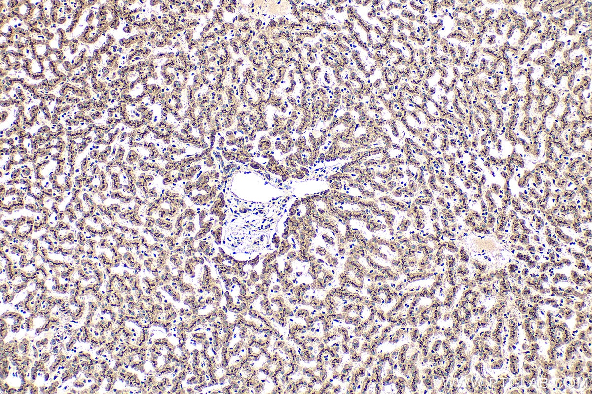
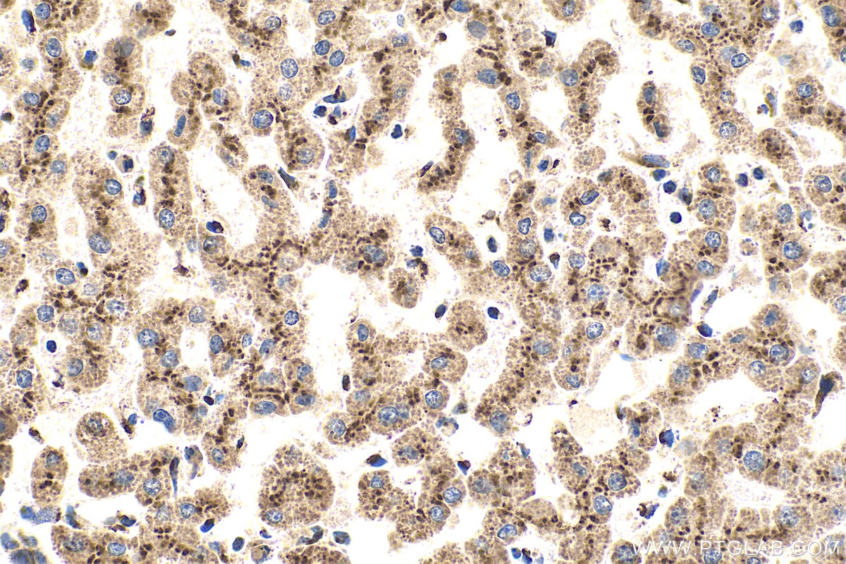
| Positive IHC detected in | human stomach cancer, human liver tissue, human stomach cancer tissue Note: suggested antigen retrieval with TE buffer pH 9.0; (*) Alternatively, antigen retrieval may be performed with citrate buffer pH 6.0 |
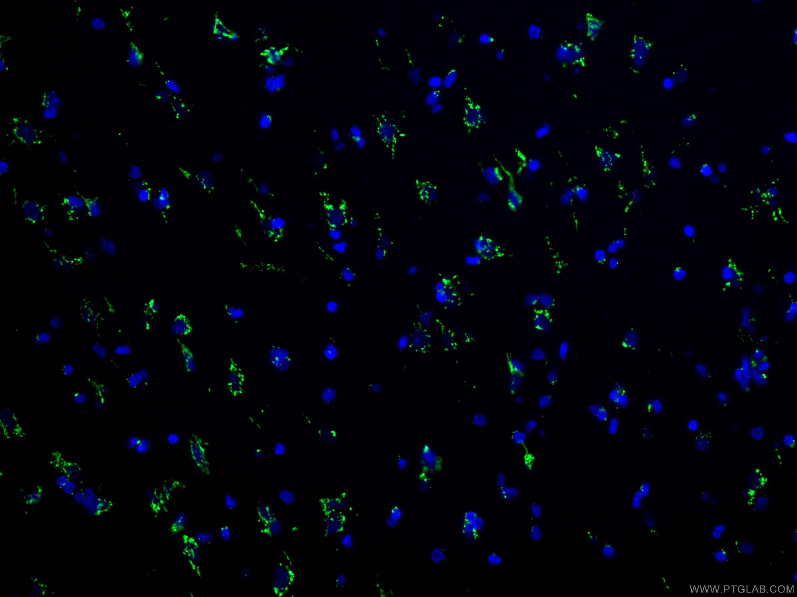
| Positive IF-P detected in | human cerebellum tissue |
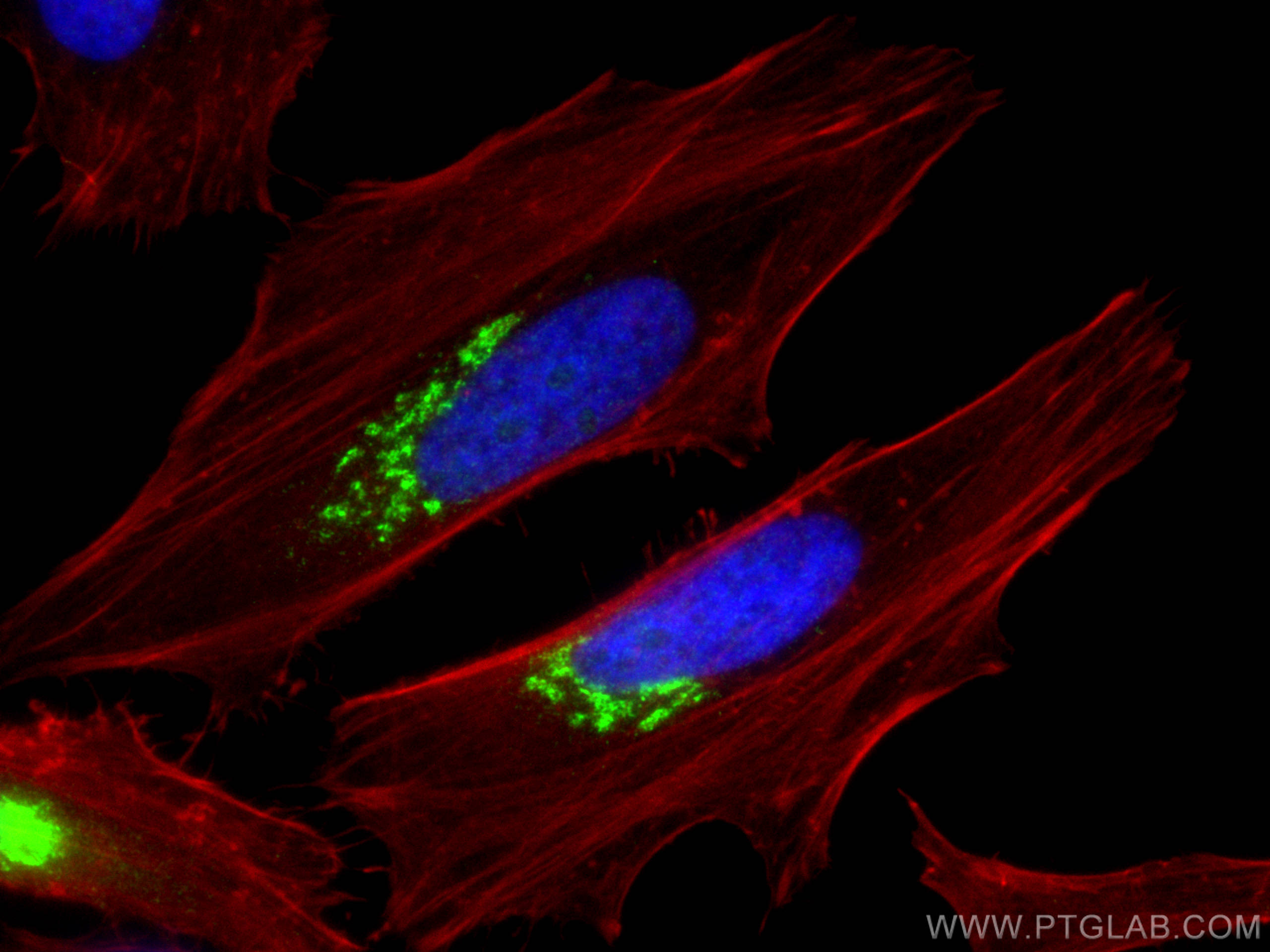
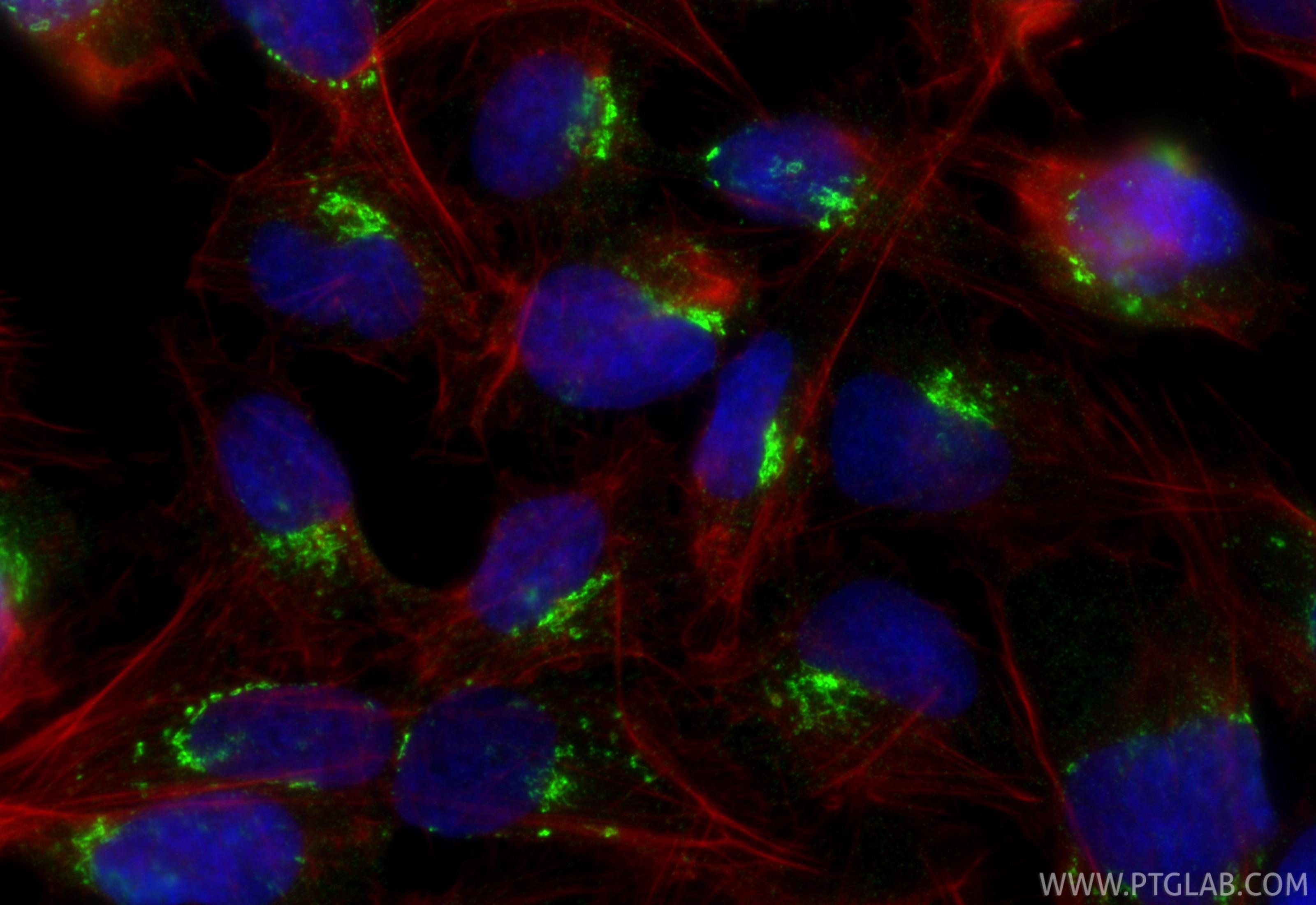
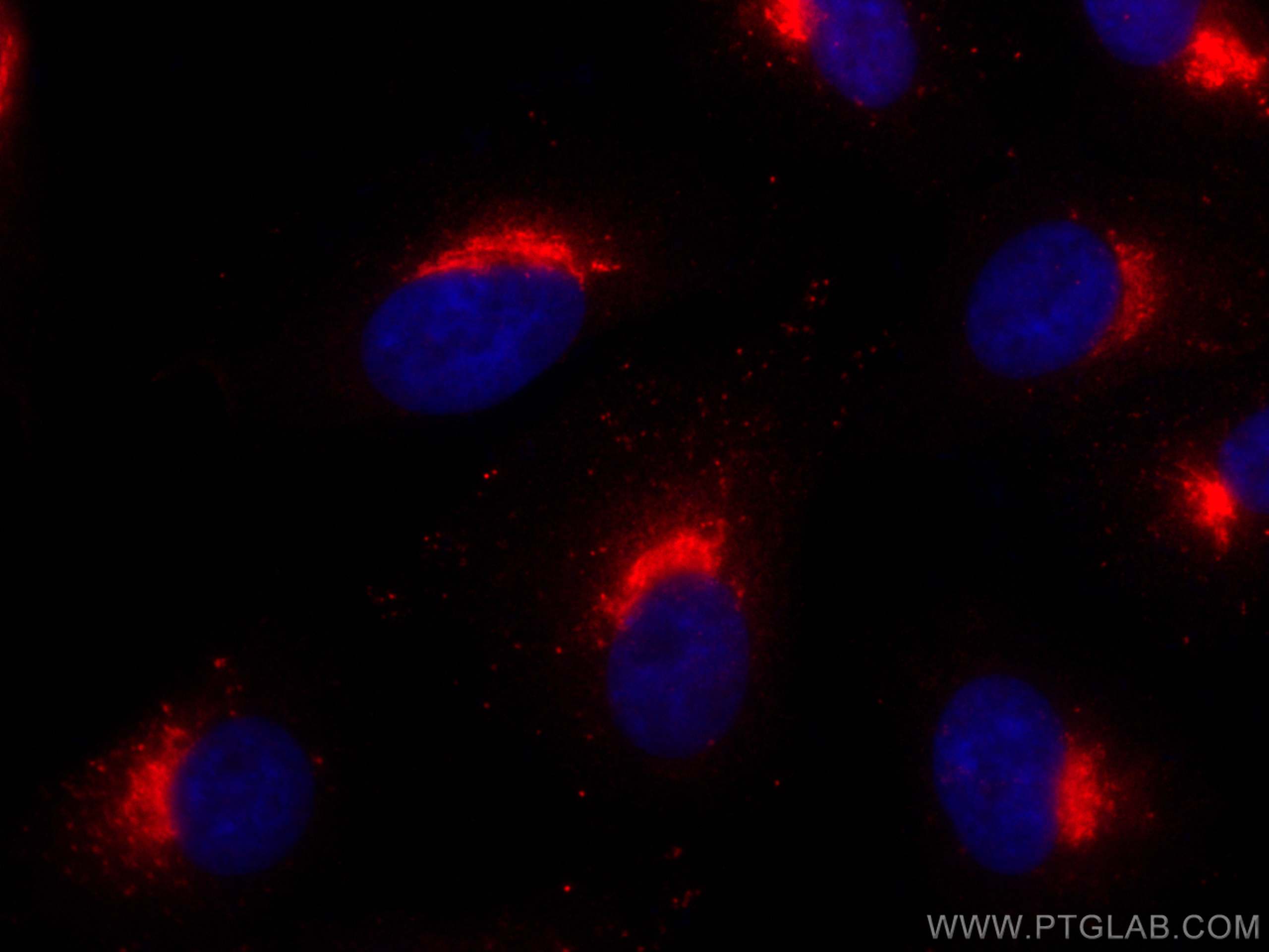
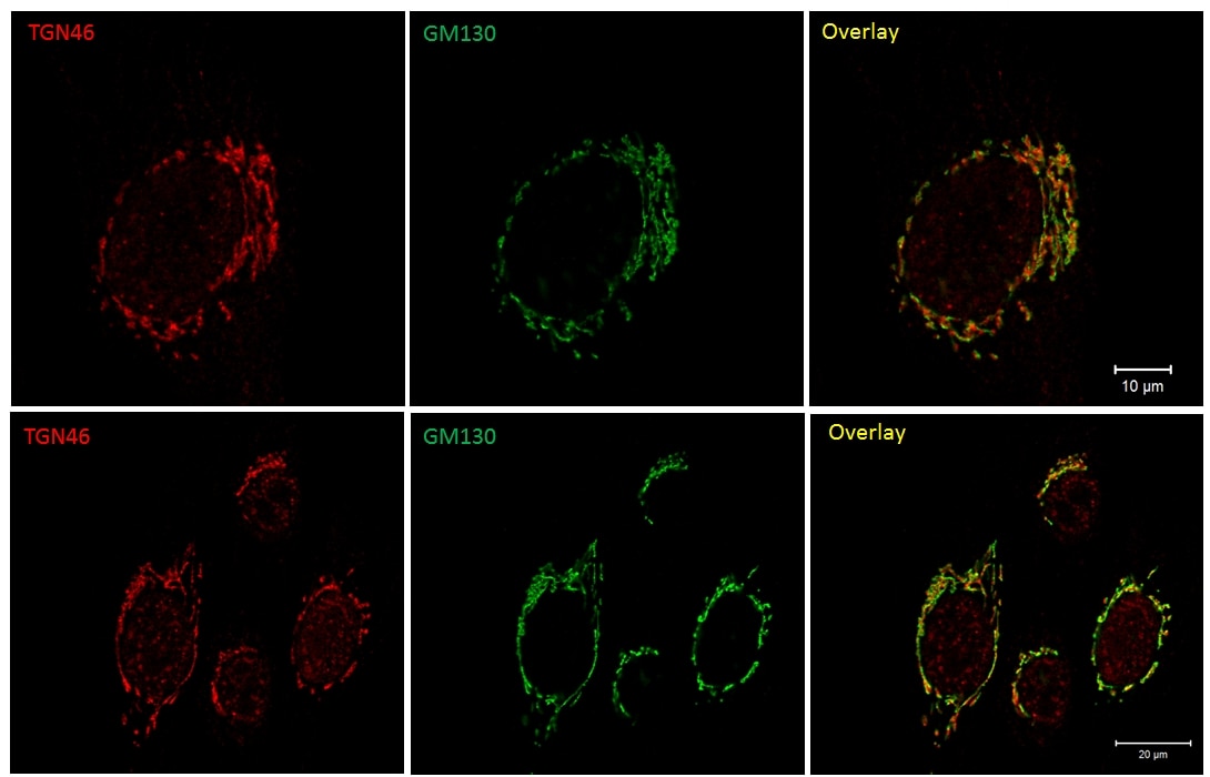
| Positive IF/ICC detected in | HeLa cells, A549 cells, U2OS cells |
Recommended dilution
| Application | Dilution |
|---|---|
| Western Blot (WB) | WB : 1:5000-1:50000 |
| Immunoprecipitation (IP) | IP : 0.5-4.0 ug for 1.0-3.0 mg of total protein lysate |
| Immunohistochemistry (IHC) | IHC : 1:400-1:1600 |
| Immunofluorescence (IF)-P | IF-P : 1:50-1:500 |
| Immunofluorescence (IF)/ICC | IF/ICC : 1:1000-1:4000 |
| It is recommended that this reagent should be titrated in each testing system to obtain optimal results. | |
| Sample-dependent, Check data in validation data gallery. | |
Product Information
13573-1-AP targets TGN46 in WB, IHC, IF/ICC, IF-P, IP, ELISA applications and shows reactivity with human samples.
| Tested Reactivity | human |
| Cited Reactivity | human, monkey, hamster |
| Host / Isotype | Rabbit / IgG |
| Class | Polyclonal |
| Type | Antibody |
| Immunogen |
CatNo: Ag4470 Product name: Recombinant human TGOLN2,TGN46 protein Source: e coli.-derived, PGEX-4T Tag: GST Domain: 25-384 aa of BC028219 Sequence: SVKQEEAGVRPSAGNVSTHPSLSQRPGGSTKSHPEPQTPKDSPSKSSAEAQTPEDTPNKSGAEAKTQKDSSNKSGAEAKTQKGSTSKSGSEAQTTKDSTSKSHPELQTPKDSTGKSGAEAQTPEDSPNRSGAEAKTQKDSPSKSGSEAQTTKDVPNKSGADGQTPKDGSSKSGAEDQTPKDVPNKSGAEKQTPKDGSNKSGAEEQGPIDGPSKSGAEEQTSKDSPNKVVPEQPSRKDHSKPISNPSDNKELPKADTNQLADKGKLSPHAFKTESGEETDLISPPQEEVKSSEPTEDVEPKEAEDDDTGPEEGSPPKEEKEKMSGSASSENREGTLSDSTGSEKDDLYPNGSGNGSAESSH Predict reactive species |
| Full Name | trans-golgi network protein 2 |
| Calculated Molecular Weight | 447 aa, 47 kDa |
| Observed Molecular Weight | 90-100 kDa |
| GenBank Accession Number | BC028219 |
| Gene Symbol | TGN46 |
| Gene ID (NCBI) | 10618 |
| RRID | AB_10597396 |
| Conjugate | Unconjugated |
| Form | Liquid |
| Purification Method | Antigen affinity purification |
| UNIPROT ID | O43493 |
| Storage Buffer | PBS with 0.02% sodium azide and 50% glycerol, pH 7.3. |
| Storage Conditions | Store at -20°C. Stable for one year after shipment. Aliquoting is unnecessary for -20oC storage. 20ul sizes contain 0.1% BSA. |
Background Information
TGN46 (TGOLN2), the human homolog of rat Tgn38, is a transmembrane glycoprotein predominantly localized to the TGN (trans-Golgi network). TGN is a major secretory pathway sorting station for proteins and lipids. TGN46 may be involved in regulating membrane traffic to and from TGN. Alternatively, spliced transcript variants encode different TGN46 isoforms. TGN46 has an apparent molecular mass of 100-150 kDa, suggesting extensive O- and N-glycosylations.
Protocols
| Product Specific Protocols | |
|---|---|
| IF protocol for TGN46 antibody 13573-1-AP | Download protocol |
| IHC protocol for TGN46 antibody 13573-1-AP | Download protocol |
| IP protocol for TGN46 antibody 13573-1-AP | Download protocol |
| WB protocol for TGN46 antibody 13573-1-AP | Download protocol |
| Standard Protocols | |
|---|---|
| Click here to view our Standard Protocols |
Publications
What published studies show
The TGN46 Polyclonal antibody (Cat# 13573-1-AP) has accumulated 59 citations across high-impact journals including Nature Genetics, Nature Cell Biology, Nature Communications, Molecular Cell, Science Advances, Cell Death & Differentiation, Autophagy, Developmental Cell, EMBO Reports, Journal of Cell Biology, PNAS, and Cell Death & Disease. Associated research fields span Golgi/TGN membrane trafficking, autophagy, neurodegeneration, cancer biology, virology, glycosylation, immune signaling, lipid metabolism, and cell death mechanisms.
| Species | Application | Title |
|---|---|---|
Nat Genet Alterations in PD-L1 succinylation shape anti-tumor immune responses in melanoma | ||
Nat Cell Biol Mitochondrial-derived vesicles drive budding-type fission of damaged lysosomes | ||
Autophagy Live imaging of intra-lysosome pH in cell lines and primary neuronal culture using a novel genetically encoded biosensor. | ||
Autophagy S-palmitoylation coordinates the trafficking of ATG9A to mediate autophagy initiation | ||
Nat Commun Rab27a regulates the transport of influenza virus membrane proteins to the plasma membrane |
Reviews
What customers say
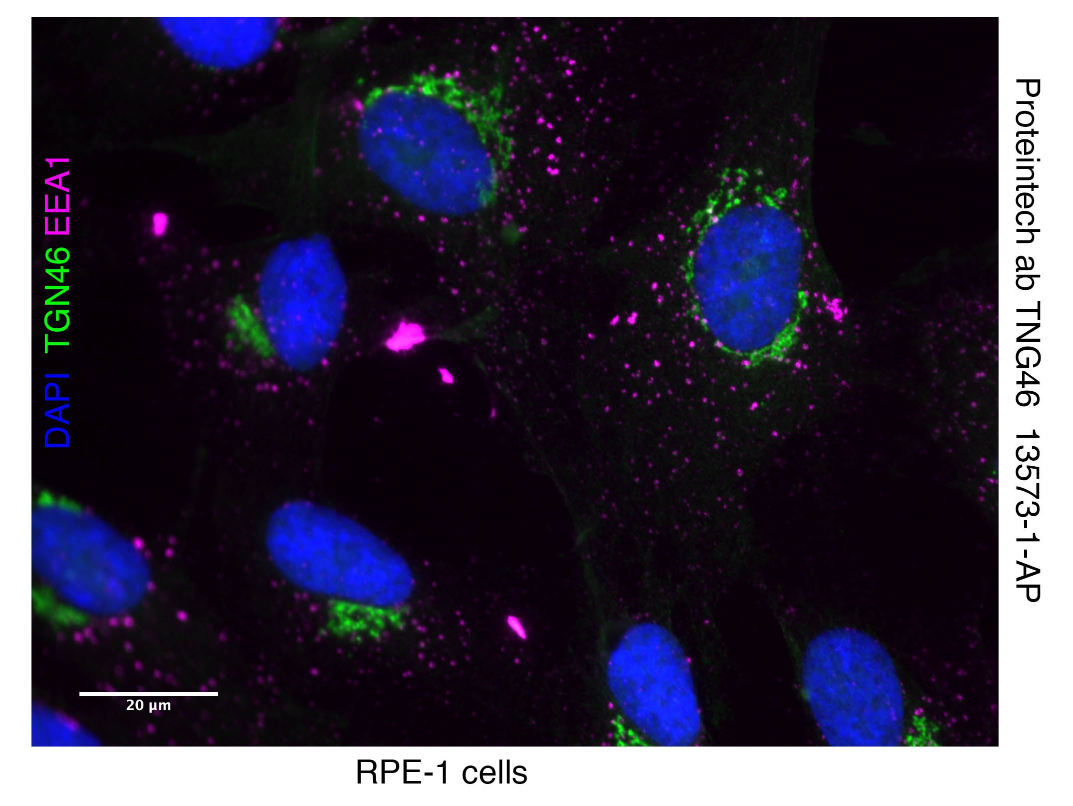
Users rate this antibody 4–5 stars across Western Blot and Immunofluorescence applications. It performs well for Golgi/TGN46 staining in HEK293T and RPE cells, with clear localization at dilutions of 1:100–1:300. For WB, it detects two bands around 85 kDa at 1:2,500. Reviewers highlight reliable Golgi staining, compatibility with co-staining (e.g., EEA1, DAPI), and straightforward protocols using standard fixation and blocking conditions. Overall, it is regarded as a dependable reagent for both IF and WB.
The reviews below have been submitted by verified Proteintech customers who received an incentive for providing their feedback.
Sammy (Verified Customer) (01-20-2024) | A good antibody for IF and also works for WB.
|
Amy (Verified Customer) (08-10-2023) | Nice Golgi staining of HEK293T cells.
|
Christine (Verified Customer) (01-27-2023) | Detects 2 bands around the 85 kDa marker, a sharp one just below and a more fuzzy one just above.
|
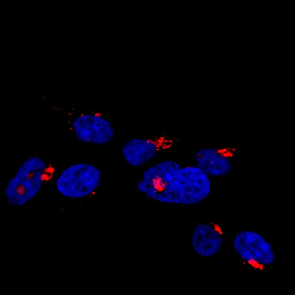
Thomas (Verified Customer) (09-18-2020) | HEK293T cells were fixed in 4% PFA for 15 mins and permeabilised in 0.1% Triton-X 100 in PBS. Cells were blocked in 1% BSA. Primary antibody solution was diluted at 1:200 in blocking solution and incubated for 1 hour. Goat anti-rabbit 647 Alexa Fluor secondary antibody (1:250 - red) and DAPI (1:2000 - blue) were incubated with cells for 1 hour.
 |
Stephen (Verified Customer) (10-01-2019) | RPE cells fixed with 4% PFA Perm. by 0.3% tx100 for 5 min blocked with 1% BSA in 1XPBS for 2 hours TGN46 antibody(green) incubated 1:300 and EEA1 antibody (purple) overnight at 4 degrees in 1%BSA in 1x PBS. Co-stained with DAPI (blue) visualize DNA
 |












