


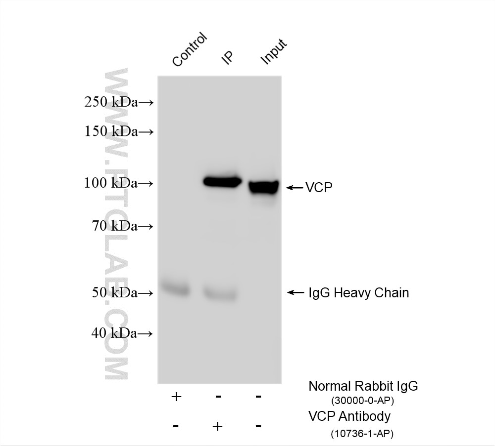


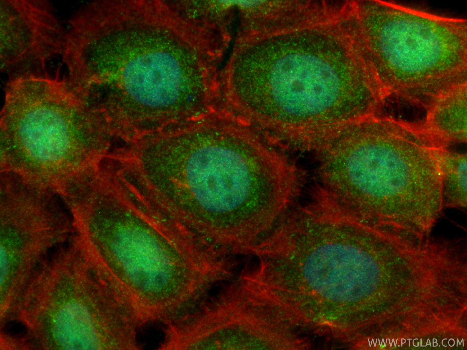

Tested Applications
| Positive WB detected in | HeLa cells, human brain tissue, rat lung tissue, mouse lung tissue, U2OS cells, C6 cells, mouse thymus tissue, mouse brain tissue, rat brain tissue, SH-SY5Y cells, NIH/3T3 cells |
| Positive IP detected in | HeLa cells, mouse brain tissue |
| Positive IHC detected in | human gliomas tissue, human colon cancer tissue, human lung cancer tissue Note: suggested antigen retrieval with TE buffer pH 9.0; (*) Alternatively, antigen retrieval may be performed with citrate buffer pH 6.0 |
| Positive IF/ICC detected in | A431 cells |
Recommended dilution
| Application | Dilution |
|---|---|
| Western Blot (WB) | WB : 1:2000-1:16000 |
| Immunoprecipitation (IP) | IP : 0.5-4.0 ug for 1.0-3.0 mg of total protein lysate |
| Immunohistochemistry (IHC) | IHC : 1:100-1:400 |
| Immunofluorescence (IF)/ICC | IF/ICC : 1:50-1:500 |
| It is recommended that this reagent should be titrated in each testing system to obtain optimal results. | |
| Sample-dependent, Check data in validation data gallery. | |
Product Information
10736-1-AP targets VCP in WB, IHC, IF/ICC, IP, CoIP, ELISA, PLA applications and shows reactivity with human, mouse, rat samples.
| Tested Reactivity | human, mouse, rat |
| Cited Reactivity | human, mouse, rat, pig |
| Host / Isotype | Rabbit / IgG |
| Class | Polyclonal |
| Type | Antibody |
| Immunogen |
CatNo: Ag1002 Product name: Recombinant human VCP protein Source: e coli.-derived, PGEX-4T Tag: GST Domain: 345-644 aa of BC007562 Sequence: GMTPSKGVLFYGPPGCGKTLLAKAIANECQANFISIKGPELLTMWFGESEANVREIFDKARQAAPCVLFFDELDSIAKARGGNIGDGGGAADRVINQILTEMDGMSTKKNVFIIGATNRPDIIDPAILRPGRLDQLIYIPLPDEKSRVAILKANLRKSPVAKDVDLEFLAKMTNGFSGADLTEICQRACKLAIRESIESEIRRERERQTNPSAMEVEEDDPVPEIRRDHFEEAMRFARRSVSDNDIRKYEMFAQTLQQSRGFGSFRFPSGNQGGAGPSQGSGGGTGGSVYTEDNDDDLYG Predict reactive species |
| Full Name | valosin-containing protein |
| Calculated Molecular Weight | 89 kDa |
| Observed Molecular Weight | 90-100 kDa |
| GenBank Accession Number | BC007562 |
| Gene Symbol | VCP |
| Gene ID (NCBI) | 7415 |
| RRID | AB_2214635 |
| Conjugate | Unconjugated |
| Form | Liquid |
| Purification Method | Antigen affinity purification |
| UNIPROT ID | P55072 |
| Storage Buffer | PBS with 0.02% sodium azide and 50% glycerol, pH 7.3. |
| Storage Conditions | Store at -20°C. Stable for one year after shipment. Aliquoting is unnecessary for -20oC storage. 20ul sizes contain 0.1% BSA. |
Background Information
VCP(Valosin-containing protein), also known as TER ATPase and 15S Mg2+-ATPase p97 subunit, belongs to the AAA ATPase family. VCP was first identified as a result of attempts to clone a putative peptide hormone called valosin. It was found that the cloned cDNA encoded a ubiquitously expressed 90 kDa cytosolic protein, termed VCP, which showed none of the characteristics of a peptide hormone precursor(PMID:1382975). Defects in VCP are the cause of inclusion body myopathy with early-onset Paget disease and frontotemporal dementia (IBMPFD) and amyotrophic lateral sclerosis type 14 with or without frontotemporal dementia (ALS14). VCP has a calculated molecular weight of 89 kDa and an apparent molecular weight of 90-100 kDa (PMID: 15732117, 1382975).
Protocols
| Product Specific Protocols | |
|---|---|
| IF protocol for VCP antibody 10736-1-AP | Download protocol |
| IHC protocol for VCP antibody 10736-1-AP | Download protocol |
| IP protocol for VCP antibody 10736-1-AP | Download protocol |
| WB protocol for VCP antibody 10736-1-AP | Download protocol |
| Standard Protocols | |
|---|---|
| Click here to view our Standard Protocols |
Publications
What published studies show
The VCP Polyclonal antibody (Cat# 10736-1-AP) has 80 citations across high-impact journals including Cell, Nature Communications, Neuron, PNAS, Science Advances, Cell Reports, EMBO Reports, and Journal of Cell Biology. Research fields span autophagy, ER-associated degradation (ERAD), DNA repair, protein quality control, stress granules, ubiquitin biology, ferroptosis, cancer (colorectal, glioblastoma, cholangiocarcinoma), and neurodegeneration (ALS, Alzheimer's). Applications are predominantly Western blot, with immunoprecipitation, IHC, and IF also represented.
| Species | Application | Title |
|---|---|---|
Cell TEX264 drives selective autophagy of DNA lesions to promote DNA repair and cell survival | ||
Cell Metab AIDA Selectively Mediates Downregulation of Fat Synthesis Enzymes by ERAD to Retard Intestinal Fat Absorption and Prevent Obesity. | ||
Exploration (Beijing) Palladium Nanoparticles Degrade Advanced Glycation End Products via Valosin-Containing Protein Mediated Autophagy to Attenuate High-Glucose/High-Fat-Induced Intervertebral Disc Degeneration | ||
Nat Commun Episodic transport of protein aggregates achieves a positive size selectivity in aggresome formation | ||
Nat Commun Analysis of Zika virus capsid-Aedes aegypti mosquito interactome reveals pro-viral host factors critical for establishing infection.
| ||
Reviews
What customers say
The single review for this antibody is highly positive, giving it a 5-star rating. The reviewer tested it on HEK293T cells, mouse tissues, and mouse neurons across Western blot, immunofluorescence, and immunoprecipitation, reporting excellent performance in all three applications. For Western blot, they used a 1:1000 dilution for low-concentration samples and 1:2000 for high-concentration samples, while immunofluorescence was performed at 1:500. Overall, the reviewer described the antibody as "amazing" and reliable across the tested conditions.
The reviews below have been submitted by verified Proteintech customers who received an incentive for providing their feedback.
Firyal (Verified Customer) (01-06-2026) | This antibody is amazing and works great in all the applications we have used it for (Western blots, IFC, and IP). For WB, we've used it at a concentration if 1:1000 in low concentration samples and 1:2000 in high concentration samples.
|






