



Tested Applications
| Positive WB detected in | mouse skeletal muscle tissue, rat skeletal muscle tissue |
| Positive IHC detected in | human liver tissue Note: suggested antigen retrieval with TE buffer pH 9.0; (*) Alternatively, antigen retrieval may be performed with citrate buffer pH 6.0 |
| Positive IF/ICC detected in | HeLa cells |
Recommended dilution
| Application | Dilution |
|---|---|
| Western Blot (WB) | WB : 1:1000-1:6000 |
| Immunohistochemistry (IHC) | IHC : 1:50-1:500 |
| Immunofluorescence (IF)/ICC | IF/ICC : 1:50-1:500 |
| It is recommended that this reagent should be titrated in each testing system to obtain optimal results. | |
| Sample-dependent, Check data in validation data gallery. | |
Product Information
19194-1-AP targets WDR45/WIPI4 in WB, IHC, IF/ICC, IP, ELISA applications and shows reactivity with human, mouse, rat samples.
| Tested Reactivity | human, mouse, rat |
| Cited Reactivity | human, mouse |
| Host / Isotype | Rabbit / IgG |
| Class | Polyclonal |
| Type | Antibody |
| Immunogen |
CatNo: Ag6741 Product name: Recombinant human WDR45 protein Source: e coli.-derived, PET28a Tag: 6*His Domain: 1-360 aa of BC000464 Sequence: MTQQPLRGVTSLRFNQDQSCFCCAMETGVRIYNVEPLMEKGHLDHEQVGSMGLVEMLHRSNLLALVGGGSSPKFSEISVLIWDDAREGKDSKEKLVLEFTFTKPVLSVRMRHDKIVIVLKNRIYVYSFPDNPRKLFEFDTRDNPKGLCDLCPSLEKQLLVFPGHKCGSLQLVDLASTKPGTSSAPFTINAHQSDIACVSLNQPGTVVASASQKGTLIRLFDTQSKEKLVELRRGTDPATLYCINFSHDSSFLCASSDKGTVHIFALKDTRLNRRSALARVGKVGPMIGQYVDSQWSLASFTVPAESACICAFGRNTSKNVNSVIAICVDGTFHKYVFTPDGNCNREAFDVYLDICDDDDF Predict reactive species |
| Full Name | WD repeat domain 45 |
| Calculated Molecular Weight | 40 kDa |
| Observed Molecular Weight | 40 kDa |
| GenBank Accession Number | BC000464 |
| Gene Symbol | WDR45 |
| Gene ID (NCBI) | 11152 |
| RRID | AB_2215404 |
| Conjugate | Unconjugated |
| Form | Liquid |
| Purification Method | Antigen affinity purification |
| UNIPROT ID | Q9Y484 |
| Storage Buffer | PBS with 0.02% sodium azide and 50% glycerol, pH 7.3. |
| Storage Conditions | Store at -20°C. Stable for one year after shipment. Aliquoting is unnecessary for -20oC storage. 20ul sizes contain 0.1% BSA. |
Background Information
WD repeat domain phosphoinositide-interacting protein 4 (WDR45) is also named as WDRX1, WDRXI4 and WIPI4, and belongs to the WD repeat PROPPIN family. WDR45 is highly conserved in mammals, and the amino acid sequences of the human, mouse, pig, bovine and horse protein have more than 97% identity (PMID:20505359). WDR45 is component of the autophagy machinery that controls the major intracellular degradation process (PMID:23435086, PMID:28561066). Together with WIPI1, WIPI2 and WIPI3/WDR45B, it forms the WIPI (WD repeat domain, phosphoinositide interacting) protein family. And this is why WDR45 is also known as WIPI4 (PMID:11814058). The best characterized molecular function of WDR45 is promoting lipid transfer together with ATG2 proteins between adjacent membranes. This function at the phagophore-ER membrane contact sites is important for autophagy (PMID:30185561, PMID:31721365). WDR45 is activated by the STK11/AMPK signaling pathway upon starvation. WDR45 is involved in autophagosome assembly downstream of WIPI2, regulating the size of forming autophagosomes (PMID:28561066).
Protocols
| Product Specific Protocols | |
|---|---|
| IF protocol for WDR45/WIPI4 antibody 19194-1-AP | Download protocol |
| IHC protocol for WDR45/WIPI4 antibody 19194-1-AP | Download protocol |
| WB protocol for WDR45/WIPI4 antibody 19194-1-AP | Download protocol |
| Standard Protocols | |
|---|---|
| Click here to view our Standard Protocols |
Publications
What published studies show
The WDR45/WIPI4 Polyclonal antibody (Cat# 19194-1-AP) has 15 citations. They appear in journals including Nature Cell Biology, Autophagy, Nature Communications, Science Advances, Lipids in Health and Disease, International Journal of Molecular Sciences, Journal of Neurochemistry, Human Gene Therapy, Human Molecular Genetics, European Journal of Medical Genetics, American Journal of Medical Genetics A, and bioRxiv. Research fields span autophagy, β-propeller protein-associated neurodegeneration, ferroptosis, exosome biogenesis, iron metabolism, lipid droplet accumulation, stress granule dynamics, and whole-body metabolism.
| Species | Application | Title |
|---|---|---|
Nat Cell Biol Loss of WIPI4 in neurodegeneration causes autophagy-independent ferroptosis | ||
Autophagy Regulation of N-Degron Recognin-Mediated Autophagy by the sars-cov-2 plpro ubiquitin deconjugase | ||
Autophagy SDC1-dependent TGM2 determines radiosensitivity in glioblastoma by coordinating EPG5-mediated fusion of autophagosomes with lysosomes | ||
Nat Commun β-propeller protein-associated neurodegeneration protein WDR45 regulates stress granule disassembly via phase separation with Caprin-1 | ||
Sci Adv Mediobasal hypothalamic FKBP51 acts as a molecular switch linking autophagy to whole-body metabolism.
|
Reviews
What customers say
Able is busy answering your previous question. Please try again later.
The reviews below have been submitted by verified Proteintech customers who received an incentive for providing their feedback.
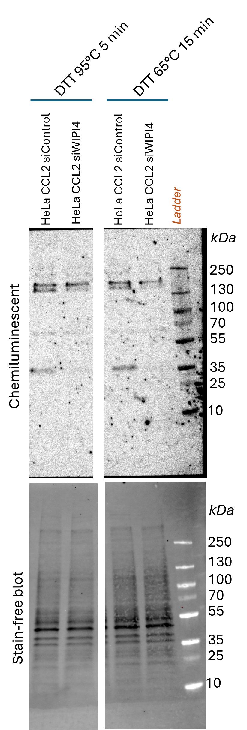
daniele (Verified Customer) (10-01-2026) | Application: Western blot Sample: HeLa (CCL-2) whole-cell lysate, siControl vs siWIPI4 Species: Human Protocol: Samples were prepared in 1X Laemmli buffer containing SDS and 100 mM DTT, then heated for either 5 min at 95°C (left) or 15 min at 60 °C (right). 20 µg of total lysate were loaded per lane and separated by SDS-PAGE (4-20% TGX Stain-free Gel, UV-activated) and transferred onto a 0.22 µm nitrocellulose membrane (stain-free blot). The membrane was blocked for 1 h in 5% non-fat milk in 1X TBS, then incubated overnight at 4 °C with the 19194-1-AP anti-WIPI4 antibody (1:1000). The next day, the membrane was washed in TBS-T (0.05% Tween-20) and incubated for 2 h with HRP-conjugated goat anti-rabbit secondary antibody (1:2500 in 5% milk in TBS-T). Signal was detected by ECL. Results: The antibody detected a strong band at approximately 37 kDa, consistent with the predicted molecular weight of WIPI4 (~40 kDa), and 2 more strong bands between 250-130 kDa. The 37 kDa and the lower band observed in the upper duplet (250-130 kDa) disappeared in siWIPI4-treated cells compared with siControl, confirming its specificity. The higher band might refere to aggregation, the other non-specific bands were observed at 70 kDa and 200 kDa, and background was moderate. Conclusion: Overall, this antibody performs well for detecting endogenous WIPI4 by western blot in HeLa cells and is suitable for validating knockdown experiments. Suggested primary antibody concentration is 1/500, since used dilution (1/1000) needed a long time exposure (10 minutes). Rating: 4/5
 |


