

Tested Applications
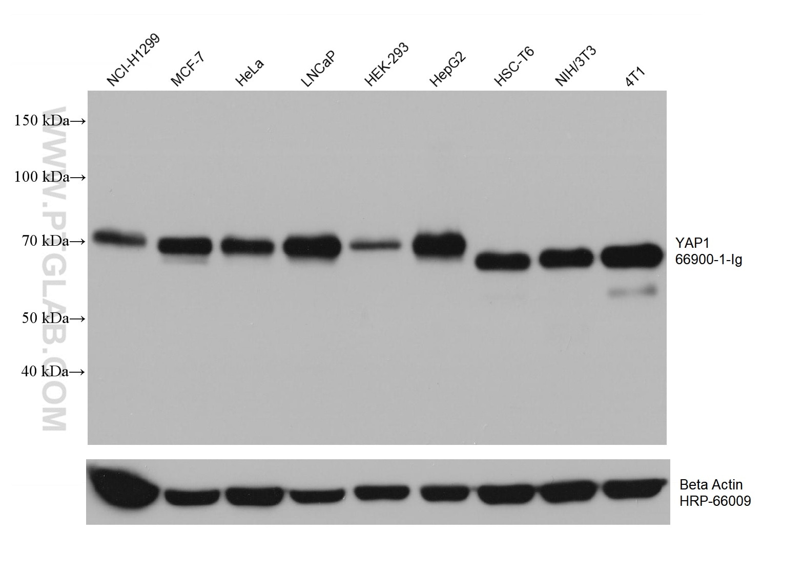
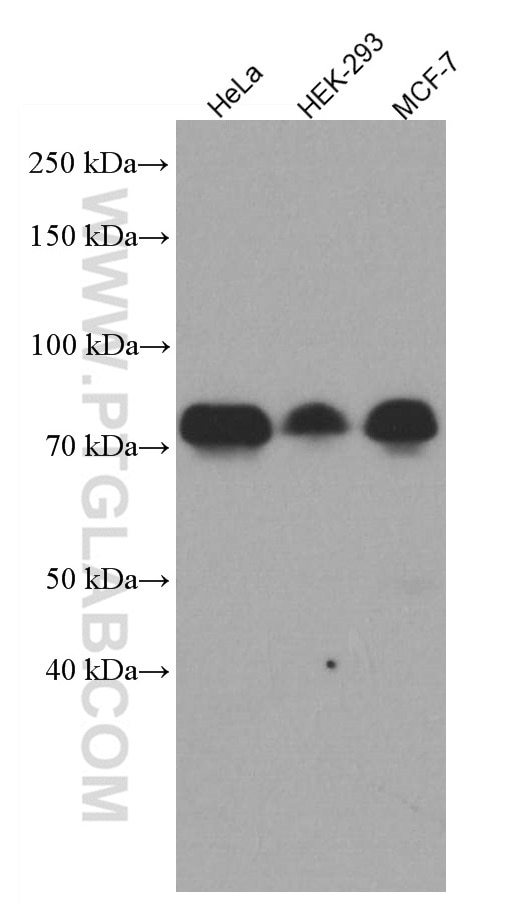
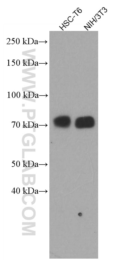
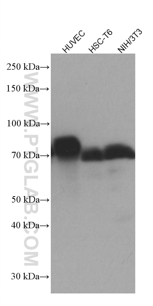
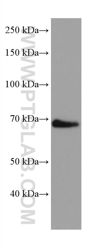
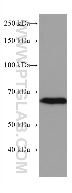
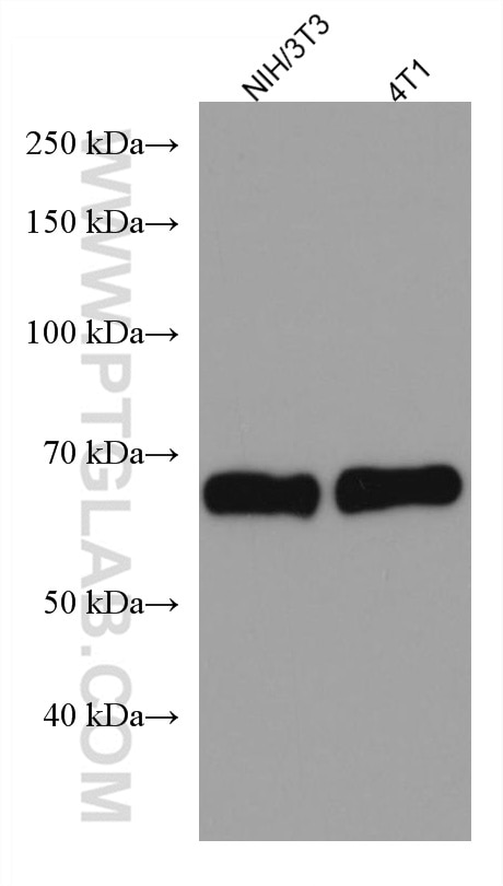
| Positive WB detected in | NCI-H1299 cells, HUVEC cells, HeLa cells, HSC-T6 cells, NIH/3T3 cells, MCF-7 cells, LNCaP cells, HEK-293 cells, HepG2 cells, NIH-3T3 cells, 4T1 cells |
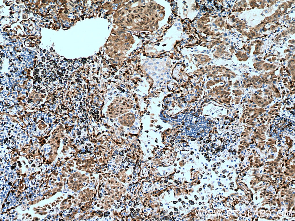
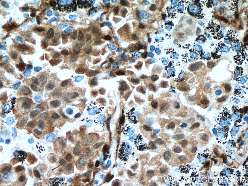
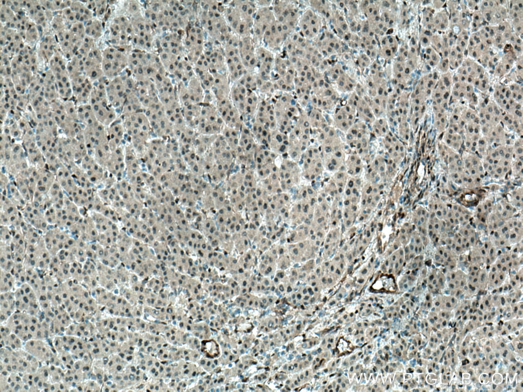
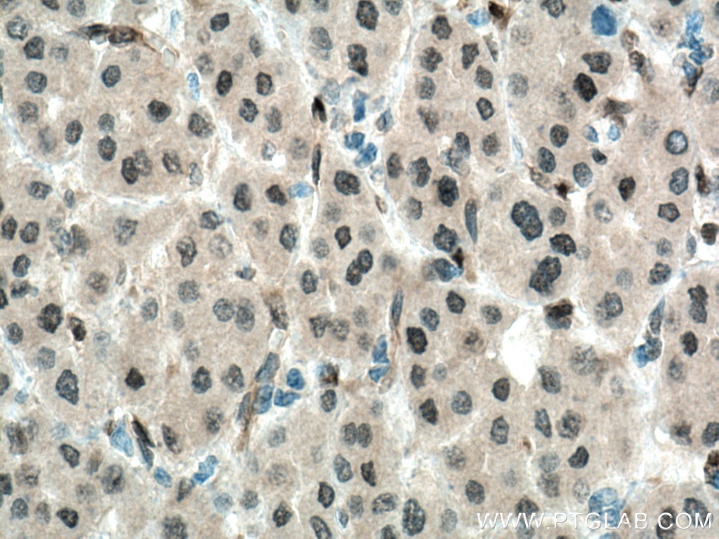
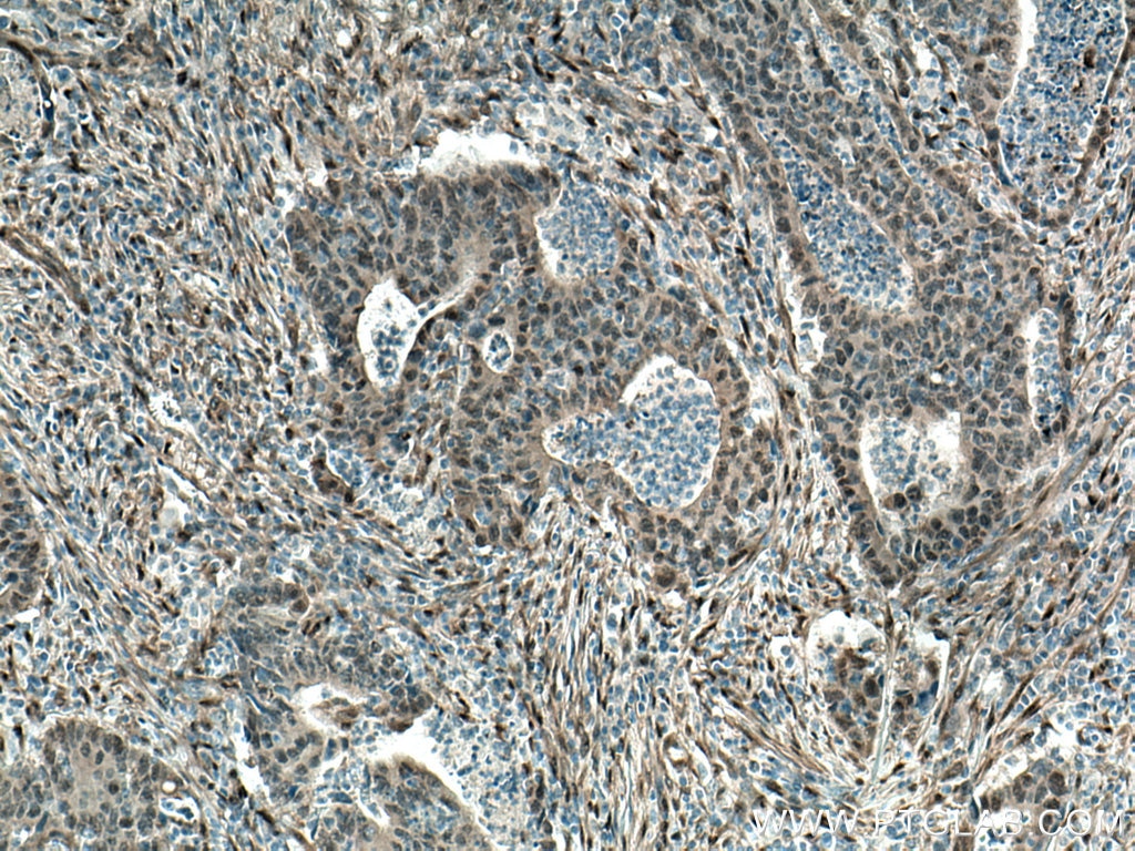
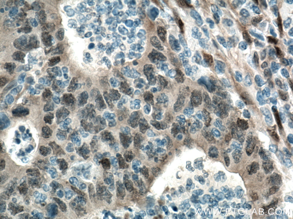
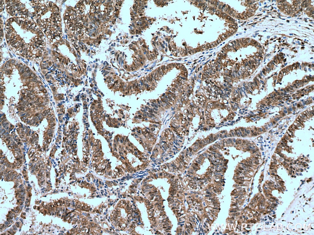
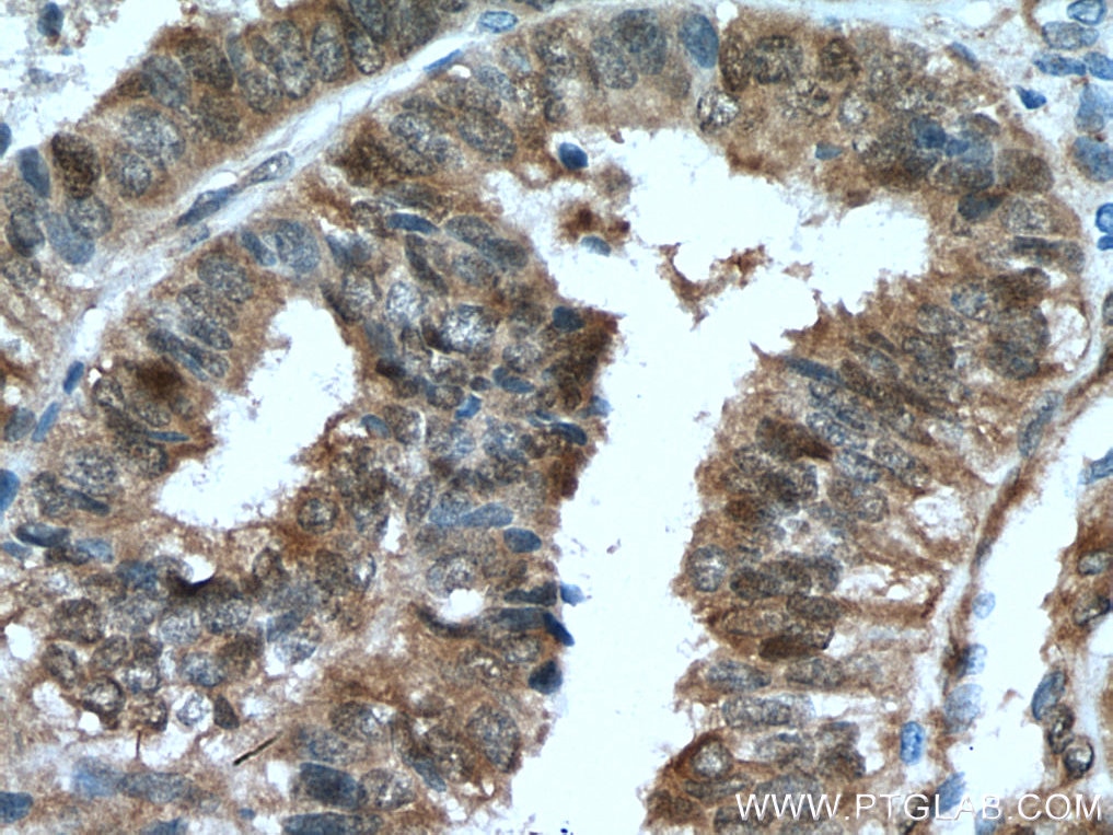
| Positive IHC detected in | human lung cancer tissue, human colon cancer tissue, human liver cancer tissue, human ovary tumor tissue Note: suggested antigen retrieval with TE buffer pH 9.0; (*) Alternatively, antigen retrieval may be performed with citrate buffer pH 6.0 |
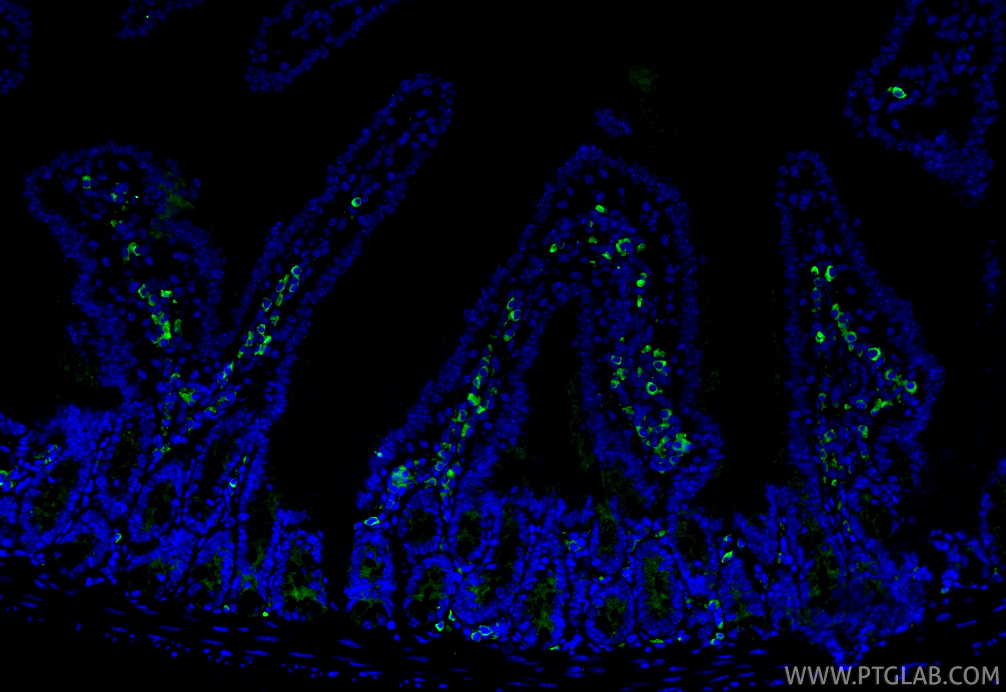
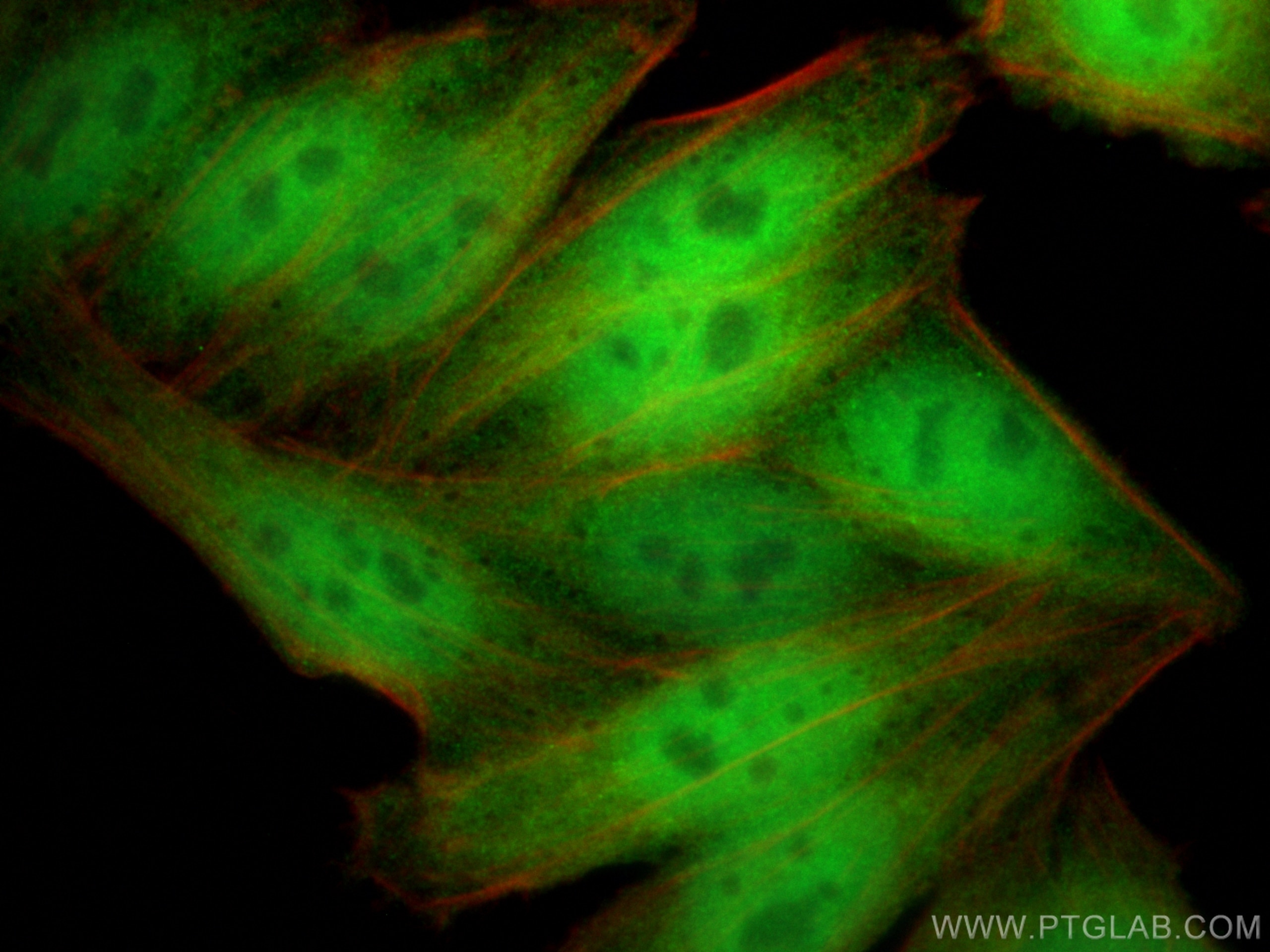
| Positive IF/ICC detected in | HepG2 cells |
Recommended dilution
| Application | Dilution |
|---|---|
| Western Blot (WB) | WB : 1:5000-1:50000 |
| Immunohistochemistry (IHC) | IHC : 1:1000-1:4000 |
| Immunofluorescence (IF)/ICC | IF/ICC : 1:400-1:1600 |
| It is recommended that this reagent should be titrated in each testing system to obtain optimal results. | |
| Sample-dependent, Check data in validation data gallery. | |
Product Information
66900-1-Ig targets YAP1 in WB, IHC, IF/ICC, IP, CoIP, ELISA applications and shows reactivity with human, mouse, rat samples.
| Tested Reactivity | human, mouse, rat |
| Cited Reactivity | human, mouse, rat, sheep |
| Host / Isotype | Mouse / IgG1 |
| Class | Monoclonal |
| Type | Antibody |
| Immunogen |
CatNo: Ag28194 Product name: Recombinant human YAP1 protein Source: e coli.-derived, PET28a Tag: 6*His Domain: 155-504 aa of BC038235 Sequence: PTAQHLRQSSFEIPDDVPLPAGWEMAKTSSGQRYFLNHIDQTTTWQDPRKAMLSQMNVTAPTSPPVQQNMMNSASGPLPDGWEQAMTQDGEIYYINHKNKTTSWLDPRLDPRFAMNQRISQSAPVKQPPPLAPQSPQGGVMGGSNSNQQQQMRLQQLQMEKERLRLKQQELLRQAMRNINPSTANSPKCQELALRSQLPTLEQDGGTQNPVSSPGMSQELRTMTTNSSDPFLNSGTYHSRDESTDSGLSMSSYSVPRTPDDFLNSVDEMDTGDTINQSTLPSQQNRFPDYLEAIPGTNVDLGTLEGDGMNIEGEELMPSLQEALSSDILNDMESVLAATKLDKESFLTWL Predict reactive species |
| Full Name | Yes-associated protein 1, 65kDa |
| Calculated Molecular Weight | 504 aa, 54 kDa |
| Observed Molecular Weight | 65-75 kDa |
| GenBank Accession Number | BC038235 |
| Gene Symbol | YAP1 |
| Gene ID (NCBI) | 10413 |
| RRID | AB_2882229 |
| Conjugate | Unconjugated |
| Form | Liquid |
| Purification Method | Protein G purification |
| UNIPROT ID | P46937 |
| Storage Buffer | PBS with 0.02% sodium azide and 50% glycerol, pH 7.3. |
| Storage Conditions | Store at -20°C. Stable for one year after shipment. Aliquoting is unnecessary for -20oC storage. 20ul sizes contain 0.1% BSA. |
Background Information
Yes-associated protein 1 (YAP1) is a transcriptional regulator which can act both as a coactivator and a corepressor and is the critical downstream regulatory target in the Hippo signaling pathway that plays a pivotal role in organ size control and tumor suppression by restricting proliferation and promoting apoptosis. The core of this pathway is composed of a kinase cascade wherein STK3/MST2 and STK4/MST1, in complex with its regulatory protein SAV1, phosphorylates and activates LATS1/2 in complex with its regulatory protein MOB1, which in turn phosphorylates and inactivates YAP1 oncoprotein and WWTR1/TAZ. Plays a key role to control cell proliferation in response to cell contact. Phosphorylation of YAP1 by LATS1/2 inhibits its translocation into the nucleus to regulate cellular genes important for cell proliferation, cell death, and cell migration. The presence of TEAD transcription factors are required for it to stimulate gene expression, cell growth, anchorage-independent growth, and epithelial mesenchymal transition (EMT) induction. Isoform 2 and isoform 3 can activate the C-terminal fragment (CTF) of ERBB4 (isoform 3).Increased expression seen in some liver and prostate cancers. Isoforms lacking the transactivation domain found in striatal neurons of patients with Huntington disease (at protein level). Phosphorylation of S381 primes YAP phosphorylation by CK1δ/ɛ, resulting in activation of a phosphodegron, thus generates a binding surface that interacts with a ubiquitin ligase, and leads to degradation by ubiquitination. (PMID: 20048001). The calcualted molecular weight of YAP1 is 54 kDa, but routinely observed at 65-75 kDa by Western Blot (PMID: 28230103, 33264286, 36255405).
Protocols
| Product Specific Protocols | |
|---|---|
| IF protocol for YAP1 antibody 66900-1-Ig | Download protocol |
| IHC protocol for YAP1 antibody 66900-1-Ig | Download protocol |
| WB protocol for YAP1 antibody 66900-1-Ig | Download protocol |
| Standard Protocols | |
|---|---|
| Click here to view our Standard Protocols |
Publications
What published studies show
Anti-YAP1 antibody (Cat# 66900-1-Ig) has accumulated 115 citations across journals including Nature, Nature Communications, Nature Cell Biology, Advanced Materials, Cell Death & Disease, Oncogene, FASEB Journal, and Science Advances. Research fields span oncology (breast, liver, prostate, colorectal, gastric, lung cancers), fibrosis (cardiac, hepatic, pulmonary, renal), mechanobiology, Hippo/YAP signaling, cardiovascular disease, neurodegeneration, bone biology, ferroptosis, and EMT. Primary applications include WB, IF, IHC, IP, and CoIP.
| Species | Application | Title |
|---|---|---|
Mol Cancer Co-transcriptional R-loops-mediated epigenetic regulation drives growth retardation and docetaxel chemosensitivity enhancement in advanced prostate cancer | ||
Adv Mater Pulling the Rug Out from Under: Biomechanical Microenvironment Remodeling for Induction of Hepatic Stellate Cell Quiescence | ||
Cancer Commun (Lond) Cytoplasmic YAP1-mediated ESCRT-III assembly promotes autophagic cell death and is ubiquitinated by NEDD4L in breast cancer | ||
Nat Cell Biol YAP maintains the dynamics of TDP-43 condensates and antagonizes TDP-43 pathological aggregates |
Reviews
What customers say
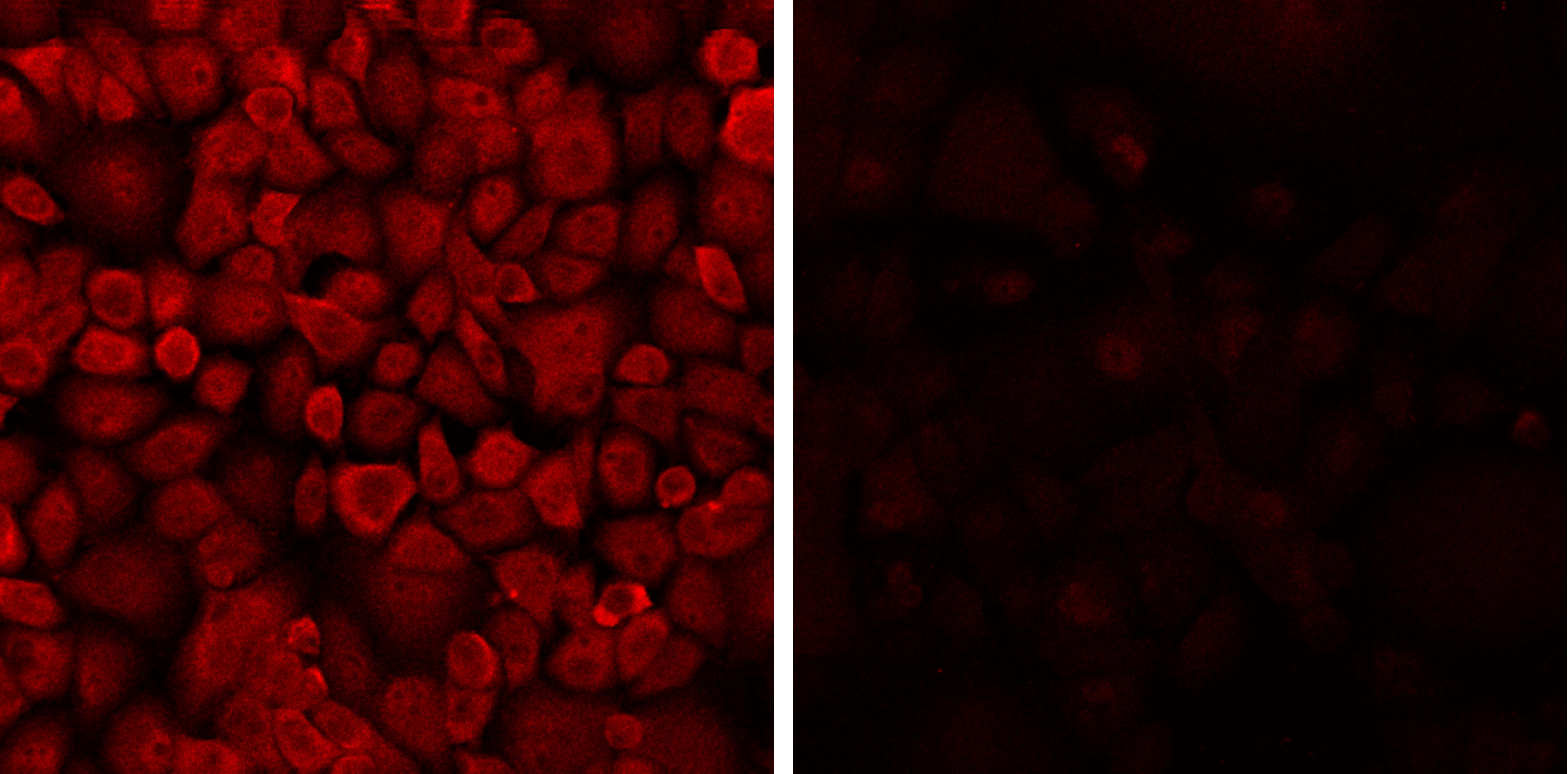
Both reviewers gave this antibody a perfect 5-star rating. One user reported successful Western blot performance on glioblastoma samples at a 1:1000 dilution. The other used it for immunofluorescence on PANC1 cells at 1:500, demonstrating clear staining in a YAP silencing experiment with fixed coverslips and an anti-mouse 488 secondary antibody. Overall, users found the antibody reliable across different applications with straightforward protocols.
The reviews below have been submitted by verified Proteintech customers who received an incentive for providing their feedback.
Udesh (Verified Customer) (11-26-2023) | Worked well at 1:1000 for WB
|
Joshua (Verified Customer) (01-29-2019) | Coverslips were fixed with 4%PFA+0.25% Triton-X-100. Primary staining was 1:500, secondary was 1:750 with rockland 488 anti-mouse overnight at 4C.Left panel: Control PANC1 cells.Right panel: PANC1 cells with YAP silencing
 |


















