

Tested Applications
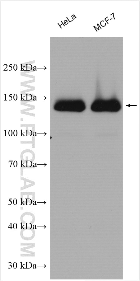
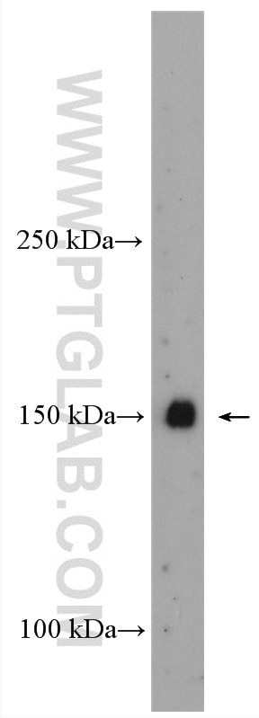
| Positive WB detected in | HeLa cells, mouse kidney tissue, MCF-7 cells |
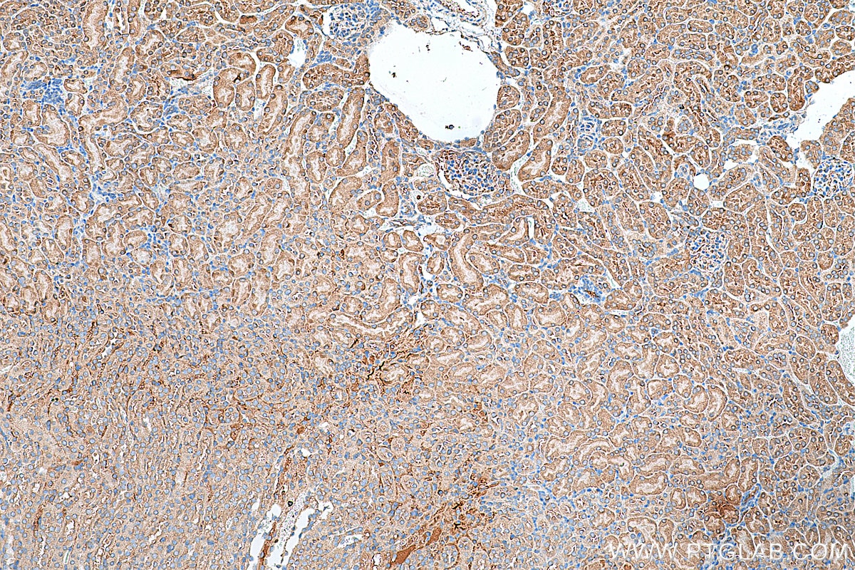
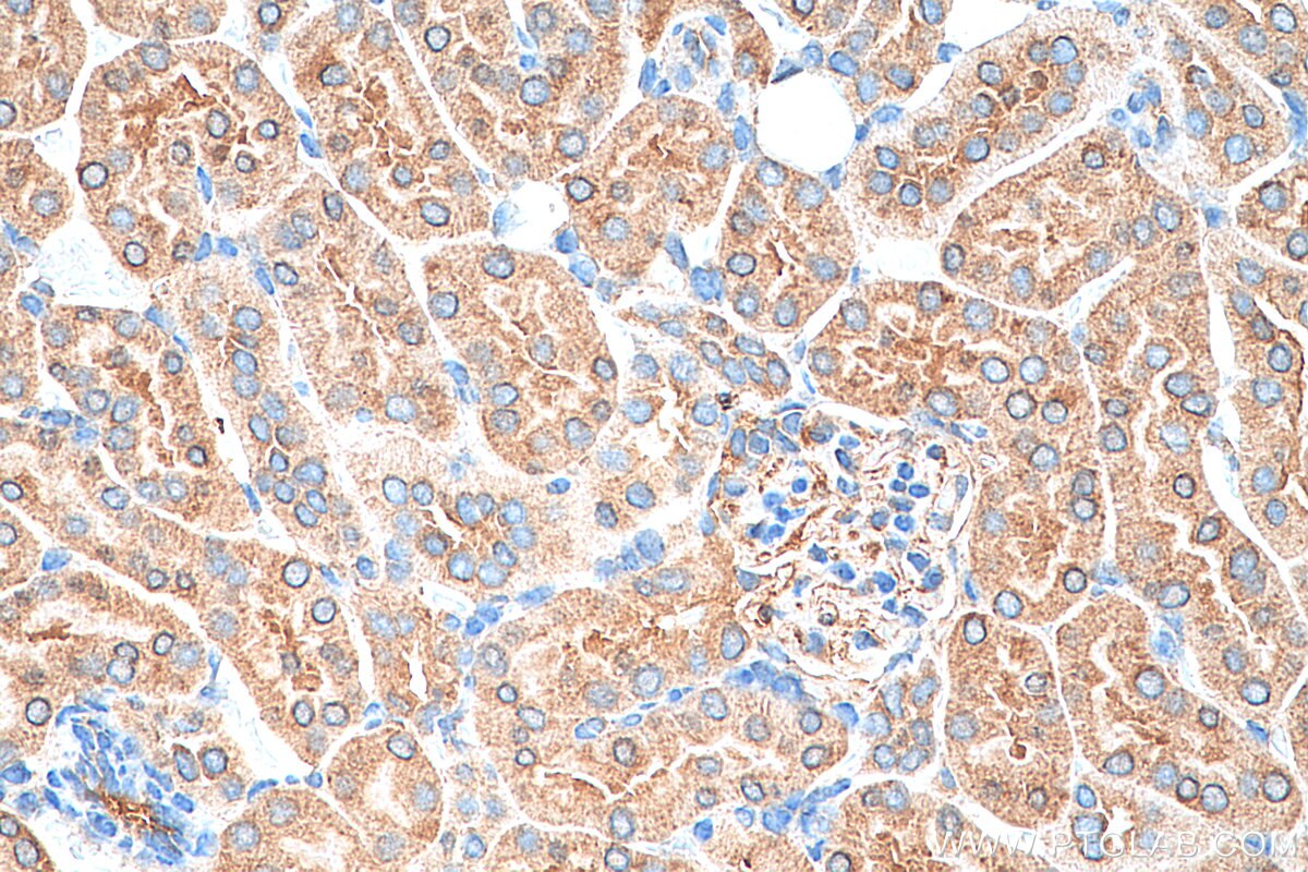
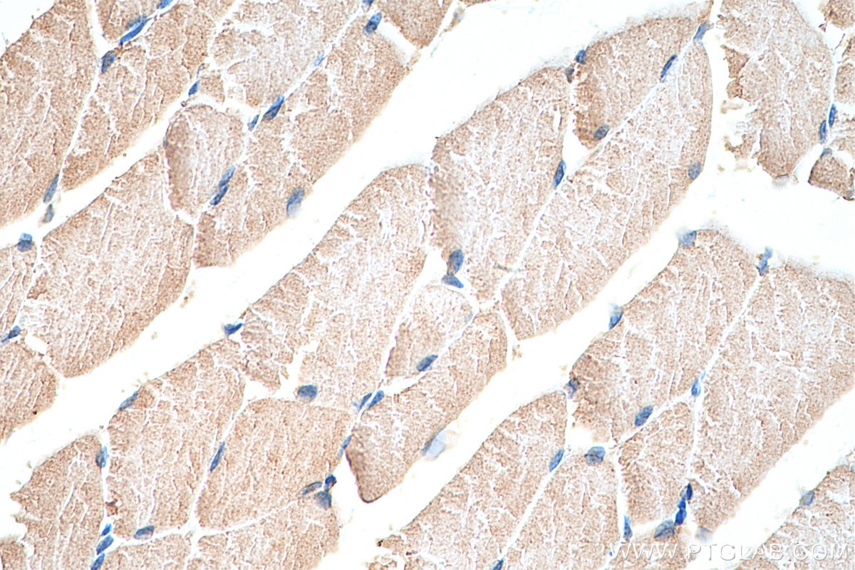
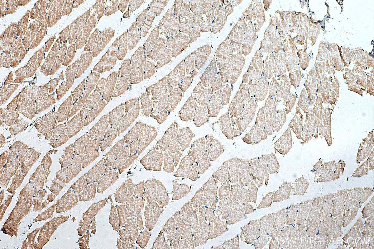
| Positive IHC detected in | mouse kidney tissue, mouse skeletal muscle tissue Note: suggested antigen retrieval with TE buffer pH 9.0; (*) Alternatively, antigen retrieval may be performed with citrate buffer pH 6.0 |
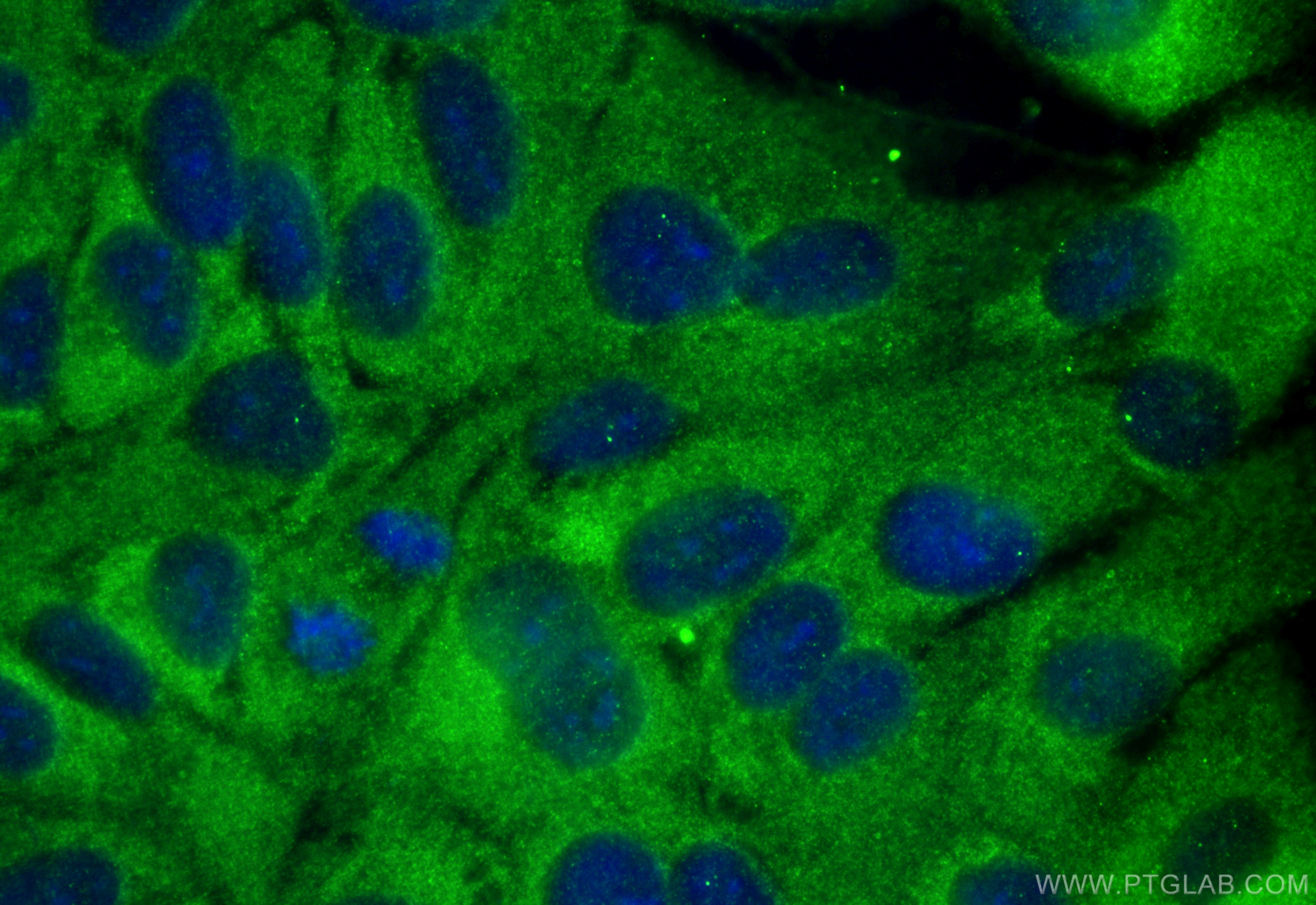
| Positive IF/ICC detected in | hTERT-RPE1 cells |
Recommended dilution
| Application | Dilution |
|---|---|
| Western Blot (WB) | WB : 1:1000-1:6000 |
| Immunohistochemistry (IHC) | IHC : 1:250-1:1000 |
| Immunofluorescence (IF)/ICC | IF/ICC : 1:50-1:500 |
| It is recommended that this reagent should be titrated in each testing system to obtain optimal results. | |
| Sample-dependent, Check data in validation data gallery. | |
Product Information
29906-1-AP targets Hamartin/TSC1 in WB, IHC, IF/ICC, IP, ELISA applications and shows reactivity with human, mouse samples.
| Tested Reactivity | human, mouse |
| Cited Reactivity | human, mouse, rat |
| Host / Isotype | Rabbit / IgG |
| Class | Polyclonal |
| Type | Antibody |
| Immunogen |
CatNo: Ag31621 Product name: Recombinant human TSC1 protein Source: e coli.-derived, PGEX-4T Tag: GST Domain: 965-1164 aa of NM_000368 Sequence: LYGRLEKDGLLKKLEEEKAEAAEAAEERLDCCNDGCSDSMVGHNEEASGHNGETKTPRPSSARGSSGSRGGGGSSSSSSELSTPEKPPHQRAGPFSSRWETTMGEASASIPTTVGSLPSSKSFLGMKARELFRNKSESQCDEDGMTSSLSESLKTELGKDLGVEAKIPLNLDGPHPSPPTPDSVGQLHIMDYNETHHEHS Predict reactive species |
| Full Name | tuberous sclerosis 1 |
| Calculated Molecular Weight | 130KD |
| Observed Molecular Weight | 140-145 kDa |
| GenBank Accession Number | NM_000368 |
| Gene Symbol | Hamartin/TSC1 |
| Gene ID (NCBI) | 7248 |
| RRID | AB_2918352 |
| Conjugate | Unconjugated |
| Form | Liquid |
| Purification Method | Antigen affinity purification |
| UNIPROT ID | Q92574 |
| Storage Buffer | PBS with 0.02% sodium azide and 50% glycerol, pH 7.3. |
| Storage Conditions | Store at -20°C. Stable for one year after shipment. Aliquoting is unnecessary for -20oC storage. 20ul sizes contain 0.1% BSA. |
Background Information
TSC1, also named as hamartin, interacts with TSC2 to form a protein complex that inhibits the nutrient-mediated or growth factor-stimulated phosphorylation of S6K1 and EIF4EBP1 by negatively regulating mTORC1 signaling (PubMed: 12271141, PubMed: 28215400). It can recruit TSC2 to HSP90AA1 and stabilizes TSC2 by preventing the interaction between TSC2 and ubiquitin ligase HERC1 (PubMed: 16464865, PubMed: 29127155). The loss of TSC1/TSC2 in mouse neurons resulted in a block in oligodendrocyte hypomyelination in vivo (PubMed: 28183733).
Protocols
| Product Specific Protocols | |
|---|---|
| IF protocol for Hamartin/TSC1 antibody 29906-1-AP | Download protocol |
| IHC protocol for Hamartin/TSC1 antibody 29906-1-AP | Download protocol |
| WB protocol for Hamartin/TSC1 antibody 29906-1-AP | Download protocol |
| Standard Protocols | |
|---|---|
| Click here to view our Standard Protocols |
Publications
What published studies show
The TSC1 antibody (Cat# 29906-1-AP) has 11 citations. It appears in journals including Nature Communications, Molecular Cell, Genes & Diseases, Phytomedicine, Journal of Translational Medicine, EMBO Molecular Medicine, British Journal of Cancer, Cell Signaling, Journal of Orthopaedic Surgery and Research, and eLife. Research fields span autophagy, mTOR/TSC1 signaling, cancer (glioblastoma, AML, breast, renal), bone metabolism, neurodegeneration, and pulmonary fibrosis, with applications in WB, IF, IHC, and IP.
| Species | Application | Title |
|---|---|---|
Nat Commun Targeting chaperone-mediated autophagy inhibits properties of glioblastoma stem cells and restores anti-tumor immunity. | ||
Genes Dis Naked cuticle homolog 2 controls the differentiation of osteoblasts and osteoclasts and ameliorates bone loss in ovariectomized mice | ||
Phytomedicine Periplocin and bufalin induce cardiotoxicity by regulating AMPK/SIRT1/PGC-1α pathway to inhibit energy metabolism and trigger autophagy. | ||
J Transl Med Polystyrene nanoplastics trigger pyroptosis in dopaminergic neurons through TSC2/TFEB-mediated disruption of autophagosome-lysosome fusion in Parkinson's disease | ||
Reviews
What customers say
Both reviewers used this antibody for Western Blot to confirm TSC1/TSC2 knockout in cell lines and gave it 5-star ratings. One validated TSC1 knockout at a 1:5,000 dilution and reported it worked well. The other confirmed TSC2 knockout in both human and mouse cells at 1:3,000, noting a few non-specific bands in mouse cells but much cleaner results in human cells. Overall, users found the antibody reliable for knockout validation with strong specificity, particularly in human cell lines.
The reviews below have been submitted by verified Proteintech customers who received an incentive for providing their feedback.
Shivali (Verified Customer) (09-13-2025) | We are working on how negative regulators of mTOR complex affect translation and used this antibody to check TSC1 knockout cell lines. Worked well
|
Parijat (Verified Customer) (08-27-2025) | I used this antibody to confirm TSC2 knockout in both human (eHap1) and mouse (IMCD3) cells and it worked great. Although in Mouse there were few non-specific bands, it was much cleaner in Human cells.
|








