Tested Applications
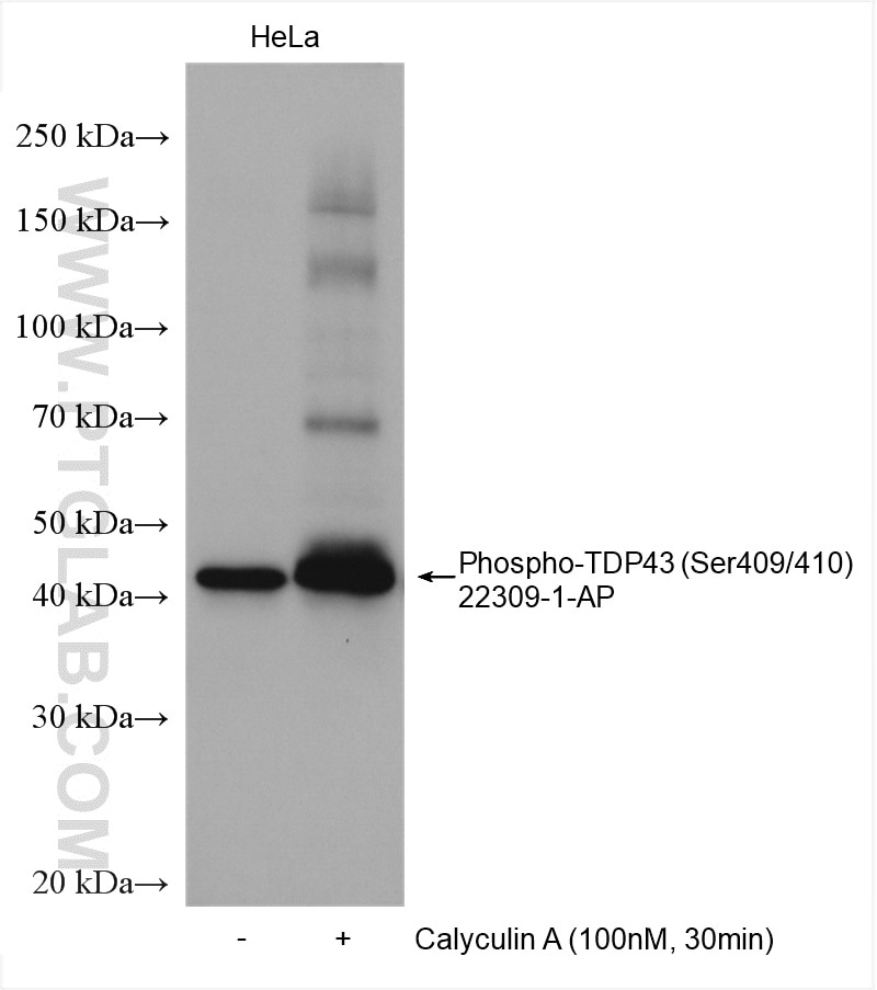
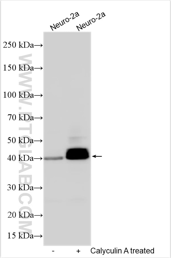
| Positive WB detected in | Neuro-2a cells, Calyculin A treated HeLa cells |
Recommended dilution
| Application | Dilution |
|---|---|
| Western Blot (WB) | WB : 1:500-1:3000 |
| It is recommended that this reagent should be titrated in each testing system to obtain optimal results. | |
| Sample-dependent, Check data in validation data gallery. | |
Published Applications
| KD/KO | See 1 publications below |
| WB | See 54 publications below |
| IHC | See 49 publications below |
| IF | See 46 publications below |
| IP | See 1 publications below |
| ELISA | See 2 publications below |
Product Information
22309-1-AP targets Phospho-TDP43 (Ser409/410) in WB, IHC, IF, IP, ELISA applications and shows reactivity with human, mouse samples.
| Tested Reactivity | human, mouse |
| Cited Reactivity | human, mouse, rat, monkey |
| Host / Isotype | Rabbit / IgG |
| Class | Polyclonal |
| Type | Antibody |
| Immunogen |
Peptide Predict reactive species |
| Full Name | TAR DNA binding protein |
| Calculated Molecular Weight | 43 kDa |
| Observed Molecular Weight | 40-50 kDa |
| GenBank Accession Number | NM_007375 |
| Gene Symbol | TDP-43 |
| Gene ID (NCBI) | 23435 |
| RRID | AB_11182943 |
| Conjugate | Unconjugated |
| Form | Liquid |
| Purification Method | Antigen affinity purification |
| UNIPROT ID | Q13148 |
| Storage Buffer | PBS with 0.02% sodium azide and 50% glycerol, pH 7.3. |
| Storage Conditions | Store at -20°C. Stable for one year after shipment. Aliquoting is unnecessary for -20oC storage. 20ul sizes contain 0.1% BSA. |
Background Information
Transactivation response (TAR) DNA-binding protein of 43 kDa (also known as TARDBP or TDP-43) was first isolated as a transcriptional inactivator binding to the TAR DNA element of the HIV-1 virus. Neumann et al. (2006) found that a hyperphosphorylated, ubiquitinated, and cleaved form of TARDBP, known as pathologic TDP-43, is the major component of the tau-negative and ubiquitin-positive inclusions that characterize amyotrophic lateral sclerosis (ALS) and the most common pathological subtype of frontotemporal lobar degeneration (FTLD-U). Various forms of TDP-43 exist, including 18-35 kDa of cleaved C-terminal fragments, 45-50 kDa phospho-protein, 55 kDa glycosylated form, 75 kDa hyperphosphorylated form, and 90-300 kDa cross-linked form. (PMID: 17023659,19823856, 21666678, 22193176).22309-1-AP is a rabbit polyclonal antibody recognizing TDP-43 only when phosphorylated at 409/410. Immunohistochemical analyses using this antibody only stain the insoluble inclusions in pathologic tissues without normal diffuse nuclear staining.
Protocols
| Product Specific Protocols | |
|---|---|
| WB protocol for Phospho-TDP43 (Ser409/410) antibody 22309-1-AP | Download protocol |
| Standard Protocols | |
|---|---|
| Click here to view our Standard Protocols |
Publications
| Species | Application | Title |
|---|---|---|
Nat Med The inhibition of TDP-43 mitochondrial localization blocks its neuronal toxicity. | ||
Nat Cell Biol Caspase-2 is a condensate-mediated deubiquitinase in protein quality control | ||
Nat Neurosci TREM2 interacts with TDP-43 and mediates microglial neuroprotection against TDP-43-related neurodegeneration. | ||
Mol Neurodegener Extracellular vesicles in TDP-43 proteinopathies: pathogenesis and biomarker potential | ||
Alzheimers Dement Entorhinal vessel density correlates with phosphorylated tau and TDP-43 pathology |
Reviews
The reviews below have been submitted by verified Proteintech customers who received an incentive for providing their feedback.
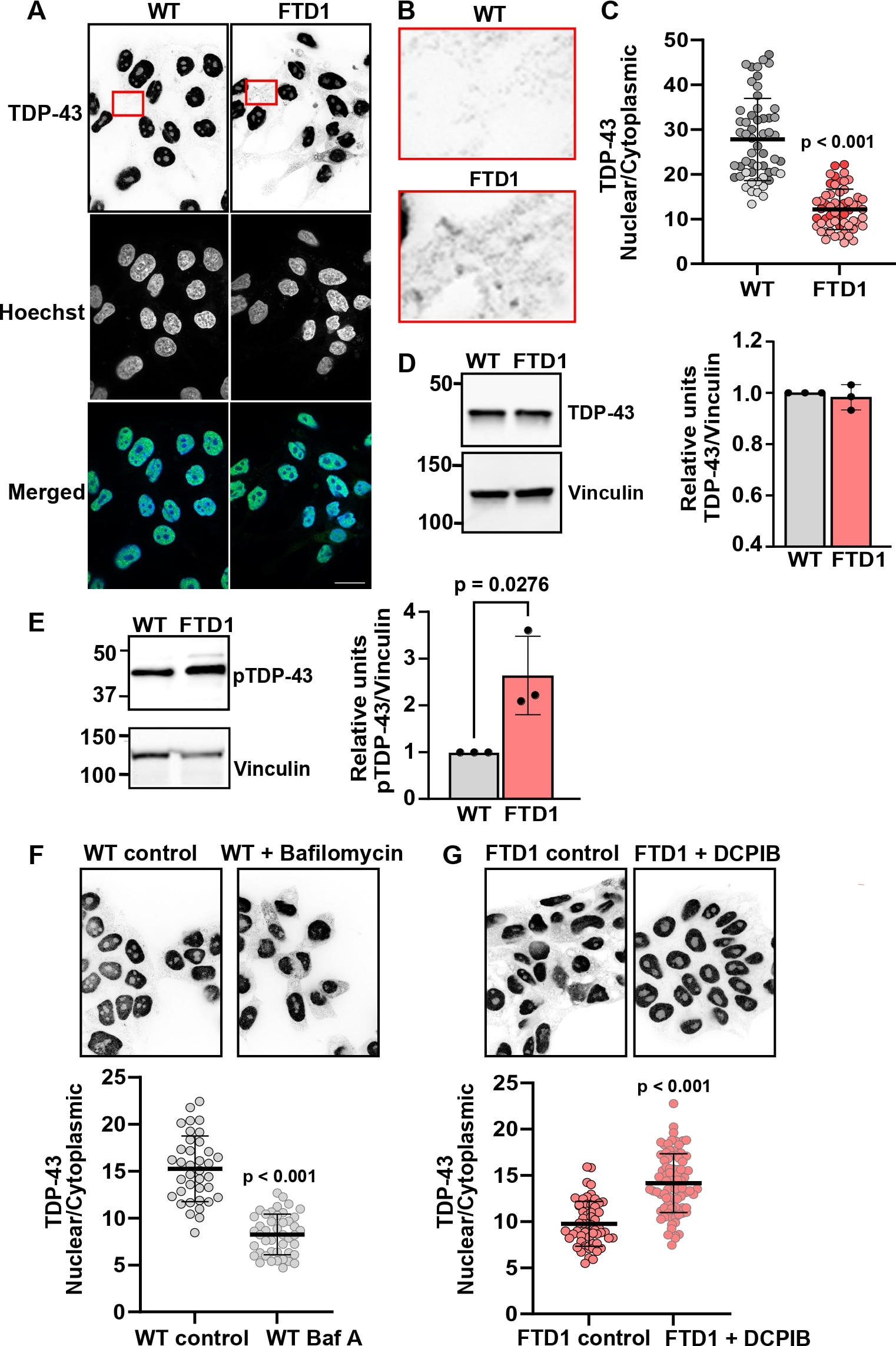
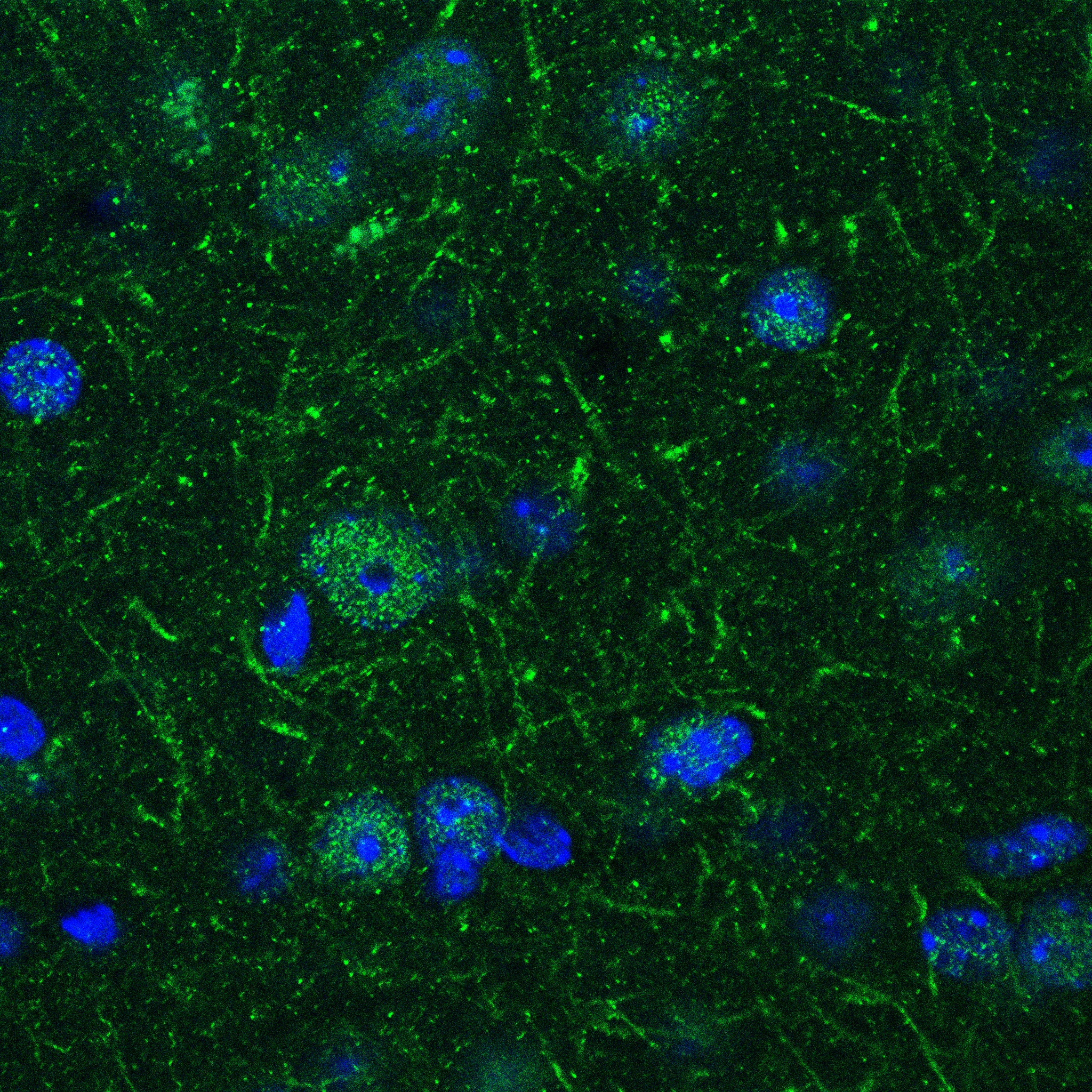
FH Sonia (Verified Customer) (10-02-2025) | We detected increased phosphorylated TDP-43 (pTDP-43) in FTD iPSCs compared with WT, determined by immunoblotting cell lysates using pTDP-43 (Ser409/410). However, our immunolabeling data with pTDP-43 were unclear since it shows strong nuclear signal.
 |
FH Rashmi (Verified Customer) (09-25-2024) | Used for WB, Highly Recommended
|
FH Vikas (Verified Customer) (07-11-2024) | Used for western blotting. Highly recommended.
|
FH Madison (Verified Customer) (06-24-2024) | This antibody Stained well in the human tissue there were very clear phospho tangles and there was not a lot of nonspecific staining in the tissue.
|
FH MANOHAR (Verified Customer) (03-06-2024) |
|
FH Shenyi (Verified Customer) (04-27-2022) | Staining with this antibody is very different from with the recombinant one (80007-1-RR) which I received as a free test vial. It showed strong signal in the nucleus while the recombinant one only showed signal in the cytoplasm. Now I'm confused: Could pTDP-43 (Ser409/410) also present in nucleus or it's the antibody being nonspecific?
 |
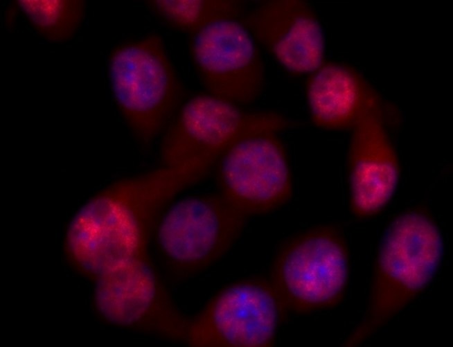
FH Azita (Verified Customer) (03-10-2020) | Immunofluorescent analysis of (4% PFA fixed) NSC-34 cells, using p- (109/110) TDP-43 antibody at 1/500 dilution and Alexa Flour conjugated Donkey anti Rabbit IgG (H+L) (Red). Bisbenzimide (Blue) labels cell nuclei.
 |
FH Laura (Verified Customer) (01-10-2020) | Works well at this concentration on HEK293T lysate.
|
FH Owen (Verified Customer) (10-30-2019) | This antibody was used to image TDP43 in tissue and cell samples. The antibody was useful to image such a difficult target.
|