

Tested Applications
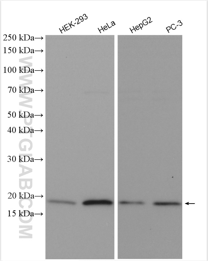
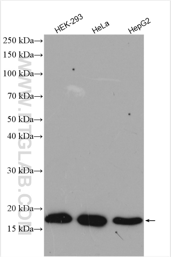
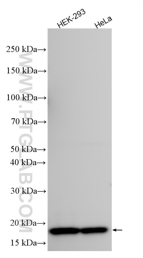
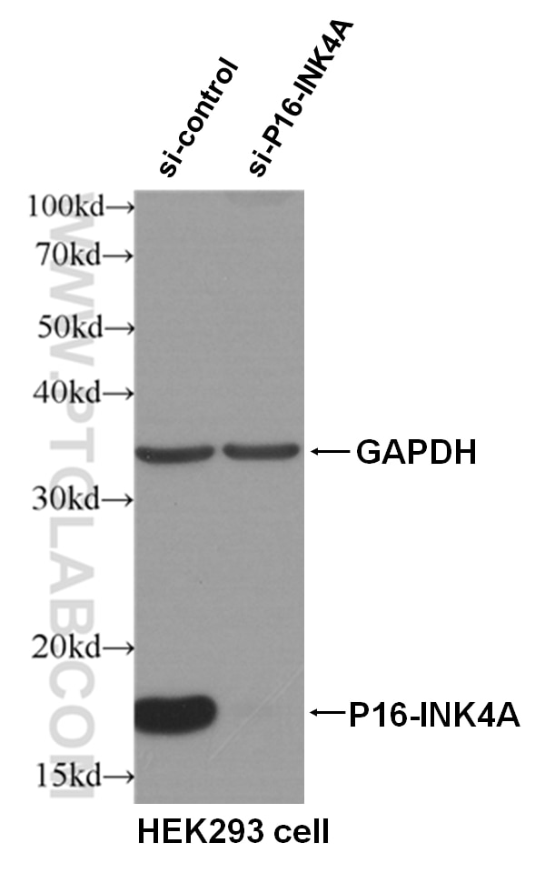
| Positive WB detected in | HEK-293 cells, HEK293 cells, HeLa cells, HepG2 cells, PC-3 cells |
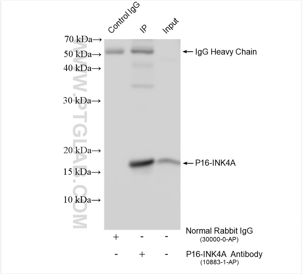
| Positive IP detected in | HEK-293 cells |
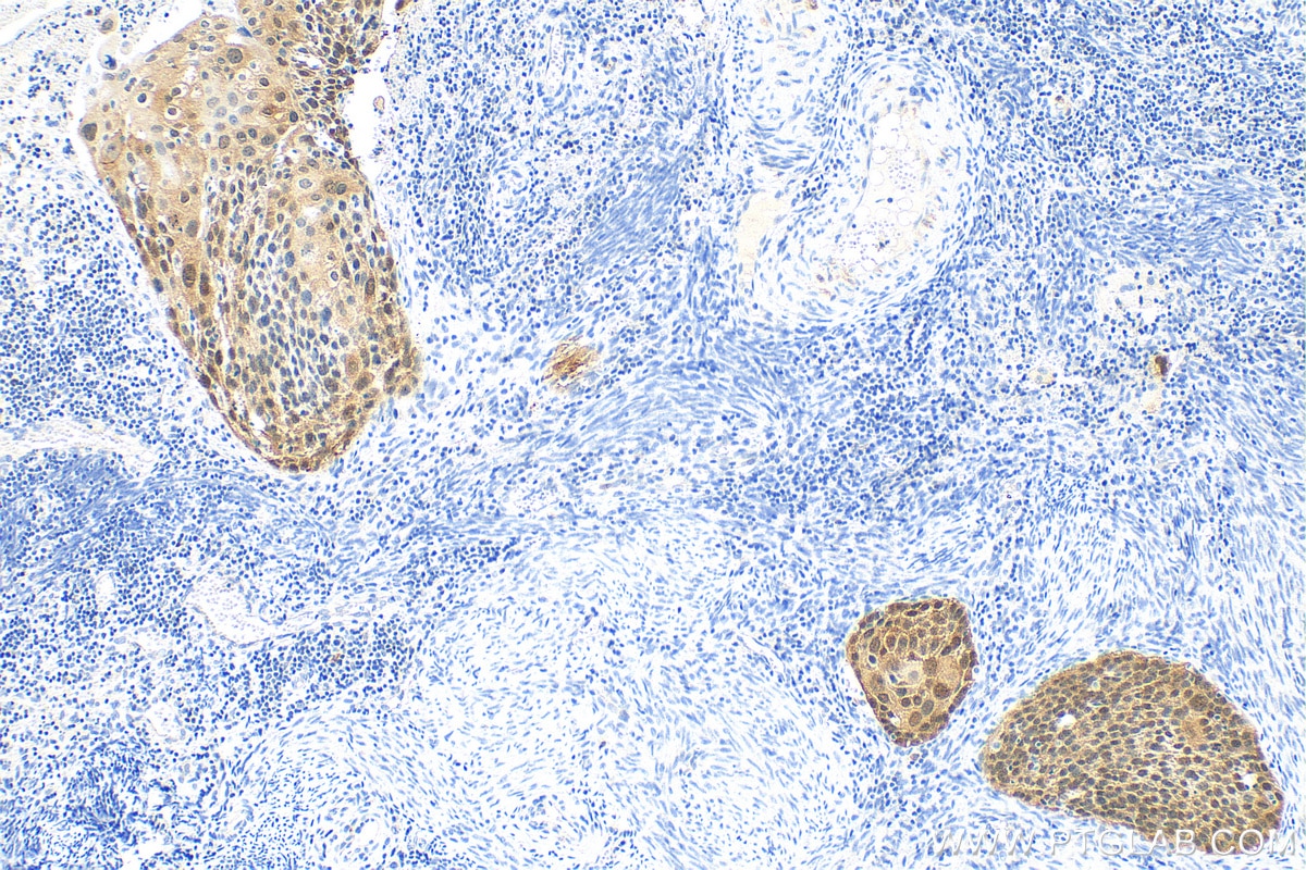
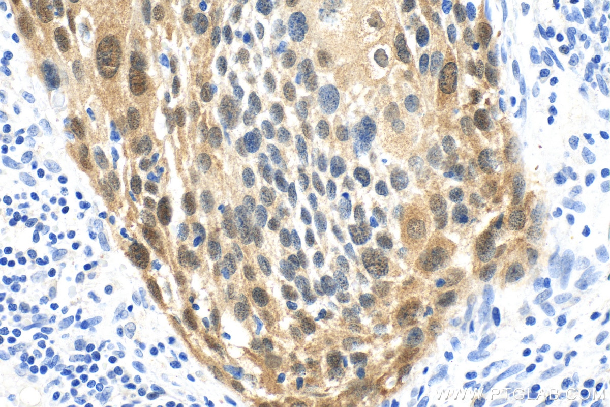
| Positive IHC detected in | human cervical cancer tissue Note: suggested antigen retrieval with TE buffer pH 9.0; (*) Alternatively, antigen retrieval may be performed with citrate buffer pH 6.0 |
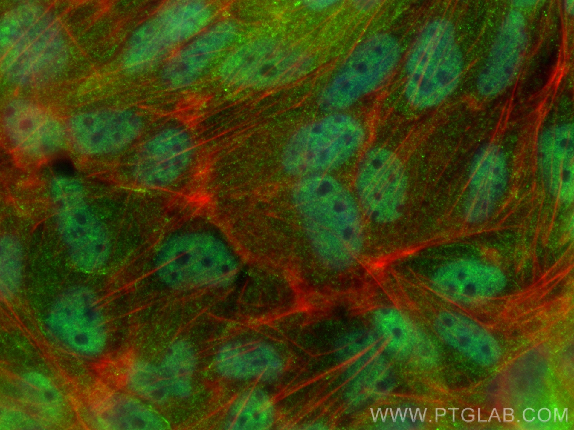
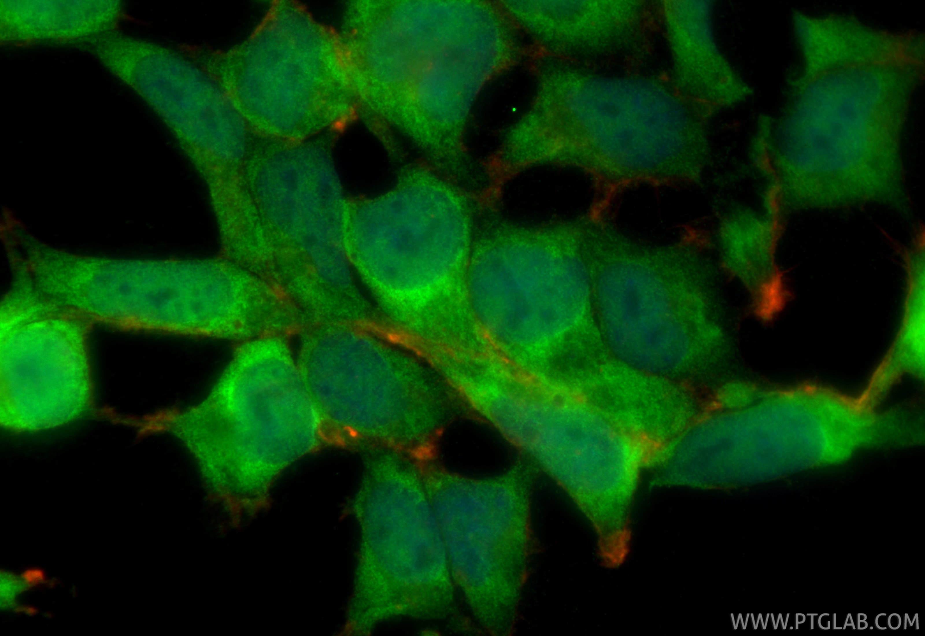
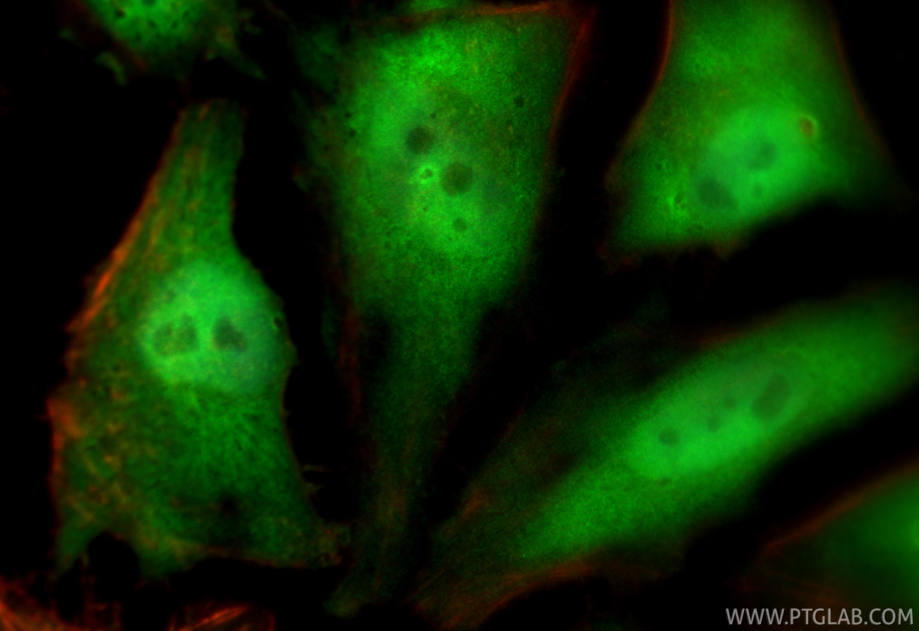
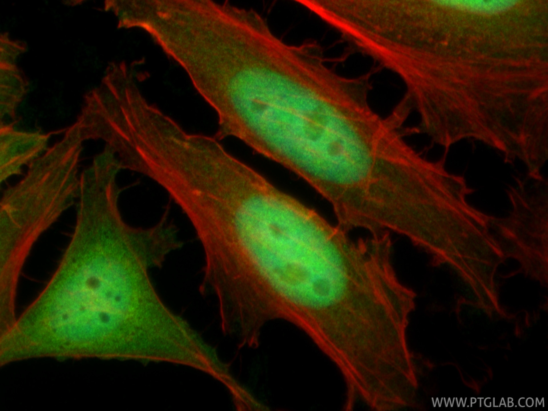
| Positive IF/ICC detected in | MDCK cells, HeLa cells, HEK-293 cells |
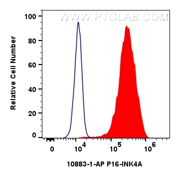
| Positive FC (Intra) detected in | HeLa cells |
Recommended dilution
| Application | Dilution |
|---|---|
| Western Blot (WB) | WB : 1:1000-1:6000 |
| Immunoprecipitation (IP) | IP : 0.5-4.0 ug for 1.0-3.0 mg of total protein lysate |
| Immunohistochemistry (IHC) | IHC : 1:1000-1:4000 |
| Immunofluorescence (IF)/ICC | IF/ICC : 1:200-1:800 |
| Flow Cytometry (FC) (INTRA) | FC (INTRA) : 0.40 ug per 10^6 cells in a 100 µl suspension |
| It is recommended that this reagent should be titrated in each testing system to obtain optimal results. | |
| Sample-dependent, Check data in validation data gallery. | |
Product Information
10883-1-AP targets CDKN2A/P16-INK4A in WB, IHC, IF/ICC, FC (Intra), IP, CoIP, ELISA applications and shows reactivity with human samples.
| Tested Reactivity | human |
| Cited Reactivity | human, pig, rabbit, canine, monkey, chicken, bovine |
| Host / Isotype | Rabbit / IgG |
| Class | Polyclonal |
| Type | Antibody |
| Immunogen |
CatNo: Ag1328 Product name: Recombinant human P16-INK4A protein Source: e coli.-derived, PGEX-4T Tag: GST Domain: 52-156 aa of BC021998 Sequence: MMMGSARVAELLLLHGAEPNCADPATLTRPVHDAAREGFLDTLVVLHRAGARLDVRDAWGRLPVDLAEELGHRDVARYLRAAAGGTRGSNHARIDAAEGPSDIPD Predict reactive species |
| Full Name | cyclin-dependent kinase inhibitor 2A |
| Calculated Molecular Weight | 16 kDa |
| Observed Molecular Weight | 16-18 kDa |
| GenBank Accession Number | BC021998 |
| Gene Symbol | CDKN2A |
| Gene ID (NCBI) | 1029 |
| RRID | AB_2078303 |
| Conjugate | Unconjugated |
| Form | Liquid |
| Purification Method | Antigen affinity purification |
| UNIPROT ID | P42771 |
| Storage Buffer | PBS with 0.02% sodium azide and 50% glycerol, pH 7.3. |
| Storage Conditions | Store at -20°C. Stable for one year after shipment. Aliquoting is unnecessary for -20oC storage. 20ul sizes contain 0.1% BSA. |
Background Information
Background
p16 is an important cell cycle regulator and acts as a tumor suppressor. It may also be referred to as one of a number of synonyms, including p16INK4a and cyclin-dependent kinase inhibitor 2A.
What is the molecular weight of P16?
16kDa. P16 is encoded by the CDKN2A gene in humans and is a chain comprising 148 amino acids.
What is the function of p16?
P16 inhibits cells from progressing from G1 into S phase, binding to cyclin-dependent kinase 4 (CDK4) and inhibiting its kinase ability, so that it cannot phosphorylate the retinoblastoma tumor suppressor (RB). Without this phosphorylation, RB does not activate downstream genes, so the G1/S checkpoint cannot be passed and the cell does not proliferate (PMID: 8259215).
What is the role of p16 in senescence?
In senescence, cells are irreversibly arrested in the cell cycle. P16 is expressed more highly in aging tissue, is associated with intrinsic cellular aging signals such as telomere shortening, and can therefore be used as a marker of senescence (PMID: 9244355; PMID: 19535234). Due to its role in cell cycle arrest, p16 drives the initiation and maintenance of a cellular senescent phenotype.
What is the role of p16 in cancer?
As a negative regulator of proliferation, p16 is a known tumor suppressor. Mutations in the CDKN2A gene that lead to inactivation of p16 protein have been associated with an increased risk of cancer and are often observed in primary tumors and in cancer cell lines (PMID: 9508208). The inactivation of p16 has been shown to be a key early stage of tumor progression. In a small number of tumor types that are caused by the human papilloma virus (HPV), p16 is in fact overexpressed when RB is inactivated, releasing p16 and causing an accumulation (PMID: 21297668).
Protocols
| Product Specific Protocols | |
|---|---|
| FC protocol for CDKN2A/P16-INK4A antibody 10883-1-AP | Download protocol |
| IF protocol for CDKN2A/P16-INK4A antibody 10883-1-AP | Download protocol |
| IHC protocol for CDKN2A/P16-INK4A antibody 10883-1-AP | Download protocol |
| IP protocol for CDKN2A/P16-INK4A antibody 10883-1-AP | Download protocol |
| WB protocol for CDKN2A/P16-INK4A antibody 10883-1-AP | Download protocol |
| Standard Protocols | |
|---|---|
| Click here to view our Standard Protocols |
Publications
What published studies show
Anti-p16 (CDKN2A) antibody (Cat# 10883-1-AP) has 659 total citations published across 70+ journals, including Nature, Science, Cell, Nature Communications, Nature Aging, Cell Metabolism, Autophagy, Redox Biology, Cancer Cell, Circulation, and Oncogene. Key research fields include cellular senescence and aging, cancer biology, cardiovascular disease, osteoarthritis, neurodegeneration, fibrosis, metabolic disorders, immunology, and regenerative medicine.
| Species | Application | Title |
|---|---|---|
Signal Transduct Target Ther Human adipose and umbilical cord mesenchymal stem cell-derived extracellular vesicles mitigate photoaging via TIMP1/Notch1 | ||
Signal Transduct Target Ther Glibenclamide targets MDH2 to relieve aging phenotypes through metabolism-regulated epigenetic modification | ||
Signal Transduct Target Ther Seryl tRNA synthetase cooperates with POT1 to regulate telomere length and cellular senescence. | ||
Cancer Cell mTORC1 Activation Blocks Braf(V600E)-Induced Growth Arrest but Is Insufficient for Melanoma Formation. | ||
Reviews
What customers say
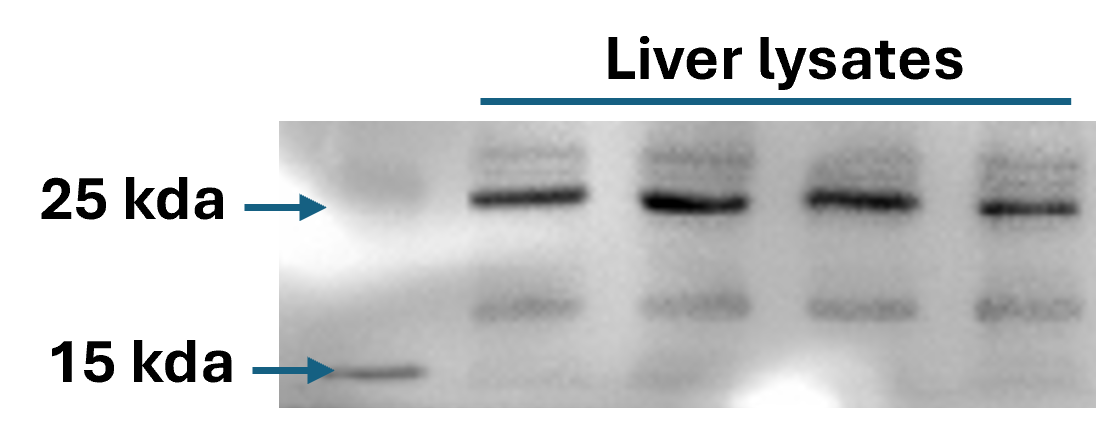
Most reviewers rate this p16 antibody highly (7 of 9 gave 5 stars). It performs well in Western Blot at 1:1000 dilution across various cell lines and tissues, producing clean bands with low background. Immunofluorescence results are described as bright and excellent. One reviewer noted the band appeared at ~20 KD rather than the expected 16 KD, though the supplier was responsive and helpful. Overall, users find it reliable and recommend it for WB and IF applications.
The reviews below have been submitted by verified Proteintech customers who received an incentive for providing their feedback.
Kamal (Verified Customer) (01-22-2026) | Antibody was diluted 1:5000 in 1X TBST.
 |
Mounika (Verified Customer) (12-25-2025) | Very good antibody, works excellently, very good precision in terms of visibility. smooth working with it!
|
Mathew (Verified Customer) (08-19-2025) | Worked well for western blots of 2DD primary foreskin fibroblast that had undergone confluence induced senescence.
|
Vikas (Verified Customer) (04-23-2024) | Highly recommended, to use 1:1000 dilution for Western bloating.
|
Vikas (Verified Customer) (04-23-2024) | Used p16 antibody for western-bolting at the dilution 1:10000, antibody worked at best. Thank you, highly recommended it.
|
Susanne (Verified Customer) (10-11-2022) | blocking: ROTIblock Incubation over night 4°C
|
Malak (Verified Customer) (04-22-2021) | Unfortunately, with this antibody, I didn't get a bad at 16 KD, however it was higher (around 20 KD). The suppliers were so professional and offered their help to optimize this antibody.
|
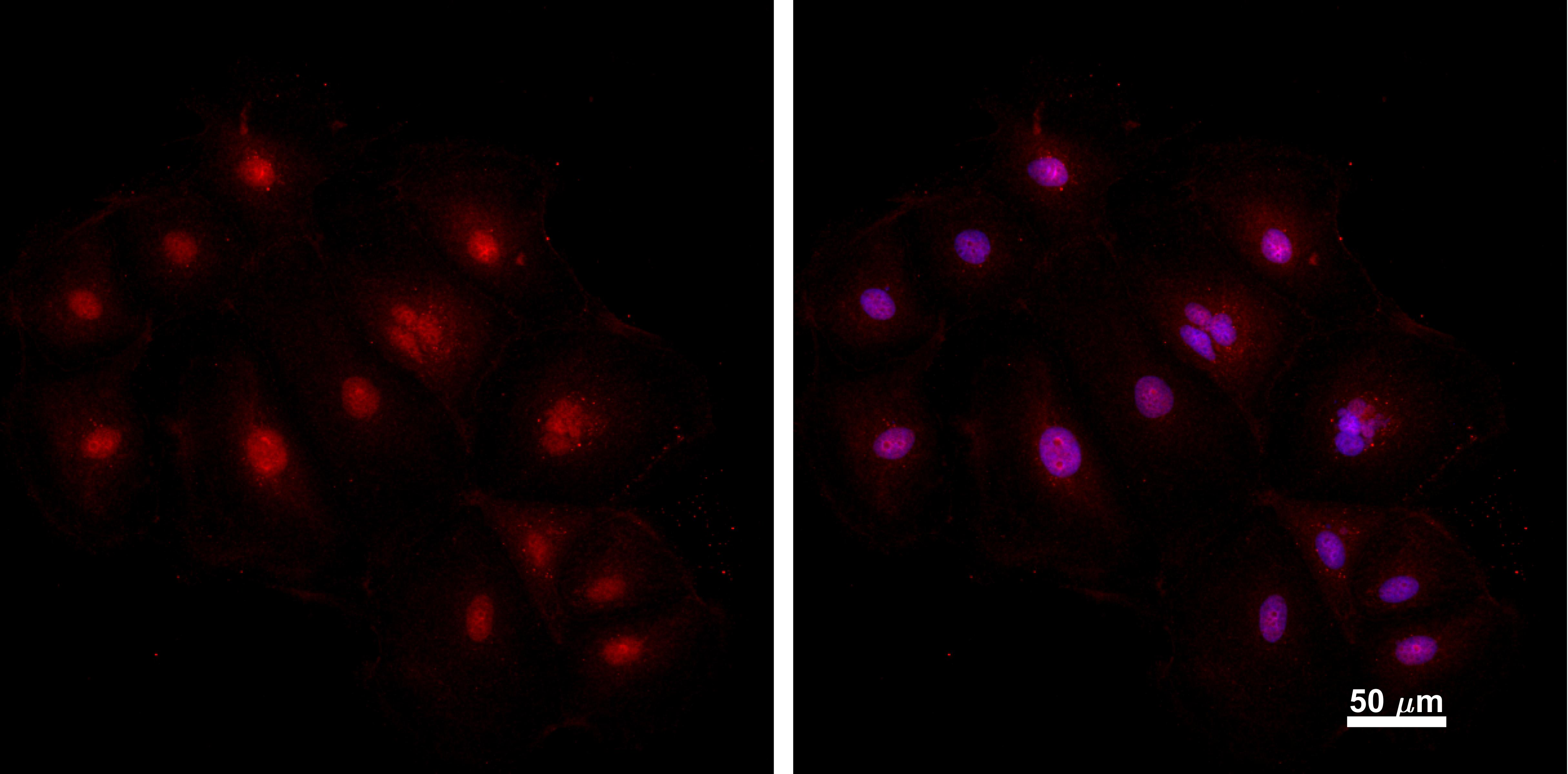
Joshua (Verified Customer) (03-12-2020) | MDCKs fixed in 4% paraformaldehyde and stained overnight at 4 C. Excellent, bright stain.
 |
Kyle (Verified Customer) (02-28-2019) | Clean bands, low background
|













