Tested Applications
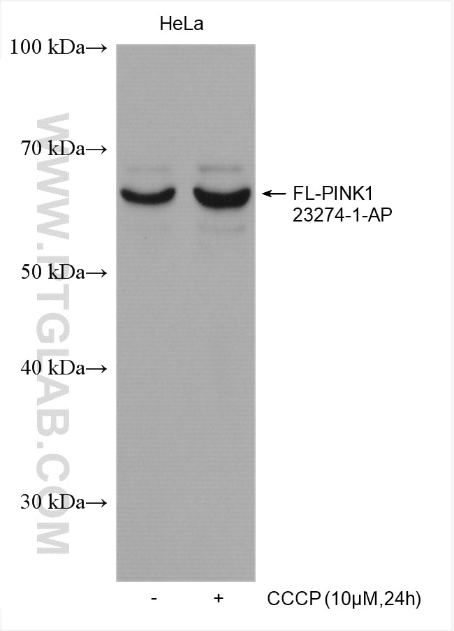
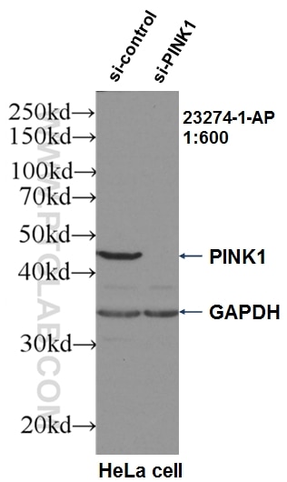
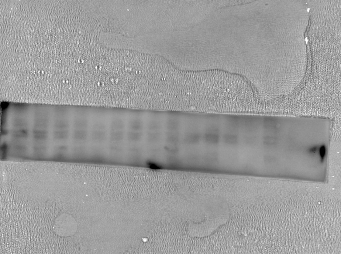
| Positive WB detected in | HEK-293 cells, HeLa cells, CCCP treated HeLa cells, PC-12 cells |
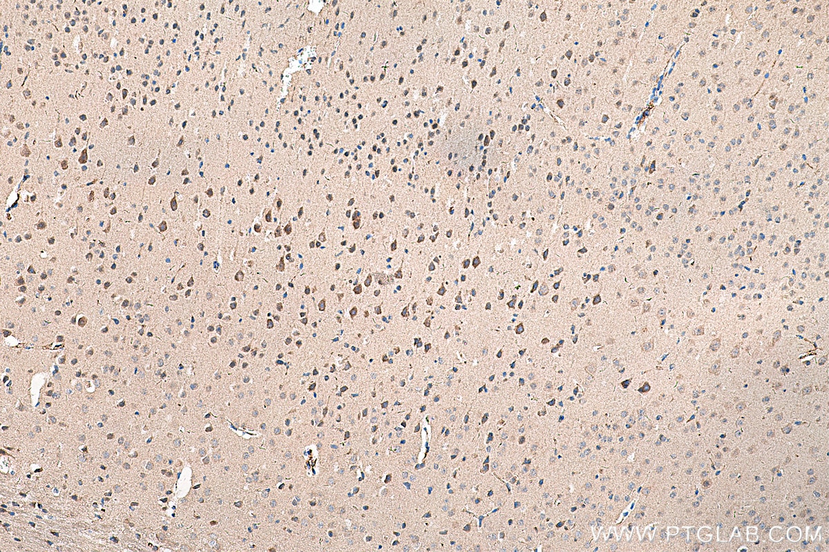
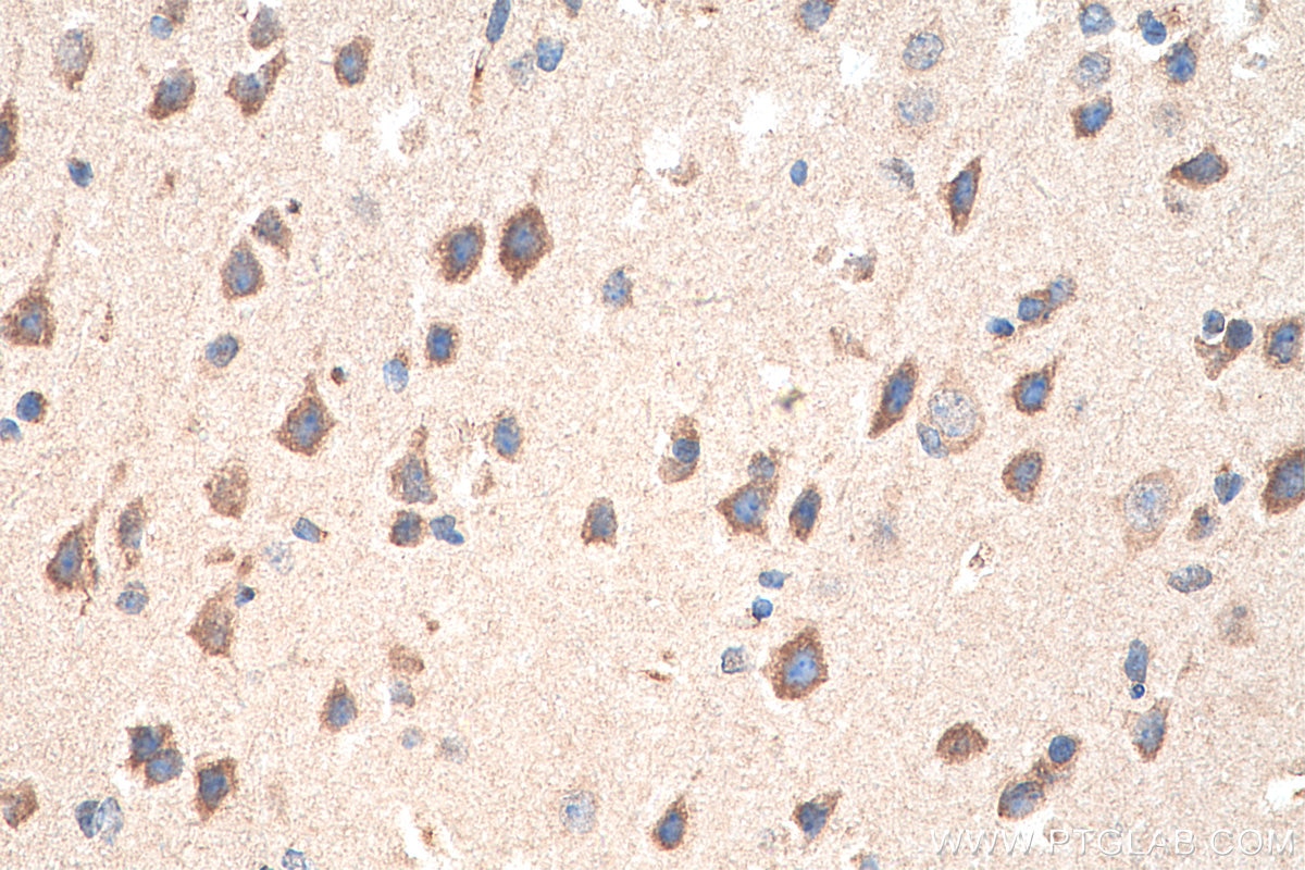
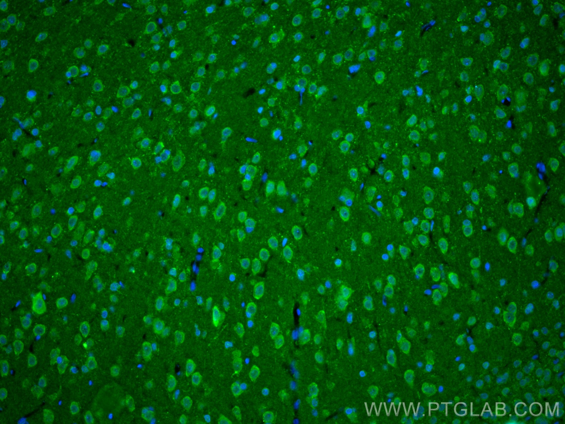
| Positive IHC detected in | mouse brain tissue Note: suggested antigen retrieval with TE buffer pH 9.0; (*) Alternatively, antigen retrieval may be performed with citrate buffer pH 6.0 |
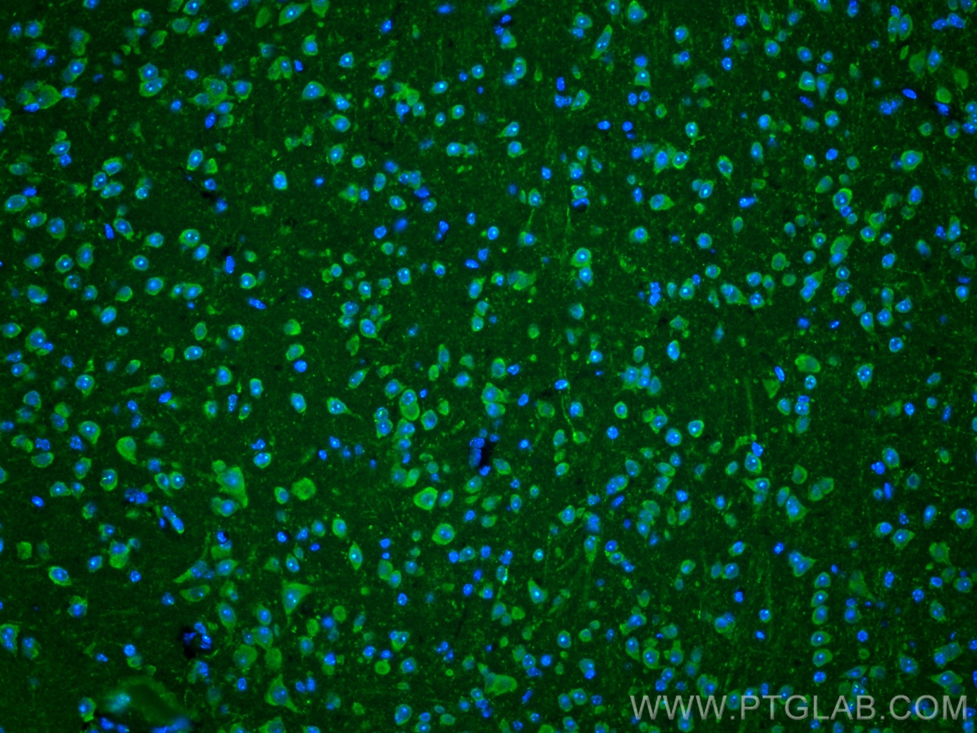
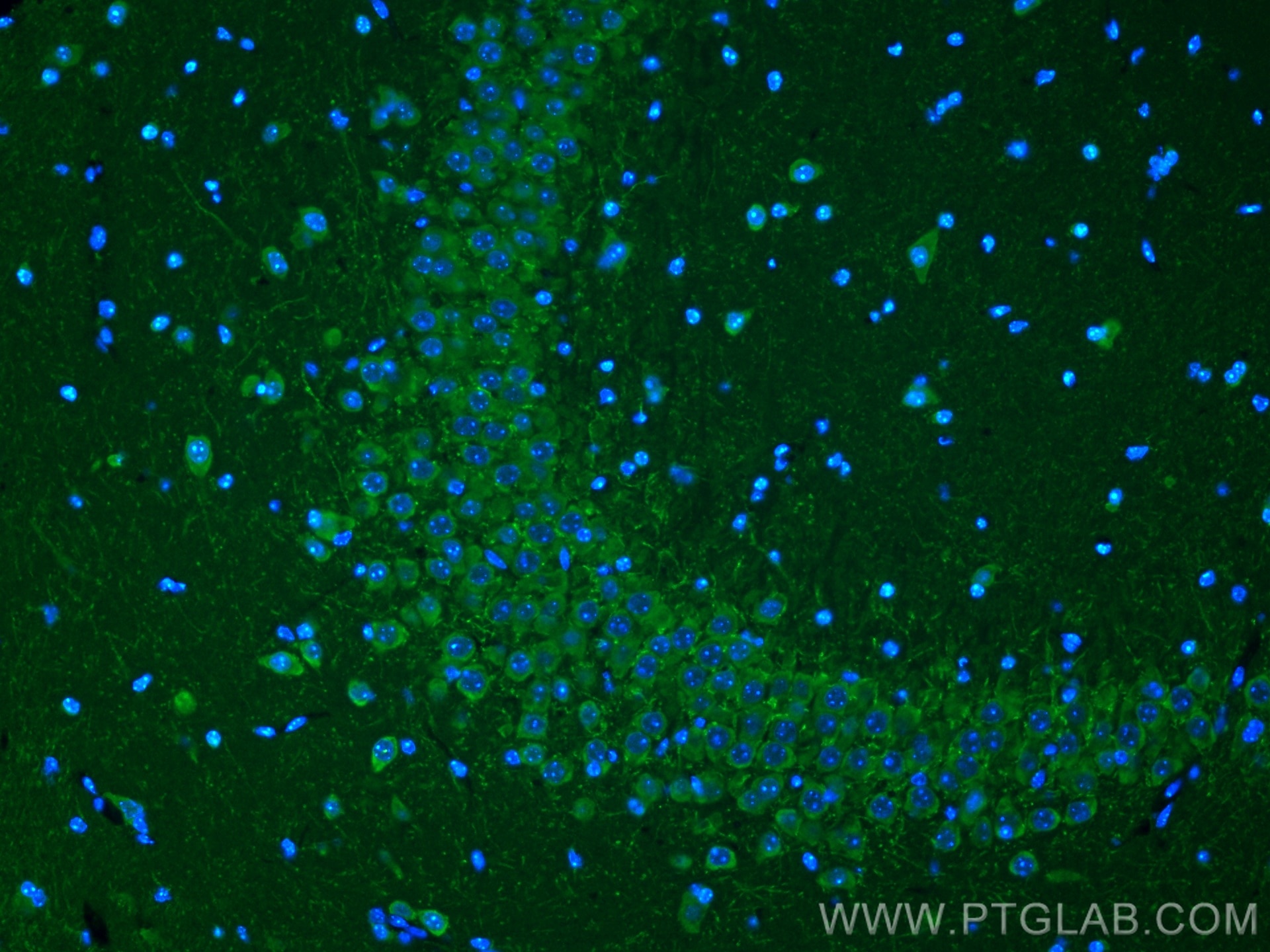
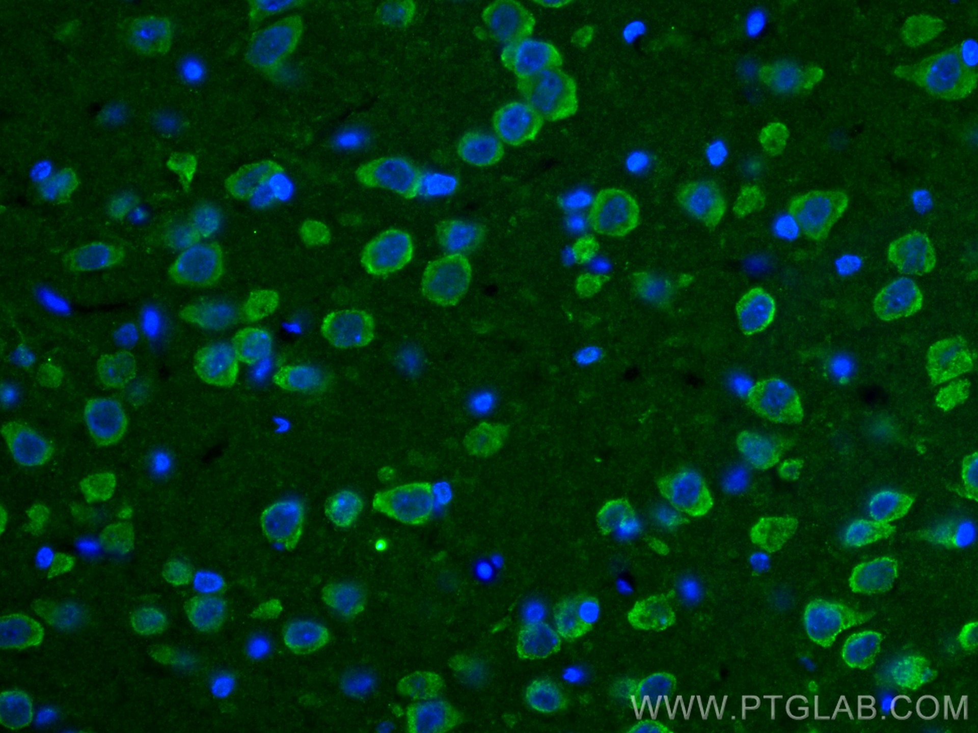
| Positive IF-P detected in | mouse brain tissue, rat brain tissue |
Recommended dilution
| Application | Dilution |
|---|---|
| Western Blot (WB) | WB : 1:1000-1:4000 |
| Immunohistochemistry (IHC) | IHC : 1:1000-1:4000 |
| Immunofluorescence (IF)-P | IF-P : 1:200-1:800 |
| It is recommended that this reagent should be titrated in each testing system to obtain optimal results. | |
| Sample-dependent, Check data in validation data gallery. | |
Published Applications
| KD/KO | See 41 publications below |
| WB | See 614 publications below |
| IHC | See 64 publications below |
| IF | See 120 publications below |
| IP | See 4 publications below |
| CoIP | See 8 publications below |
| ChIP | See 1 publications below |
Product Information
23274-1-AP targets PINK1 in WB, IHC, IF-P, IP, CoIP, ChIP, ELISA applications and shows reactivity with human, mouse, rat samples.
| Tested Reactivity | human, mouse, rat |
| Cited Reactivity | human, mouse, rat, pig, rabbit, monkey, zebrafish, sheep, goat, duck |
| Host / Isotype | Rabbit / IgG |
| Class | Polyclonal |
| Type | Antibody |
| Immunogen |
CatNo: Ag19825 Product name: Recombinant human PINK1 protein Source: e coli.-derived, PGEX-4T Tag: GST Domain: 336-581 aa of BC028215 Sequence: PRLAAMMLLQLLEGVDHLVQQGIAHRDLKSDNILVELDPDGCPWLVIADFGCCLADESIGLQLPFSSWYVDRGGNGCLMAPEVSTARPGPRAVIDYSKADAWAVGAIAYEIFGLVNPFYGQGKAHLESRSYQEAQLPALPESVPPDVRQLVRALLQREASKRPSARVAANVLHLSLWGEHILALKNLKLDKMVGWLLQQSAATLLANRLTEKCCVETKMKMLFLANLECETLCQAALLLCSWRAAL Predict reactive species |
| Full Name | PTEN induced putative kinase 1 |
| Calculated Molecular Weight | 581 aa, 63 kDa |
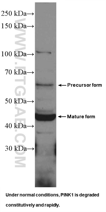
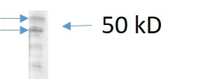
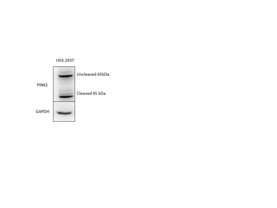
| Observed Molecular Weight | 65 kDa, 45 kDa |
| GenBank Accession Number | BC028215 |
| Gene Symbol | PINK1 |
| Gene ID (NCBI) | 65018 |
| RRID | AB_2879244 |
| Conjugate | Unconjugated |
| Form | Liquid |
| Purification Method | Antigen Affinity purified |
| UNIPROT ID | Q9BXM7 |
| Storage Buffer | PBS with 0.02% sodium azide and 50% glycerol, pH 7.3. |
| Storage Conditions | Store at -20°C. Stable for one year after shipment. Aliquoting is unnecessary for -20oC storage. 20ul sizes contain 0.1% BSA. |
Background Information
PINK1 is a mitochondrial serine/threonine-protein kinase that protects cells from stress-induced mitochondrial dysfunction. The precursor of PINK1 (65 kDa) is synthesized in the cytosol and is imported into the outer membrane of mitochondria. PINK1 is further transferred into the inner membrane. The full-length PINK1 can be proteolytically processed into 52-55 kDa and 45-46 kDa forms (PMID: 18221368; 25108683; 18031932). The half-life of the mature form of PINK1 is very short and it was proposed that the proteasome is involved in its degradation (PMID: 23472196). The gene of PINK1 maps to chromosome 1p36.12. Two alternatively spliced variants exist, the shorter isoform (30 kDa) produced by alternative splicing. Mutations in the PINK1 gene cause autosomal recessive early-onset Parkinson's disease.
Protocols
| Product Specific Protocols | |
|---|---|
| IF protocol for PINK1 antibody 23274-1-AP | Download protocol |
| IHC protocol for PINK1 antibody 23274-1-AP | Download protocol |
| WB protocol for PINK1 antibody 23274-1-AP | Download protocol |
| Standard Protocols | |
|---|---|
| Click here to view our Standard Protocols |
Publications
| Species | Application | Title |
|---|---|---|
Nat Cell Biol Ammonia-induced lysosomal and mitochondrial damage causes cell death of effector CD8+ T cells | ||
Bioact Mater NIR-responsive bio-system with sequential antibacterial and immunomodulatory effects for the treatment of periodontitis | ||
Adv Sci (Weinh) WAC Facilitates Mitophagy-mediated MSC Osteogenesis and New Bone Formation via Protecting PINK1 from Ubiquitination-Dependent Degradation | ||
Autophagy KDM4A-induced tumor senescence enhances the efficacy of immunotherapy by inhibiting AGT-PHB1 axis-mediated mitophagy in colorectal cancer |
Reviews
The reviews below have been submitted by verified Proteintech customers who received an incentive for providing their feedback.
FH MALLIKARJUNA (Verified Customer) (10-17-2025) | GOOD QUALITY AB FOR WB
|
FH Javier (Verified Customer) (09-09-2025) | Very good for Western Blotting.
|
FH aidan (Verified Customer) (04-10-2024) | Good results. Would try higher dilution next time (1:500) on precast gel. Used handcast gel overnight incubation at 4 degrees. Blocked 1 hr 5% milk
 |
FH Nin (Verified Customer) (02-27-2023) | It is okay to probe PINK1 with two bands although there are some weak non-specific bands.
 |
FH Yahir Alberto (Verified Customer) (11-12-2021) | The antibody works fine with overnight incubation at 4 °C.
 |
FH Tanusree (Verified Customer) (12-03-2019) | This antibody works good in western blotting analysis using mouse tissues.
|
FH HONGXUE (Verified Customer) (08-19-2019) | The specific is not good. You can detect many bands using WB.
 |