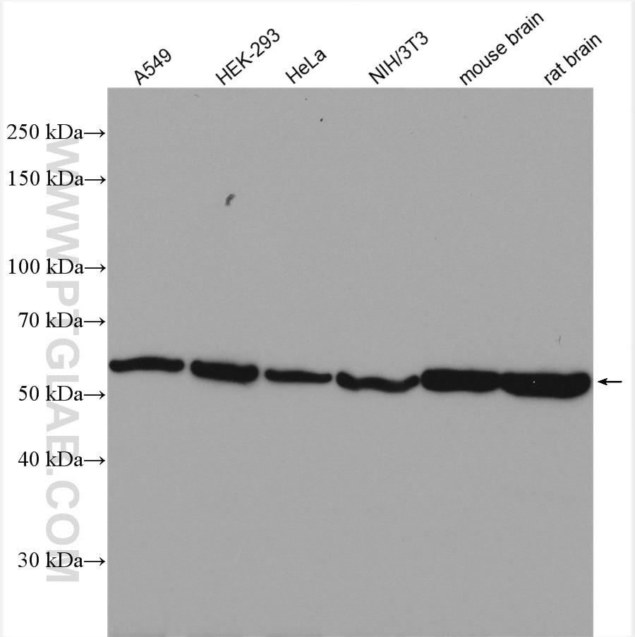
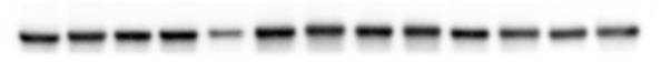
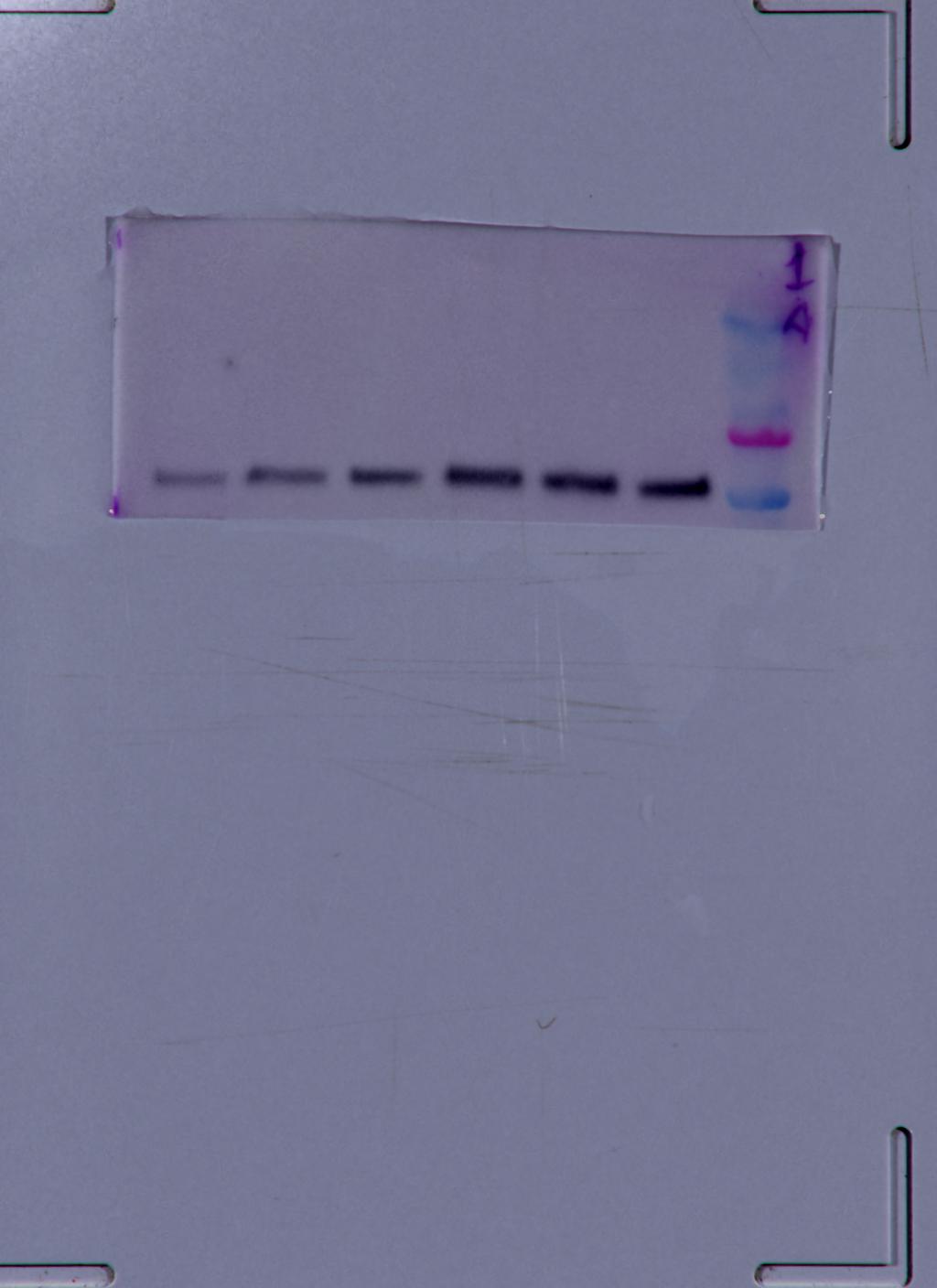
at dilution of 1:10000 incubated at room temperature for 1.5 hours.")
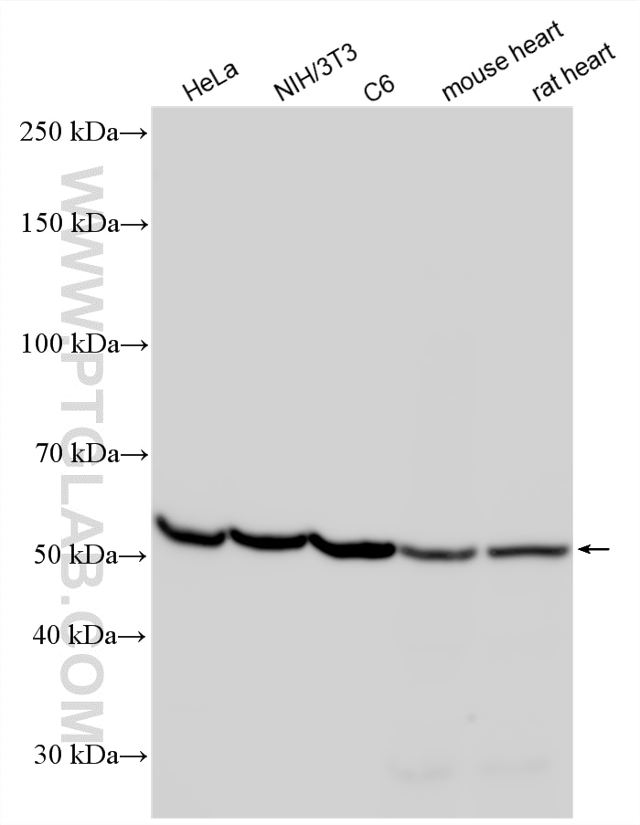
 at dilution of 1:10000 incubated at room temperature for 1.5 hours.")
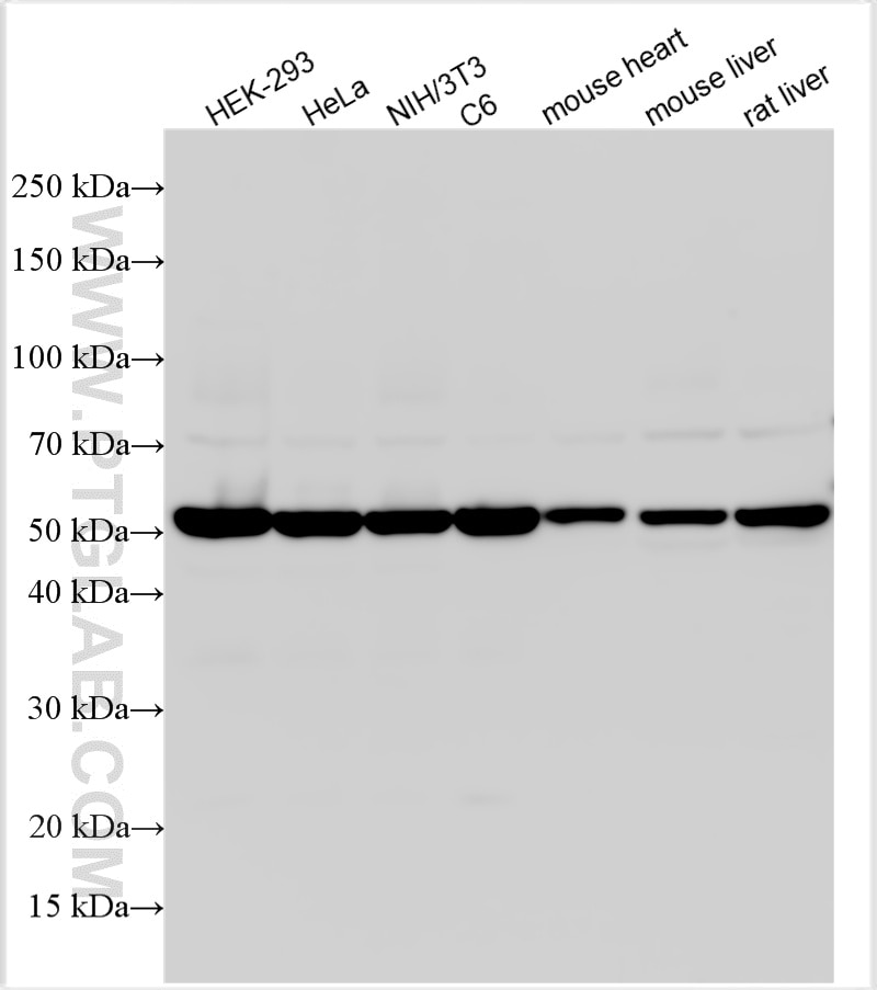
 at dilution of 1:15000 incubated at room temperature for 1.5 hours.")
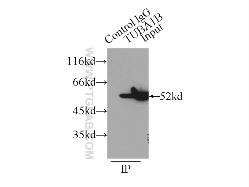
 with HeLa cells lysate 5000ug.")
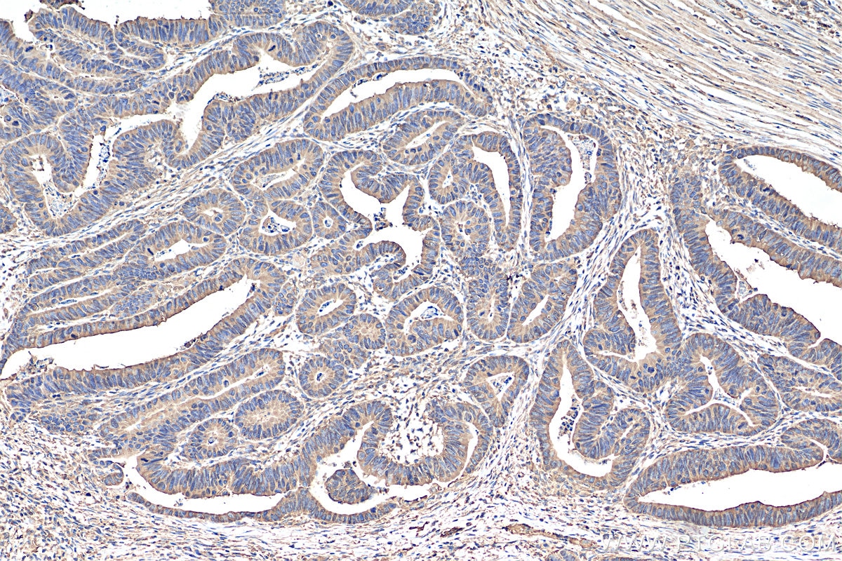
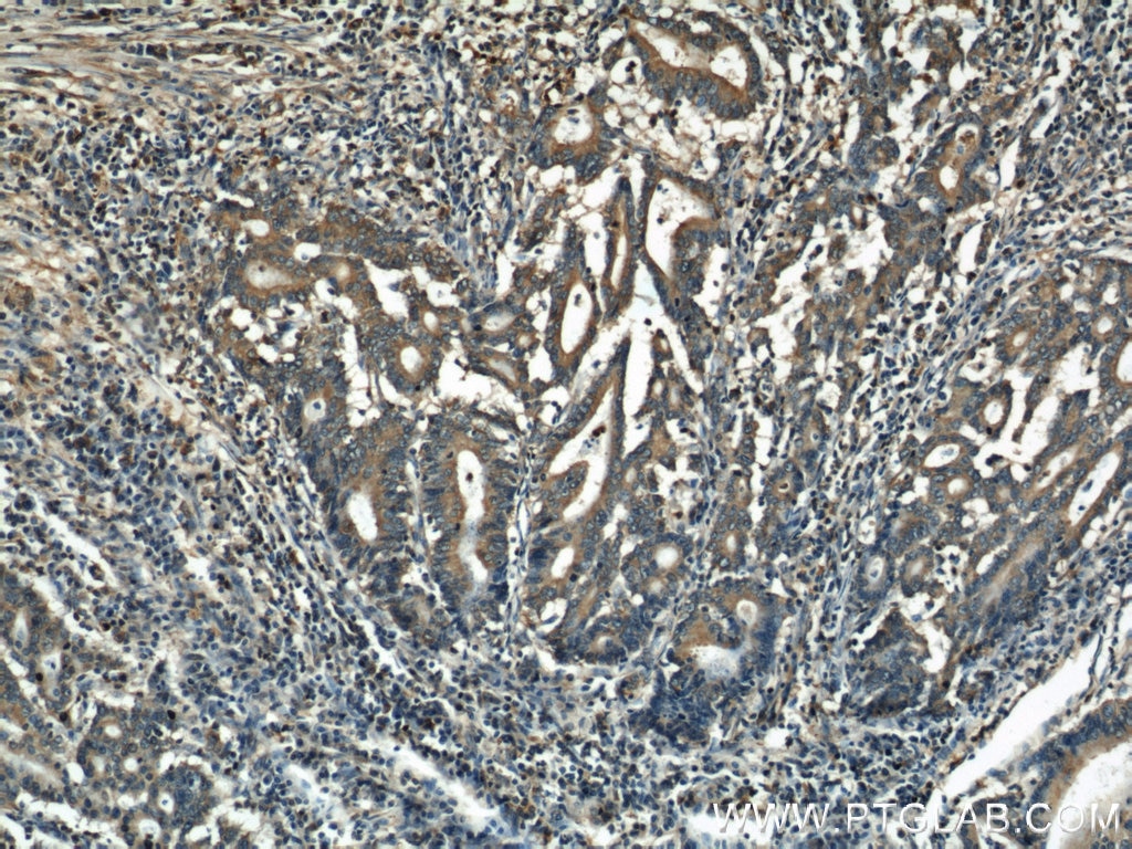
 at dilution of 1:200 (under 10x lens). Heat mediated antigen retrieval with Tris-EDTA buffer (pH 9.0).")
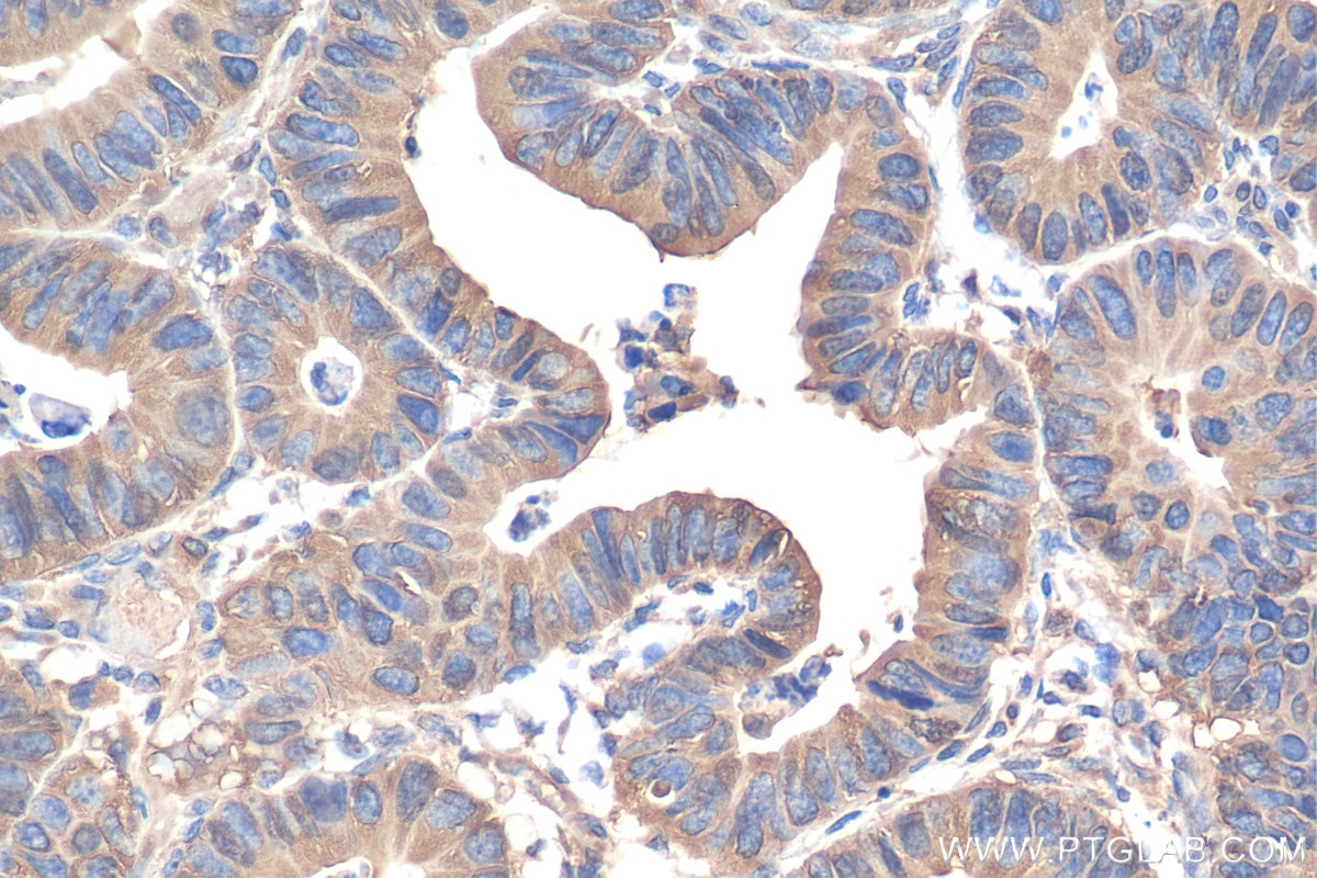
 at dilution of 1:200 (under 40x lens). Heat mediated antigen retrieval with Tris-EDTA buffer (pH 9.0).")
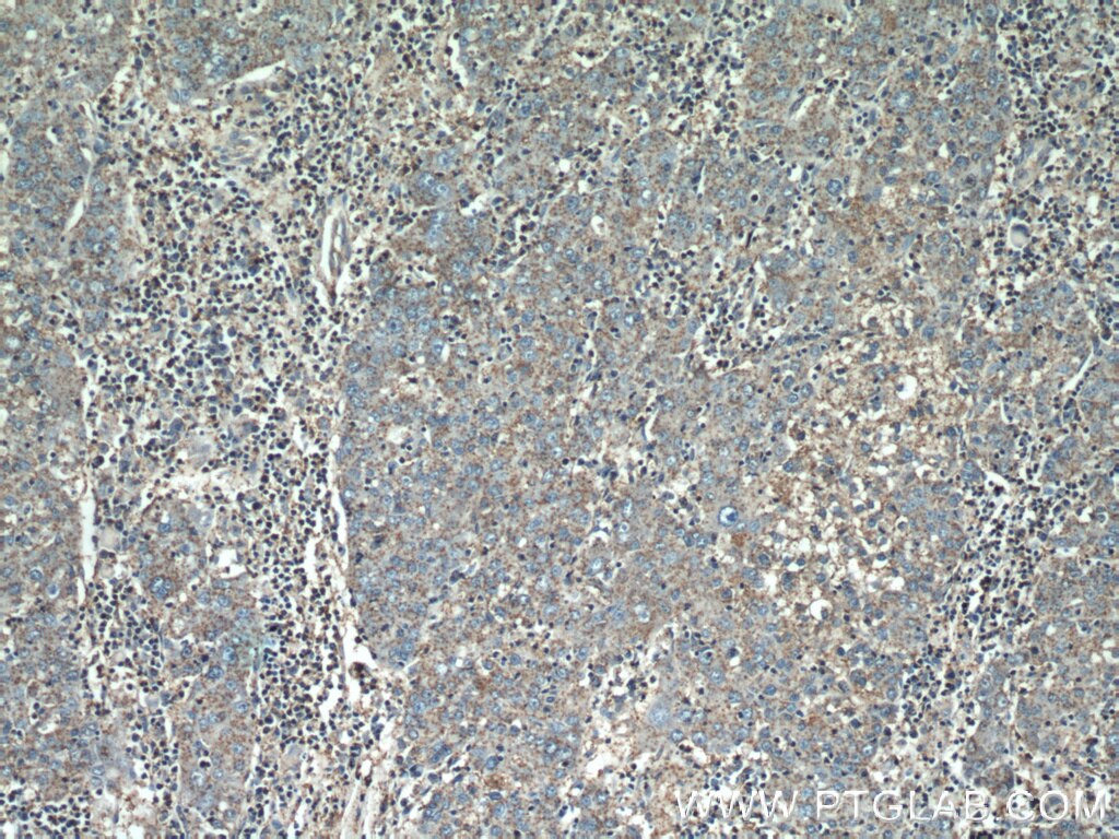
 at dilution of 1:50 (under 10x lens).")
 at dilution of 1:50 (under 10x lens).")
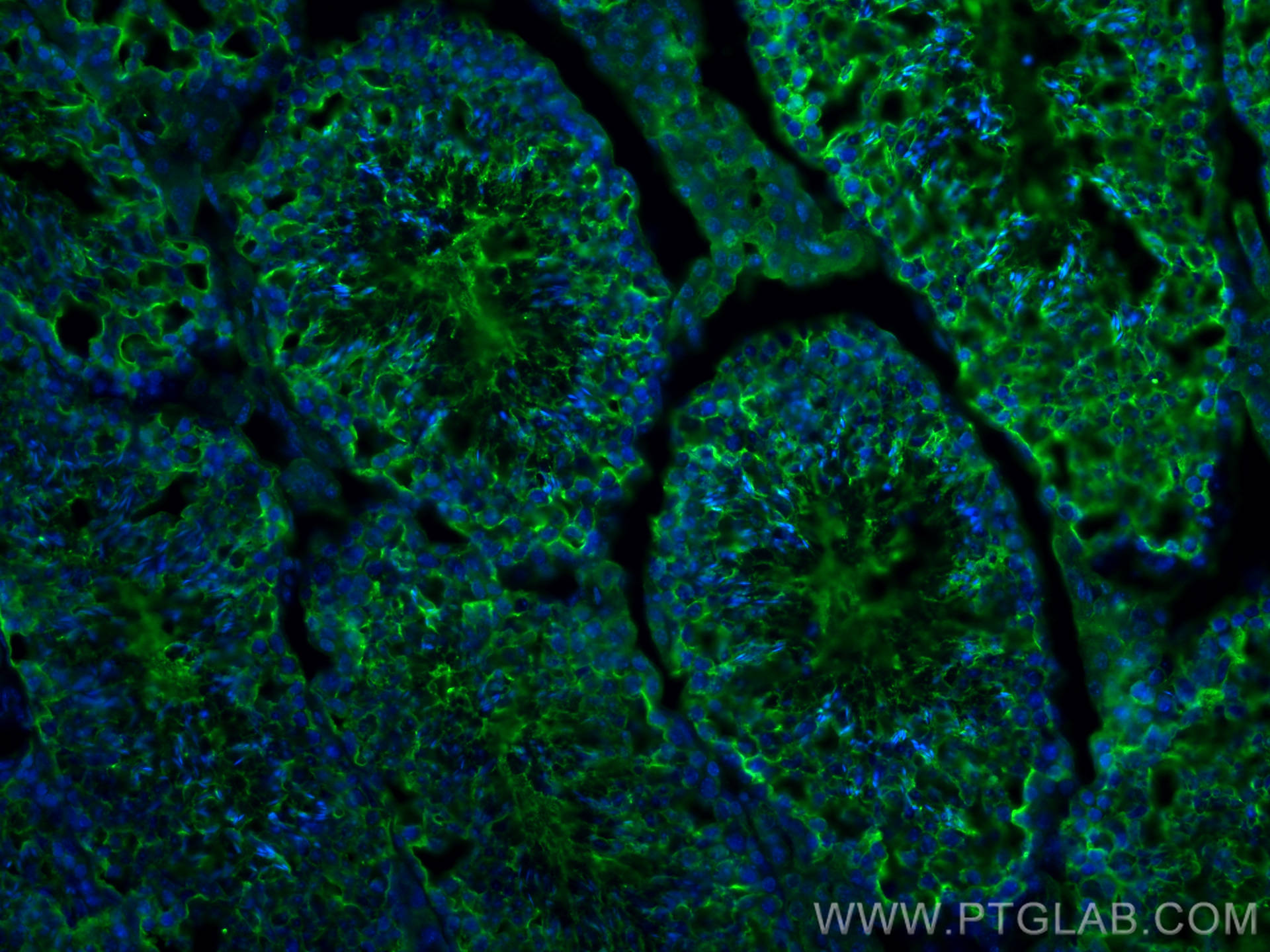
 fixed frozen OCT-embedded mouse testis tissue using Alpha Tubulin antibody (11224-1-AP) at dilution of 1:200 and CoraLite®488-Conjugated AffiniPure Goat Anti-Rabbit IgG(H+L).")
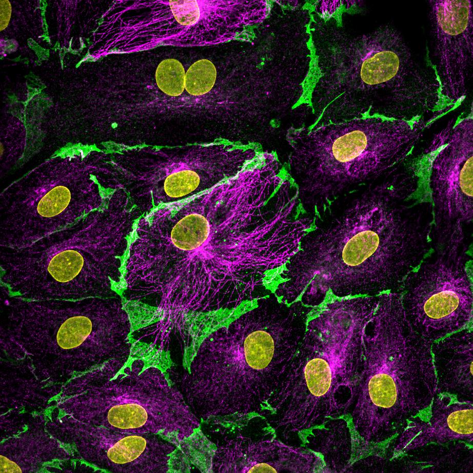
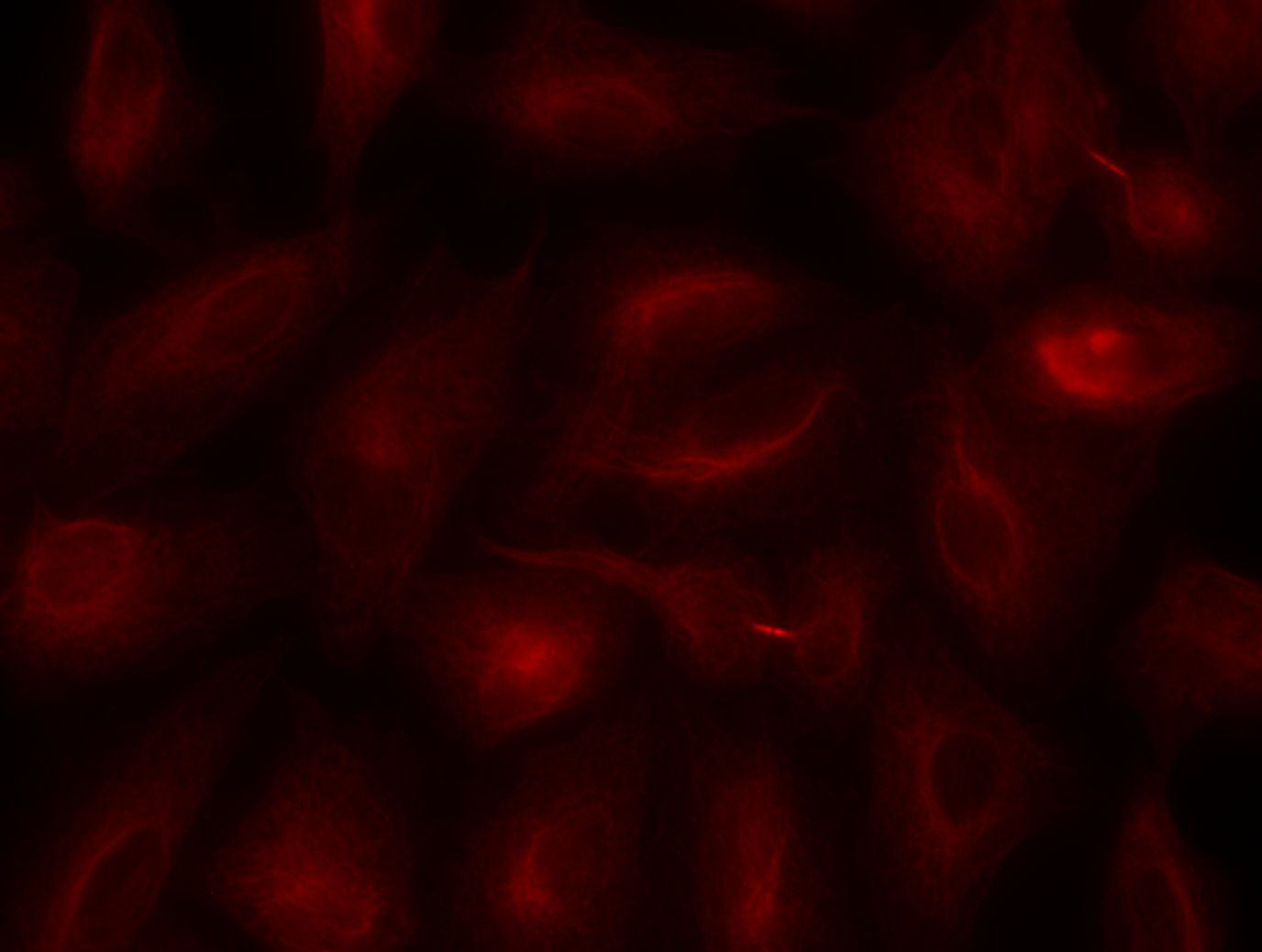
 and alpha tubulin (magenta, Cat. 11224-1-AP, 1:200) with DAPI as a counterstain for visualizing the nucleus (yellow). CD31 is an endothelial cell adhesion protein and was conjugated to CoraLite-488 fluorescent dye to allow for direct visualization. Alpha tubulin is a cytoskeletal protein important in cell division and migration and was visualized with an Alexa Fluor 568 secondary antibody. Cells were fixed with 4% paraformaldehyde. Image credit: @Immunofluorescence on Instagram")
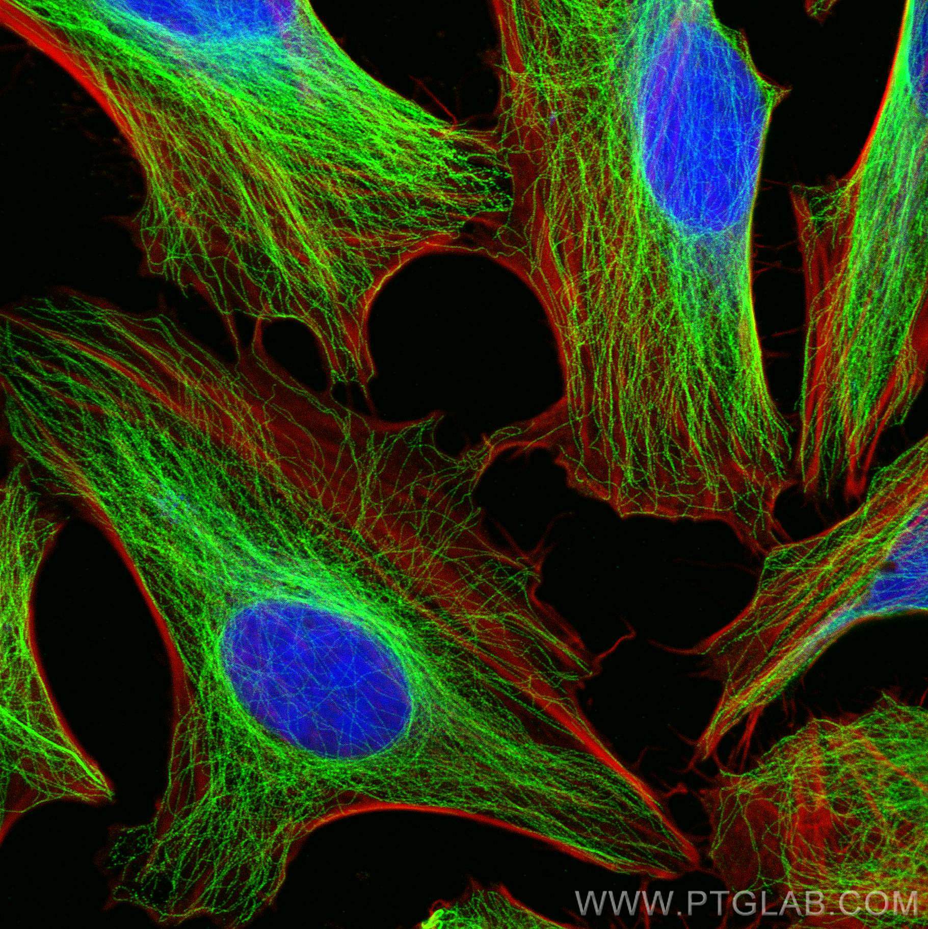
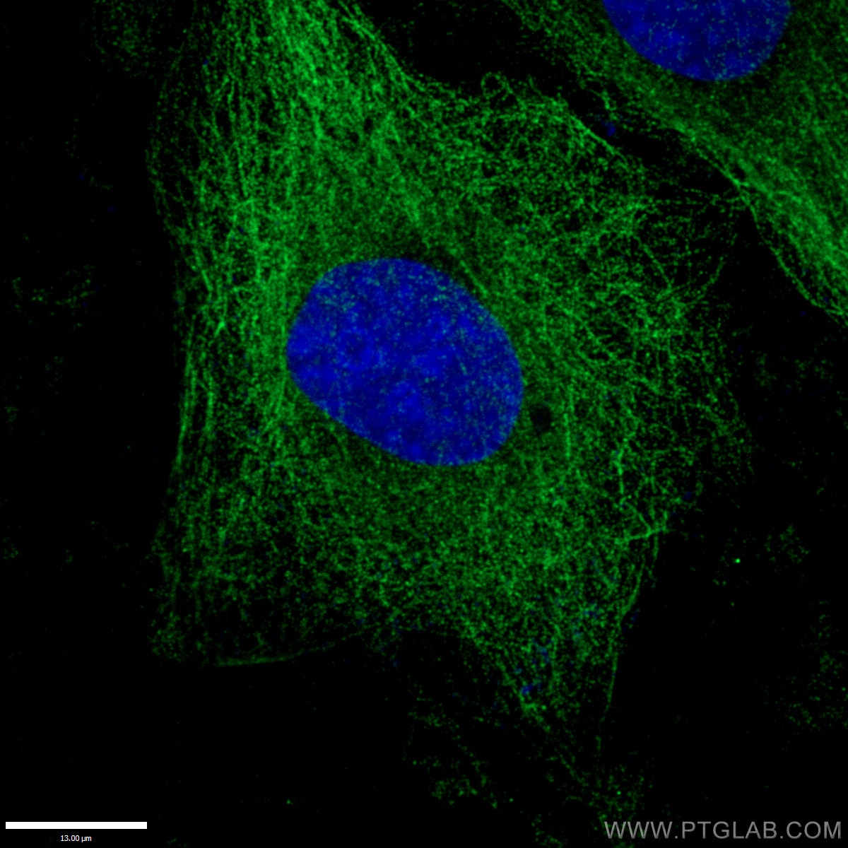
 fixed HeLa cells using Alpha Tubulin antibody (11224-1-AP) at dilution of 1:200 and CoraLite®488-Conjugated AffiniPure Goat Anti-Rabbit IgG(H+L), CL594-Phalloidin (red).")
 fixed MCF-7 cells using 11224-1-AP (alpha Tubulin antibody) at dilution of 1:200 and CoraLite488-Conjugated AffiniPure Goat Anti-Rabbit IgG(H+L).")
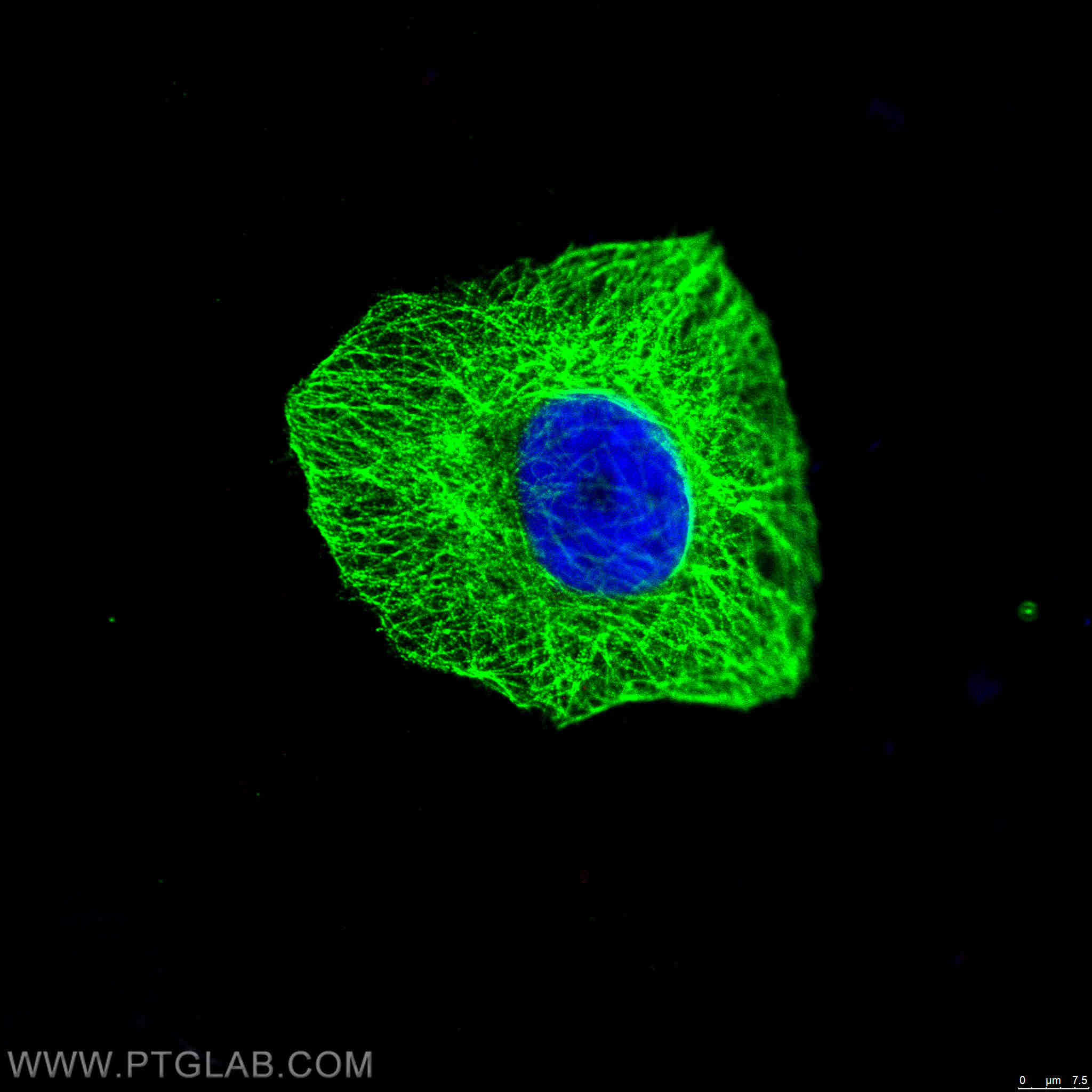
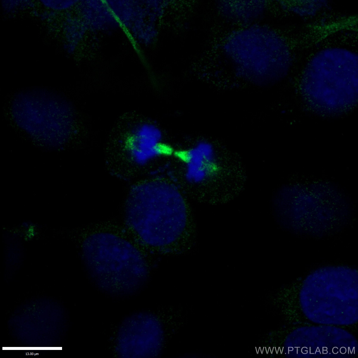
 fixed HepG2 cells using 11224-1-AP (alpha Tubulin antibody) at dilution of 1:50 and Alexa Fluor 488-conjugated AffiniPure Goat Anti-Rabbit IgG(H+L).")
 fixed HepG2 cells using 11224-1-AP (alpha Tubulin antibody) at dilution of 1:50 and Alexa Fluor 488-conjugated AffiniPure Goat Anti-Rabbit IgG(H+L).")
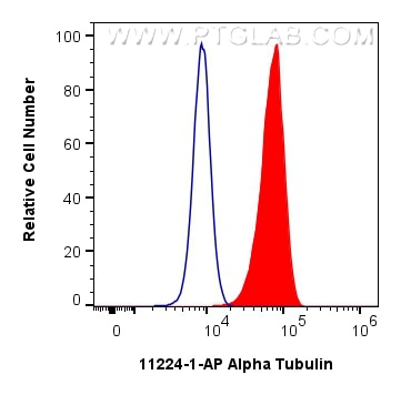
 and CoraLite®488-Conjugated Goat Anti-Rabbit IgG(H+L) (SA00013-2)(red), or 0.5 ug rabbit IgG isotype control (blue). Cells were fixed with 4% PFA and permeabilized with Flow Cytometry Perm Buffer.")
"Alpha Tubulin Antibodies" Comparison
View side-by-side comparison of Alpha Tubulin antibodies from other vendors to find the one that best suits your research needs.
Tested Applications
| Positive WB detected in | HEK-293 cells, A549 cells, HeLa cells, NIH/3T3 cells, rat brain, C6 cells, mouse heart tissue, mouse liver tissue, rat liver tissue, rat heart tissue |
| Positive IP detected in | HeLa cells |
| Positive IHC detected in | human colon cancer tissue, human liver cancer tissue, human endometrial cancer tissue Note: suggested antigen retrieval with TE buffer pH 9.0; (*) Alternatively, antigen retrieval may be performed with citrate buffer pH 6.0 |
| Positive IF-Fro detected in | mouse testis tissue |
| Positive IF/ICC detected in | HeLa cells, HepG2 cells, MCF-7 cells, Lymphatic endothelial cells cells |
| Positive FC (Intra) detected in | HEK-293 cells |
Recommended dilution
| Application | Dilution |
|---|---|
| Western Blot (WB) | WB : 1:5000-1:50000 |
| Immunoprecipitation (IP) | IP : 0.5-4.0 ug for 1.0-3.0 mg of total protein lysate |
| Immunohistochemistry (IHC) | IHC : 1:50-1:500 |
| Immunofluorescence (IF)-FRO | IF-FRO : 1:50-1:500 |
| Immunofluorescence (IF)/ICC | IF/ICC : 1:50-1:500 |
| Flow Cytometry (FC) (INTRA) | FC (INTRA) : 0.50 ug per 10^6 cells in a 100 µl suspension |
| It is recommended that this reagent should be titrated in each testing system to obtain optimal results. | |
| Sample-dependent, Check data in validation data gallery. | |
Published Applications
| WB | See 1715 publications below |
| IHC | See 4 publications below |
| IF | See 105 publications below |
| IP | See 11 publications below |
| CoIP | See 1 publications below |
Product Information
11224-1-AP targets Alpha Tubulin in WB, IHC, IF/ICC, IF-Fro, FC (Intra), IP, CoIP, ELISA applications and shows reactivity with human, mouse, rat samples.
| Tested Reactivity | human, mouse, rat |
| Cited Reactivity | human, mouse, rat, rabbit, monkey, chicken, zebrafish, hamster, goat, p. lividus |
| Host / Isotype | Rabbit / IgG |
| Class | Polyclonal |
| Type | Antibody |
| Immunogen |
CatNo: Ag1727 Product name: Recombinant human Tubulin-Alpha protein Source: e coli.-derived, PGEX-4T Tag: GST Domain: 1-451 aa of BC009314 Sequence: MRECISIHVGQAGVQIGNACWELYCLEHGIQPDGQMPSDKTIGGGDDSFNTFFSETGAGKHVPRAVFVDLEPTVIDEVRTGTYRQLFHPEQLITGKEDAANNYARGHYTIGKEIIDLVLDRIRKLADQCTGLQGFLVFHSFGGGTGSGFTSLLMERLSVDYGKKSKLEFSIYPAPQVSTAVVEPYNSILTTHTTLEHSDCAFMVDNEAIYDICRRNLDIERPTYTNLNRLISQIVSSITASLRFDGALNVDLTEFQTNLVPYPRIHFPLATYAPVISAEKAYHEQLSVAEITNACFEPANQMVKCDPRHGKYMACCLLYRGDVVPKDVNAAIATIKTKRSIQFVDWCPTGFKVGINYQPPTVVPGGDLAKVQRAVCMLSNTTAIAEAWARLDHKFDLMYAKRAFVHWYVGEGMEEGEFSEAREDMAALEKDYEEVGVDSVEGEGEEEGEEY Predict reactive species |
| Full Name | tubulin, alpha 1b |
| Calculated Molecular Weight | 50 kDa |
| Observed Molecular Weight | 50-55 kDa |
| GenBank Accession Number | BC009314 |
| Gene Symbol | Alpha Tubulin |
| Gene ID (NCBI) | 10376 |
| RRID | AB_2210206 |
| Conjugate | Unconjugated |
| Form | Liquid |
| Purification Method | Antigen affinity purification |
| UNIPROT ID | P68363 |
| Storage Buffer | PBS with 0.02% sodium azide and 50% glycerol, pH 7.3. |
| Storage Conditions | Store at -20°C. Stable for one year after shipment. Aliquoting is unnecessary for -20oC storage. 20ul sizes contain 0.1% BSA. |
Background Information
What is the function of alpha tubulin?
Alpha-tubulin belongs to a large superfamily of tubulin proteins. There are a number of different subtypes that have a molecular weight of ~50kDa and are able to bind to beta-tubulin, forming a heterodimer that polymerises to microtubules as part of the cytoskeleton. These maintain cell structure, provide platforms for intracellular transport and are also involved in cell division.
Where is alpha-tubulin expressed?
Alpha tubulin is highly conserved and is present in nearly all eukaryotic cells as one of the building blocks of microtubules. The ubiquitous nature of this protein has led to its common use as a control protein for many tissue types as well as highlighting the structure of the cytoskeleton.
What are the post-translational modifications of alpha tubulin?
The function and properties of microtubules are drastically affected by the post-translational modifications undergone by tubulin, which may occur to the tubulin dimer directly or to the polymerised mictotubule. For example, the first modification to be identified was detyrosination1, as most alpha-tubulins have a tyrosine at their terminus. This process affects microtubules more than dimers and leads to patches of detyronisation along the structure, regulating protein interactions and allowing subcellular compartments to be defined.2,3 Polyglutamylation also occurs on several sites within the carboxy-terminal tails. However, to date, the most-studied alpha tubulin modification is related to acetylation of lysine 40 (K40).
1. Gundersen, G. G., Khawaja, S. & Bulinski, J. C. Postpolymerization detyrosination of alpha-tubulin: a mechanism for subcellular differentiation of microtubules. J. Cell Biol. 105, 251-64 (1987).
2. Galjart, N. Plus-End-Tracking Proteins and Their Interactions at Microtubule Ends. Curr. Biol. 20, R528-R537 (2010).
3. Jiang, K. & Akhmanova, A. Microtubule tip-interacting proteins: a view from both ends. Curr. Opin. Cell Biol. 23, 94-101 (2011).
Protocols
| Product Specific Protocols | |
|---|---|
| FC protocol for Alpha Tubulin antibody 11224-1-AP | Download protocol |
| IF protocol for Alpha Tubulin antibody 11224-1-AP | Download protocol |
| IHC protocol for Alpha Tubulin antibody 11224-1-AP | Download protocol |
| IP protocol for Alpha Tubulin antibody 11224-1-AP | Download protocol |
| WB protocol for Alpha Tubulin antibody 11224-1-AP | Download protocol |
| Standard Protocols | |
|---|---|
| Click here to view our Standard Protocols |
Publications
| Species | Application | Title |
|---|---|---|
Nat Biotechnol Magnify is a universal molecular anchoring strategy for expansion microscopy | ||
Cell Res SARS-CoV-2 Z-RNA activates the ZBP1-RIPK3 pathway to promote virus-induced inflammatory responses | ||
Nature Activity-based E3 ligase profiling uncovers an E3 ligase with esterification activity. | ||
Mol Cancer A positive feedback circuit driven by m6A-modified circular RNA facilitates colorectal cancer liver metastasis |
Reviews
The reviews below have been submitted by verified Proteintech customers who received an incentive for providing their feedback.
FH Will (Verified Customer) (02-27-2026) | Works great for blots, very reproducible.
|
FH Corre (Verified Customer) (09-19-2025) | Very clear and specific band
 |
FH Harvey (Verified Customer) (09-10-2025) | Housekeeper for mouse tissue western blot
|
FH Patrick (Verified Customer) (07-16-2025) | Used for a flotation gradient and worked fine as a cytosolic marker. Even though we needed a longer exposure and incubation with ECL compared to actin.
|
FH Samantha (Verified Customer) (05-10-2024) | Bands were faint but still visible when changes in contrast were applied. Some samples had big bands due to the large amounts present in the samples
|
FH Kamal (Verified Customer) (02-15-2024) | One of the best antibody to detect alpha-tubulin. Works well when diluted in 1X TBST.
|
FH Brogan (Verified Customer) (11-09-2022) | Used as a control for C elegans western blot
|
FH WEI (Verified Customer) (03-03-2022) | Good antibody for detect tubulin proteins
|
FH Maria (Verified Customer) (06-19-2021) | a Tubulin in human primary fibroblasts. 10 ug of total protein with DTT. 4-15 TGX gel. 3% BSA in PBST 1h RT. 1:1000 primary ab in BSA 3% in PBST (0.1% Tween) O/N 4ºC. Secondary ab 1:5000 1h RT.
 |
FH Azita (Verified Customer) (06-17-2021) | Immunocytochemistry labelling of (4% PFA) fixed SH-SY5Y cells by Alpha Tubulin Polyclonal antibody at dilution of 1:100 showed strong labelling.
|
FH Chao (Verified Customer) (03-12-2020) | Good sharp clear band in western blot.
|
FH Wei (Verified Customer) (01-30-2020) | Very good, sharp band for internal control.The protein is better than GAPDH for quantification in heart diseases.
|
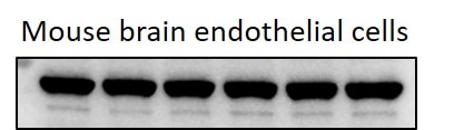
FH KARTHIKEYAN (Verified Customer) (11-20-2019) | I used this antibody in mouse brain endothelial cell line, it worked really well, i would definitely recommend this product.
 |
FH Pooja (Verified Customer) (11-05-2019) | This antibody works very well as loading control. Even lower dilution can be use.
|
FH Yuan (Verified Customer) (11-02-2019) | Very nice staining for A549 cells with 1:200 dilution
 |
FH Kishor (Verified Customer) (01-30-2019) | I got good results for Western Blotting (1:1000) and IF (1:300)
 |
FH kk (Verified Customer) (11-21-2018) | Excellent product, single and sharp band in all my blots
|