- Featured Product
- KD/KO Validated
Lamin B1 Monoklonaler Antikörper
Lamin B1 Monoklonal Antikörper für WB, IHC, IF/ICC, IF-P, FC (Intra), IP, ELISA
Wirt / Isotyp
Maus / IgG1
Getestete Reaktivität
human, Maus, Ratte und mehr (6)
Anwendung
WB, IHC, IF/ICC, IF-P, FC (Intra), IP, CoIP, ELISA
Konjugation
Unkonjugiert
CloneNo.
3C10G12
Kat-Nr. : 66095-1-Ig
Synonyme
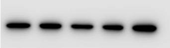
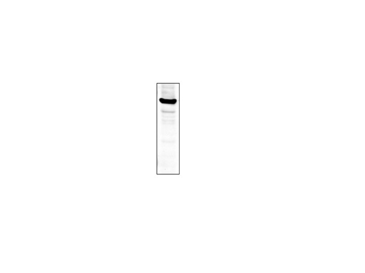
 at dilution of 1:100000 incubated at room temperature for 1.5 hours.")
 at dilution 1:20000.")
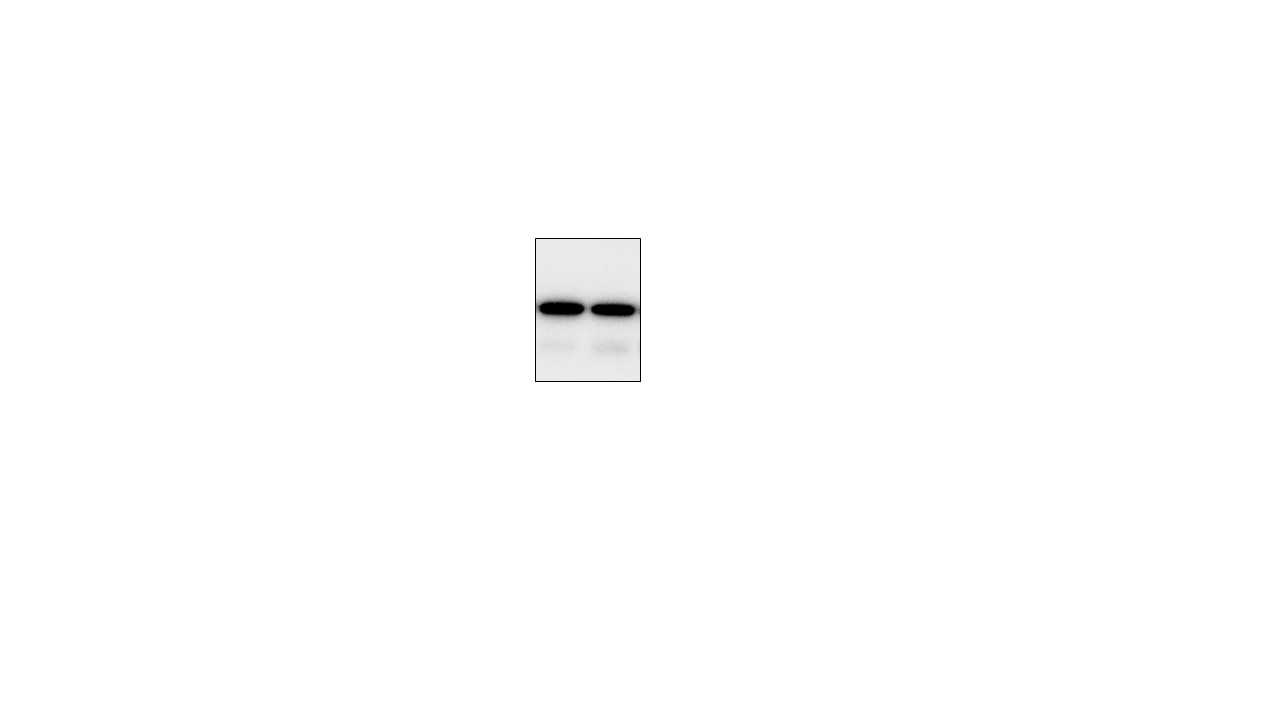
 with sh-Control and sh-Lamin B1 transfected HeLa cells.")
 with HeLa cells lysate 3560ug.")
 at dilution of 1:1000 (under 10x lens. Heat mediated antigen retrieval with Tris-EDTA buffer (pH 9.0).")
 at dilution of 1:1000 (under 40x lens. Heat mediated antigen retrieval with Tris-EDTA buffer (pH 9.0).")
 at dilution of 1:1000 (under 40x lens. Heat mediated antigen retrieval with Tris-EDTA buffer (pH 9.0).")
 fixed mouse eye tissue using Lamin B1 antibody (66095-1-Ig, Clone: 3C10G12 ) at dilution of 1:400 and CoraLite®488-Conjugated AffiniPure Goat Anti-Mouse IgG(H+L).")
 fixed HepG2 cells using 66095-1-Ig (Lamin B1 antibody) at dilution of 1:500 and CoraLite488-Conjugated AffiniPure Goat Anti-Mouse IgG(H+L).")
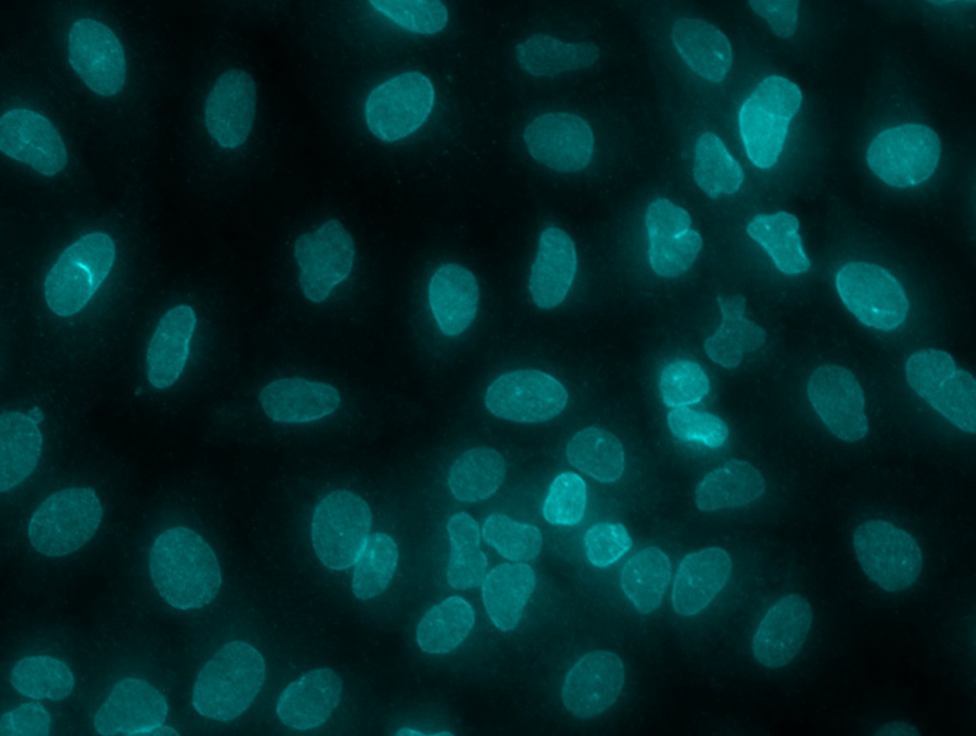
 fixed HeLa cells using 66095-1-Ig (Lamin B1 antibody) at dilution of 1:200 and Alexa Fluor 488-Conjugated AffiniPure Goat Anti-Mouse IgG(H+L).")
 and CoraLite®488-Conjugated AffiniPure Goat Anti-Mouse IgG(H+L) at dilution 1:1000 (red), or 0.4 ug Mouse IgG1 Isotype Control (MOPC-21) (65124-1-Ig, Clone: MOPC-21) (blue). Cells were fixed and permeabilized with Transcription Factor Staining Buffer Kit (PF00011).")
"Lamin B1 Antibodies" Comparison
View side-by-side comparison of Lamin B1 antibodies from other vendors to find the one that best suits your research needs.
Geprüfte Anwendungen
| Erfolgreiche Detektion in WB | NCI-H1299-Zellen, HEK-293-Zellen, HeLa-Zellen, HepG2-Zellen, Jurkat-Zellen, K-562-Zellen, Multizellen/-gewebe, NIH/3T3-Zellen, PC-12-Zellen |
| Erfolgreiche IP | HeLa-Zellen |
| Erfolgreiche Detektion in IHC | humanes Pankreaskarzinomgewebe, humanes Mammakarzinomgewebe Hinweis: Antigendemaskierung mit TE-Puffer pH 9,0 empfohlen. (*) Wahlweise kann die Antigendemaskierung auch mit Citratpuffer pH 6,0 erfolgen. |
| Erfolgreiche Detektion in IF-P | Maus-Augengewebe |
| Erfolgreiche Detektion in IF/ICC | HepG2-Zellen, HeLa-Zellen |
| Erfolgreiche Detektion in FC (Intra) | HeLa-Zellen |
Empfohlene Verdünnung
| Anwendung | Verdünnung |
|---|---|
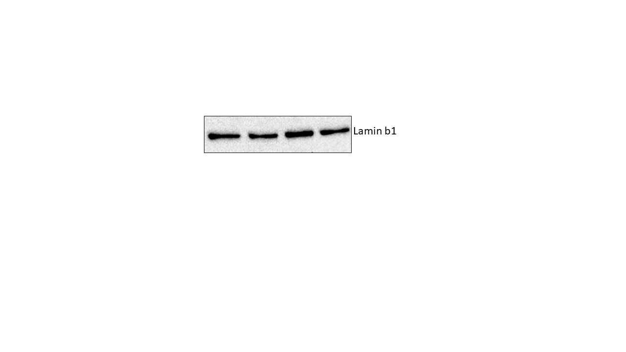
| Western Blot (WB) | WB : 1:20000-1:100000 |
| Immunpräzipitation (IP) | IP : 0.5-4.0 ug for 1.0-3.0 mg of total protein lysate |
| Immunhistochemie (IHC) | IHC : 1:500-1:2000 |
| Immunfluoreszenz (IF)-P | IF-P : 1:200-1:800 |
| Immunfluoreszenz (IF)/ICC | IF/ICC : 1:250-1:1000 |
| Durchflusszytometrie (FC) (INTRA) | FC (INTRA) : 0.40 ug per 10^6 cells in a 100 µl suspension |
| It is recommended that this reagent should be titrated in each testing system to obtain optimal results. | |
| Sample-dependent, check data in validation data gallery | |
Veröffentlichte Anwendungen
| KD/KO | See 1 publications below |
| WB | See 389 publications below |
| IHC | See 3 publications below |
| IF | See 33 publications below |
| IP | See 4 publications below |
| CoIP | See 2 publications below |
Produktinformation
66095-1-Ig bindet in WB, IHC, IF/ICC, IF-P, FC (Intra), IP, CoIP, ELISA Lamin B1 und zeigt Reaktivität mit human, Maus, Ratten
| Getestete Reaktivität | human, Maus, Ratte |
| In Publikationen genannte Reaktivität | human, hamster, Huhn, Hund, Kaninchen, Maus, Ratte, Rind, Zebrafisch |
| Wirt / Isotyp | Maus / IgG1 |
| Klonalität | Monoklonal |
| Typ | Antikörper |
| Immunogen | Lamin B1 fusion protein Ag20522 |
| Vollständiger Name | lamin B1 |
| Berechnetes Molekulargewicht | 66 kDa |
| Beobachtetes Molekulargewicht | 66-70 kDa |
| GenBank-Zugangsnummer | BC012295 |
| Gene symbol | Lamin B1 |
| Gene ID (NCBI) | 4001 |
| Konjugation | Unkonjugiert |
| Form | Liquid |
| Reinigungsmethode | Protein-G-Reinigung |
| Lagerungspuffer | PBS with 0.02% sodium azide and 50% glycerol |
| Lagerungsbedingungen | Bei -20°C lagern. Nach dem Versand ein Jahr lang stabil Aliquotieren ist bei -20oC Lagerung nicht notwendig. 20ul Größen enthalten 0,1% BSA. |
Hintergrundinformationen
Lamins are components of the nuclear lamina, a fibrous layer on the nucleoplasmic side of the inner nuclear membrane, which is thought to provide a framework for the nuclear envelope and may also interact with chromatin. The nuclear lamina consists of a two-dimensional matrix of proteins located next to the inner nuclear membrane. The lamin family of proteins make up the matrix and are highly conserved in evolution. During mitosis, the lamina matrix is reversibly disassembled as the lamin proteins are phosphorylated. Vertebrate lamins consist of two types, A and B. This gene encodes one of the two B type proteins, B1. This protein is not suitable for samples where the nuclear envelope has been removed.
Protokolle
| PRODUKTSPEZIFISCHE PROTOKOLLE | |
|---|---|
| WB protocol for Lamin B1 antibody 66095-1-Ig | Protokoll herunterladen |
| IHC protocol for Lamin B1 antibody 66095-1-Ig | Protokoll herunterladenl |
| IF protocol for Lamin B1 antibody 66095-1-Ig | Protokoll herunterladen |
| IP protocol for Lamin B1 antibody 66095-1-Ig | Protokoll herunterladen |
| FC protocol for Lamin B1 antibody 66095-1-Ig | Download protocol |
| STANDARD-PROTOKOLLE | |
|---|---|
| Klicken Sie hier, um unsere Standardprotokolle anzuzeigen |
Publikationen
| Species | Application | Title |
|---|---|---|
Nat Cell Biol Ceramide-rich microdomains facilitate nuclear envelope budding for non-conventional exosome formation. | ||
J Hepatol OGDHL silencing promotes hepatocellular carcinoma by reprogramming glutamine metabolism. | ||
Cell Rep Med Management of prostate cancer by targeting 3βHSD1 after enzalutamide and abiraterone treatment. | ||
Nat Commun ARF1 prevents aberrant type I interferon induction by regulating STING activation and recycling | ||
Nat Commun FOXP3+ regulatory T cell perturbation mediated by the IFNγ-STAT1-IFITM3 feedback loop is essential for anti-tumor immunity | ||
J Clin Invest FAM117B promotes gastric cancer growth and drug resistance by targeting the KEAP1/NRF2 signaling pathway |
Rezensionen
The reviews below have been submitted by verified Proteintech customers who received an incentive for providing their feedback.
FH Echo (Verified Customer) (10-03-2024) | Quality great
 |
FH PK (Verified Customer) (08-14-2024) | Excellent
 |
FH S (Verified Customer) (12-12-2022) |
 |
FH WEI (Verified Customer) (03-08-2022) | Specfic bands around predicted size with higher non-specific bands
|
FH P. (Verified Customer) (05-15-2021) | Good antibody!
 |
FH Eiko (Verified Customer) (11-18-2020) | Used RIPA buffer for western sample preparation, and 5% Skim milk TBS-T was used for blocking and antibody dilution.Since I could see clear lamin B1 signal, I am happy to use this antibody as one of my internal controls.
|
FH Tom (Verified Customer) (10-09-2020) | I observed a discrete ~70kDa Lamin B1 band using this antibody.
|
FH Yuan (Verified Customer) (12-13-2019) | Did staining for human A549 cell.The Lamin B antibody had high background in nucleus. Please see attached image.
 |
FH Shubham (Verified Customer) (03-14-2019) | good
 |

