- Phare
- Validé par KD/KO
Anticorps Polyclonal de lapin anti-MTCO2
MTCO2 Polyclonal Antibody for WB, IHC, IF/ICC, FC (Intra), IP, ELISA
Hôte / Isotype
Lapin / IgG
Réactivité testée
Humain et plus (4)
Applications
WB, IHC, IF/ICC, FC (Intra), IP, CoIP, ELISA
Conjugaison
Non conjugué
N° de cat : 55070-1-AP
Synonymes
Galerie de données de validation
 at dilution of 1:6000 incubated at room temperature for 1.5 hours.")
) at dilution of 1:4000 incubated at room temperature for 1 hours.")
 with 143B and 143B.TK.ρ0 cell. 143B is wild-type cell line, 143B.TK.ρ0 is mtDNA deletion cell line.")
 with HeLa cells lysate 1880 ug.")
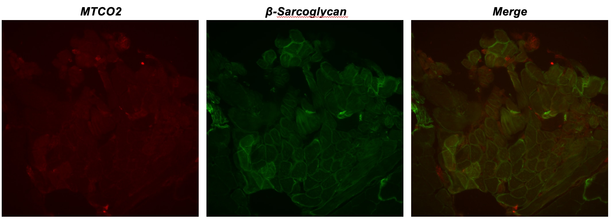
 at dilution of 1:8000 (under 40x lens). Heat mediated antigen retrieval with Tris-EDTA buffer (pH 9.0).")
 at dilution of 1:8000 (under 10x lens). Heat mediated antigen retrieval with Tris-EDTA buffer (pH 9.0).")
 fixed HepG2 cells using MTCO2 antibody (55070-1-AP) at dilution of 1:200 and CoraLite®488-Conjugated AffiniPure Goat Anti-Rabbit IgG(H+L), CL594-phalloidin (red).")
 and CoraLite®488-Conjugated AffiniPure Goat Anti-Rabbit IgG(H+L) at dilution 1:1000 (red), or 0.4 ug Control Antibody. Cells were fixed with 4% PFA and permeabilized with Flow Cytometry Perm Buffer (PF00011-C).")
Applications testées
| Résultats positifs en WB | cellules A431, cellules HeLa, cellules MCF-7, échantillons divers |
| Résultats positifs en IP | cellules HeLa, |
| Résultats positifs en IHC | tissu de cancer du côlon humain, il est suggéré de démasquer l'antigène avec un tampon de TE buffer pH 9.0; (*) À défaut, 'le démasquage de l'antigène peut être 'effectué avec un tampon citrate pH 6,0. |
| Résultats positifs en IF/ICC | cellules HepG2, |
| Résultats positifs en FC (Intra) | cellules A431, |
Dilution recommandée
| Application | Dilution |
|---|---|
| Western Blot (WB) | WB : 1:2000-1:12000 |
| Immunoprécipitation (IP) | IP : 0.5-4.0 ug for 1.0-3.0 mg of total protein lysate |
| Immunohistochimie (IHC) | IHC : 1:4000-1:16000 |
| Immunofluorescence (IF)/ICC | IF/ICC : 1:50-1:500 |
| Flow Cytometry (FC) (INTRA) | FC (INTRA) : 0.40 ug per 10^6 cells in a 100 µl suspension |
| It is recommended that this reagent should be titrated in each testing system to obtain optimal results. | |
| Sample-dependent, check data in validation data gallery | |
Applications publiées
| KD/KO | See 2 publications below |
| WB | See 140 publications below |
| IHC | See 10 publications below |
| IF | See 6 publications below |
| IP | See 1 publications below |
| CoIP | See 1 publications below |
Informations sur le produit
55070-1-AP cible MTCO2 dans les applications de WB, IHC, IF/ICC, FC (Intra), IP, CoIP, ELISA et montre une réactivité avec des échantillons Humain
| Réactivité | Humain |
| Réactivité citée | rat, Humain, singe, souris, Hamster |
| Hôte / Isotype | Lapin / IgG |
| Clonalité | Polyclonal |
| Type | Anticorps |
| Immunogène | Peptide |
| Nom complet | cytochrome c oxidase II |
| Masse moléculaire calculée | 26 kDa |
| Poids moléculaire observé | 23-26 kDa |
| Numéro d’acquisition GenBank | YP_003024029 |
| Symbole du gène | MTCO2 |
| Identification du gène (NCBI) | 4513 |
| Conjugaison | Non conjugué |
| Forme | Liquide |
| Méthode de purification | Purification par affinité contre l'antigène |
| Tampon de stockage | PBS with 0.02% sodium azide and 50% glycerol |
| Conditions de stockage | Stocker à -20°C. Stable pendant un an après l'expédition. L'aliquotage n'est pas nécessaire pour le stockage à -20oC Les 20ul contiennent 0,1% de BSA. |
Informations générales
MTCO2, also named as COII, COXII, and COX2, belongs to the cytochrome c oxidase subunit 2 family. It is the component of the respiratory chain that catalyzes the reduction of oxygen to water. Subunits 1-3 form the functional core of the enzyme complex. Subunit 2 transfers the electrons from cytochrome c via its binuclear copper A center to the bimetallic center of the catalytic subunit 1. Defects in COX2 are a cause of mitochondrial complex IV deficiency (MT-C4D). The antibody is specific to MTCO2.
Protocole
| Product Specific Protocols | |
|---|---|
| WB protocol for MTCO2 antibody 55070-1-AP | Download protocol |
| IHC protocol for MTCO2 antibody 55070-1-AP | Download protocol |
| IF protocol for MTCO2 antibody 55070-1-AP | Download protocol |
| IP protocol for MTCO2 antibody 55070-1-AP | Download protocol |
| FC protocol for MTCO2 antibody 55070-1-AP | Download protocol |
| Standard Protocols | |
|---|---|
| Click here to view our Standard Protocols |
Publications
| Species | Application | Title |
|---|---|---|
Cell Mitophagy in Intestinal Epithelial Cells Triggers Adaptive Immunity during Tumorigenesis. | ||
Cell Metab Proteome Imbalance of Mitochondrial Electron Transport Chain in Brown Adipocytes Leads to Metabolic Benefits. | ||
Cell Metab Malic enzyme 2 connects the Krebs cycle intermediate fumarate to mitochondrial biogenesis. | ||
Immunity Excessive Polyamine Generation in Keratinocytes Promotes Self-RNA Sensing by Dendritic Cells in Psoriasis. | ||
Mol Cell Hepatic micropeptide modulates mitochondrial RNA processing machinery in hepatocellular carcinoma |
Avis
The reviews below have been submitted by verified Proteintech customers who received an incentive for providing their feedback.
FH Matthieu (Verified Customer) (09-24-2025) | Band is clearly visible at the correct size
|
FH Manon (Verified Customer) (09-24-2025) | vey nice band
|
FH Emilie (Verified Customer) (09-24-2025) | Bands appeared sharp and well defined under the chosen dilution.
|
FH Vignesh (Verified Customer) (09-03-2025) | It's a good product with reduced or no nonspecific bands.
|
FH Lenie (Verified Customer) (07-18-2025) | The IF staining and WB with the followed protocol of ProteinTech did not work on zebrafish tissue, even it describes on the site it should. Is there a possibility to send a working protocol about this Abies for zebrafish? Thanks. The bond which shows is a human patient sample.
 |
FH Baptiste (Verified Customer) (05-23-2025) | Working perfectly
 |
FH Edward (Verified Customer) (11-15-2022) | Used with 12 ug protein lysate MTCO2 works quite well, both with ECL detection and fluorescent secondary antibodies. Sometimes a bit fainter with ECL.
|
FH Maria (Verified Customer) (03-11-2021) | MTCO2 in treated Human Primary Fibroblasts. 10 ug of total protein. 12% gel. 1:1000 primary ab in BSA 3% in PBST O/N 4ºC incubation. Secondary ab HRP 1:5000 1h RT.
 |

