Anticorps Monoclonal anti-COXIV

COXIV Monoclonal Antibody for FC, IF, IHC, WB, ELISA

Hôte / Isotype

Mouse / IgG1

Réactivité testée

Humain et plus (2)

Applications

WB, IHC, IF, FC, ELISA

Conjugaison

Non conjugué

CloneNo.

2A7B2

N° de cat : 66110-1-Ig

Synonymes

Cell and organelle markers, COX IV 1, COX4, COX4I1, COXIV, Cytochrome c oxidase polypeptide IV, Cytochrome c oxidase subunit 4 isoform 1 mitochondrial, Mitochondria Marker, Mitochondrial Marker, Mitochondrion Marker


Filter:
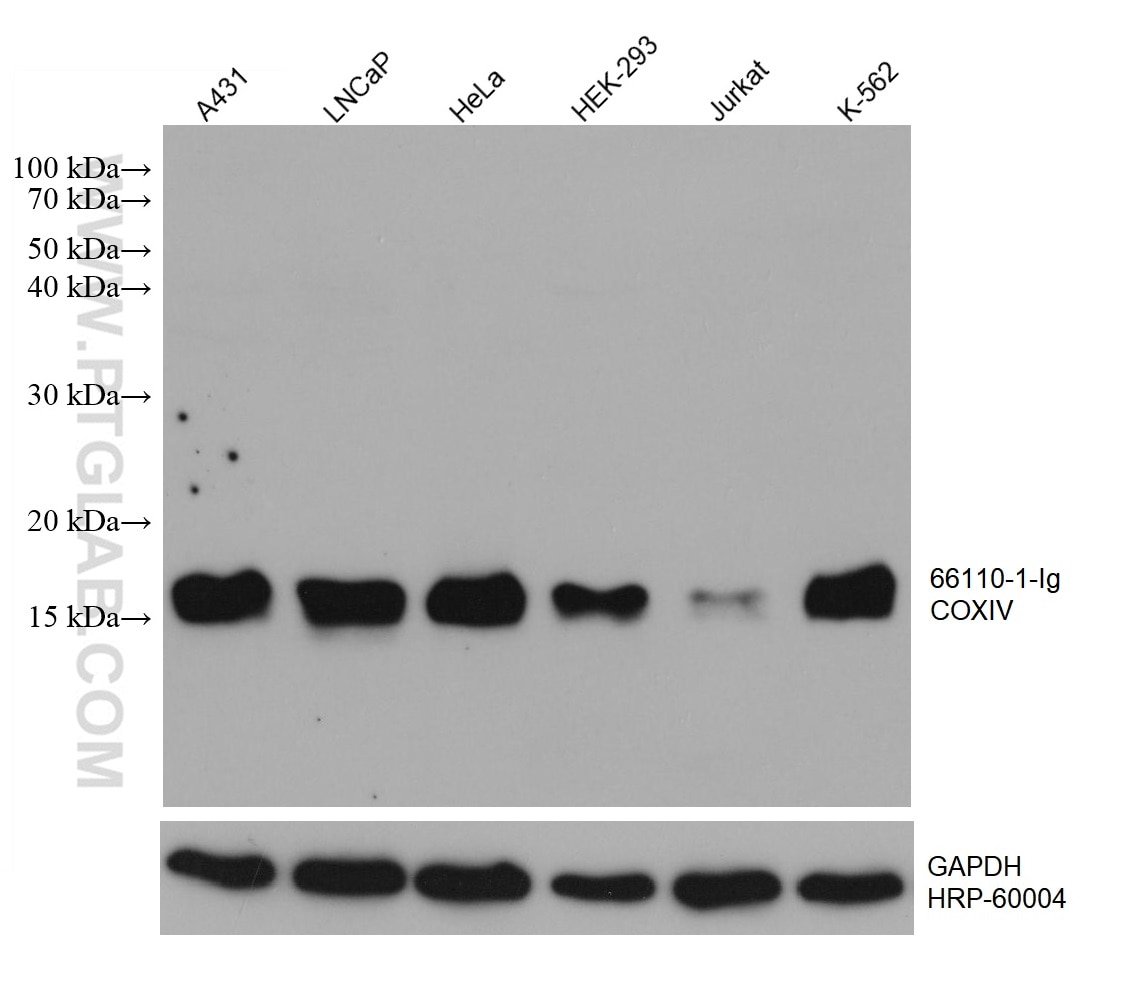
  • Western Blot (WB) analysis of various lysates using COXIV Monoclonal antibody (66110-1-Ig)

    WB analysis using 66110-1-Ig

    Various lysates were subjected to SDS PAGE followed by western blot with 66110-1-Ig (COXIV antibody) at dilution of 1:20000 incubated at room temperature for 1.5 hours. The membrane was stripped and reblotted with HRP-conjugated GAPDH Monoclonal antibody (HRP-60004) as loading control.

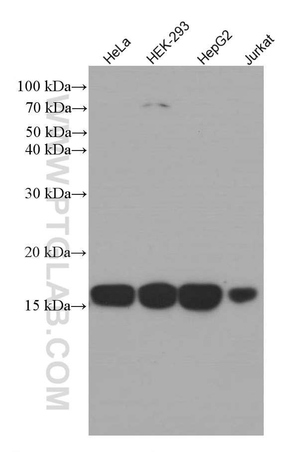
  • Western Blot (WB) analysis of various lysates using COXIV Monoclonal antibody (66110-1-Ig)

    WB analysis using 66110-1-Ig

    Various lysates were subjected to SDS PAGE followed by western blot with 66110-1-Ig (COXIV antibody) at dilution of 1:3000 incubated at room temperature for 1.5 hours.

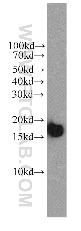
  • Western Blot (WB) analysis of HeLa cells using COXIV Monoclonal antibody (66110-1-Ig)

    WB analysis of HeLa using 66110-1-Ig

    HeLa cells were subjected to SDS PAGE followed by western blot with 66110-1-Ig (COXIV antibody) at dilution of 1:2000 incubated at room temperature for 1.5 hours.

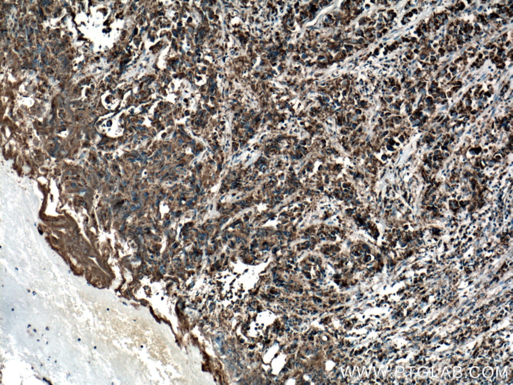
  • Immunohistochemistry (IHC) staining of human prostate cancer tissue using COXIV Monoclonal antibody (66110-1-Ig)

    IHC staining of human prostate cancer using 66110-1-Ig

    Immunohistochemical analysis of paraffin-embedded human prostate cancer tissue slide using 66110-1-Ig (COXIV antibody) at dilution of 1:500 (under 10x lens. Heat mediated antigen retrieval with Tris-EDTA buffer (pH 9.0).

  • Immunohistochemistry (IHC) staining of human prostate cancer tissue using COXIV Monoclonal antibody (66110-1-Ig)

    IHC staining of human prostate cancer using 66110-1-Ig

    Immunohistochemical analysis of paraffin-embedded human prostate cancer tissue slide using 66110-1-Ig (COXIV antibody) at dilution of 1:500 (under 40x lens. Heat mediated antigen retrieval with Tris-EDTA buffer (pH 9.0).

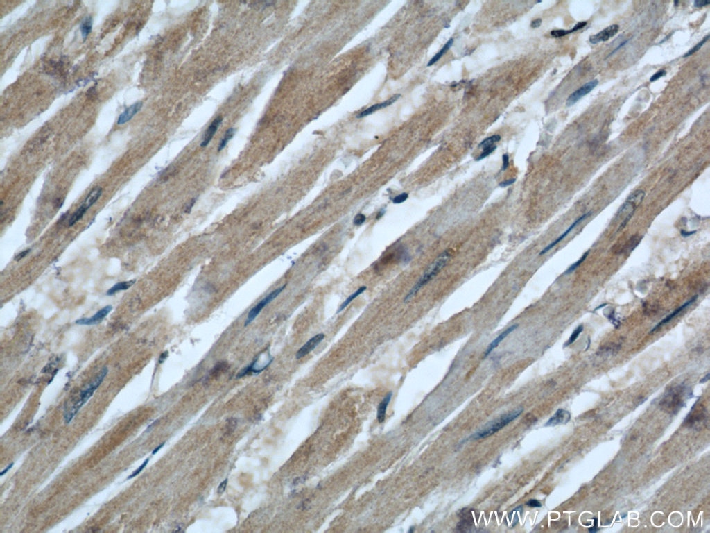
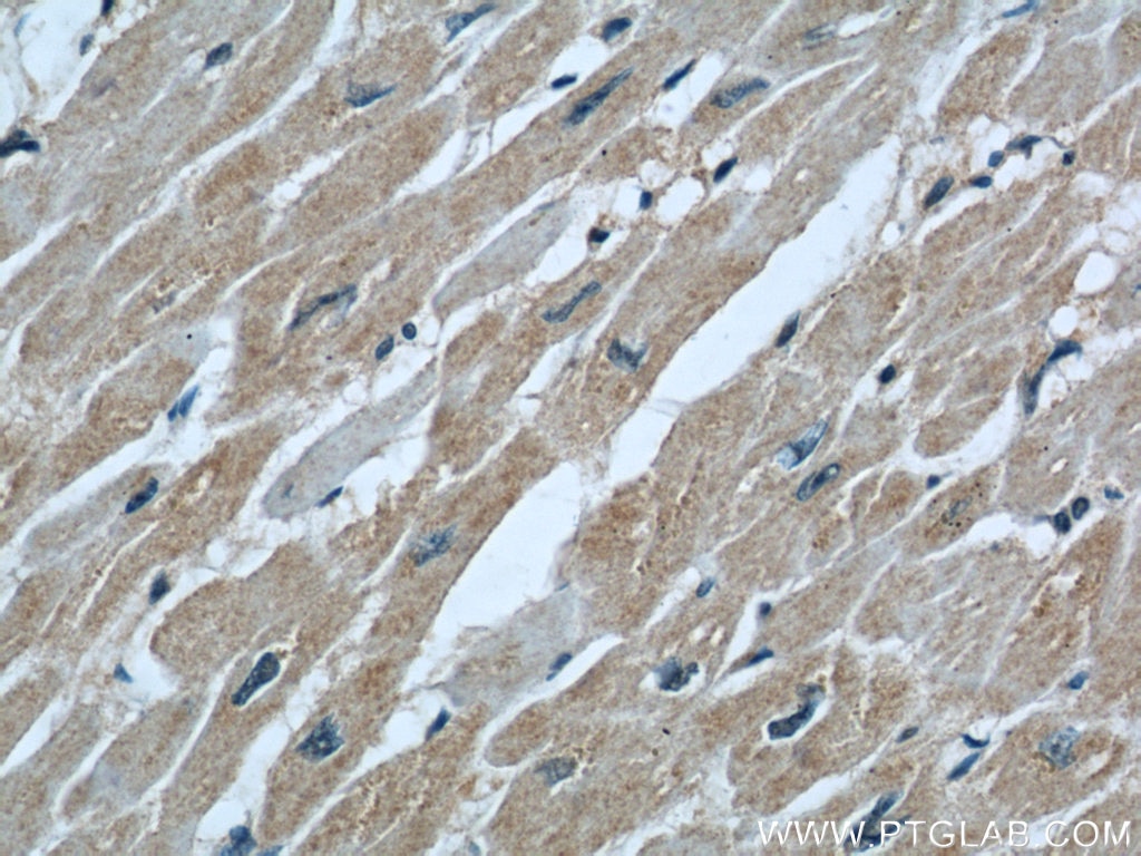
  • Immunohistochemistry (IHC) staining of human heart tissue using COXIV Monoclonal antibody (66110-1-Ig)

    IHC staining of human heart using 66110-1-Ig

    Immunohistochemical analysis of paraffin-embedded human heart using 66110-1-Ig(COXIV antibody) at dilution of 1:200 (under 40x lens).

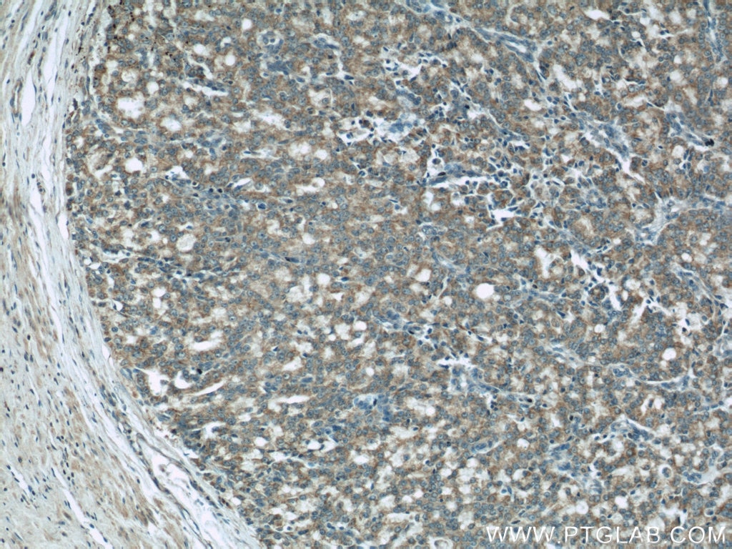
  • Immunohistochemistry (IHC) staining of human prostate cancer tissue using COXIV Monoclonal antibody (66110-1-Ig)

    IHC staining of human prostate cancer using 66110-1-Ig

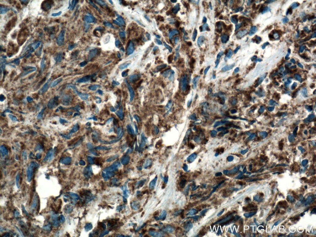
    Immunohistochemical analysis of paraffin-embedded human prostate cancer using 66110-1-Ig(COXIV antibody) at dilution of 1:200 (under 10x lens).

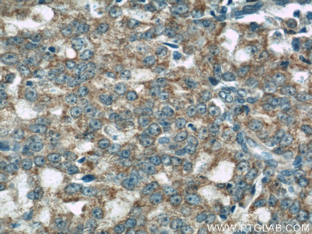
  • Immunohistochemistry (IHC) staining of human prostate cancer tissue using COXIV Monoclonal antibody (66110-1-Ig)

    IHC staining of human prostate cancer using 66110-1-Ig

    Immunohistochemical analysis of paraffin-embedded human prostate cancer using 66110-1-Ig(COXIV antibody) at dilution of 1:200 (under 40x lens).

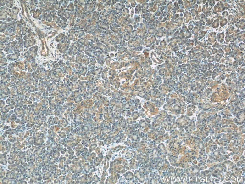
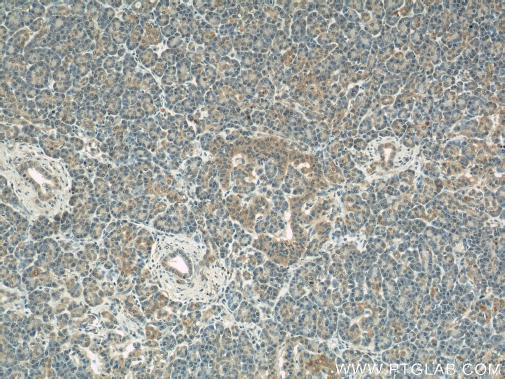
  • Immunohistochemistry (IHC) staining of human pancreas tissue using COXIV Monoclonal antibody (66110-1-Ig)

    IHC staining of human pancreas using 66110-1-Ig

    Immunohistochemical analysis of paraffin-embedded human pancreas using 66110-1-Ig(COXIV antibody) at dilution of 1:100 (under 10x lens).

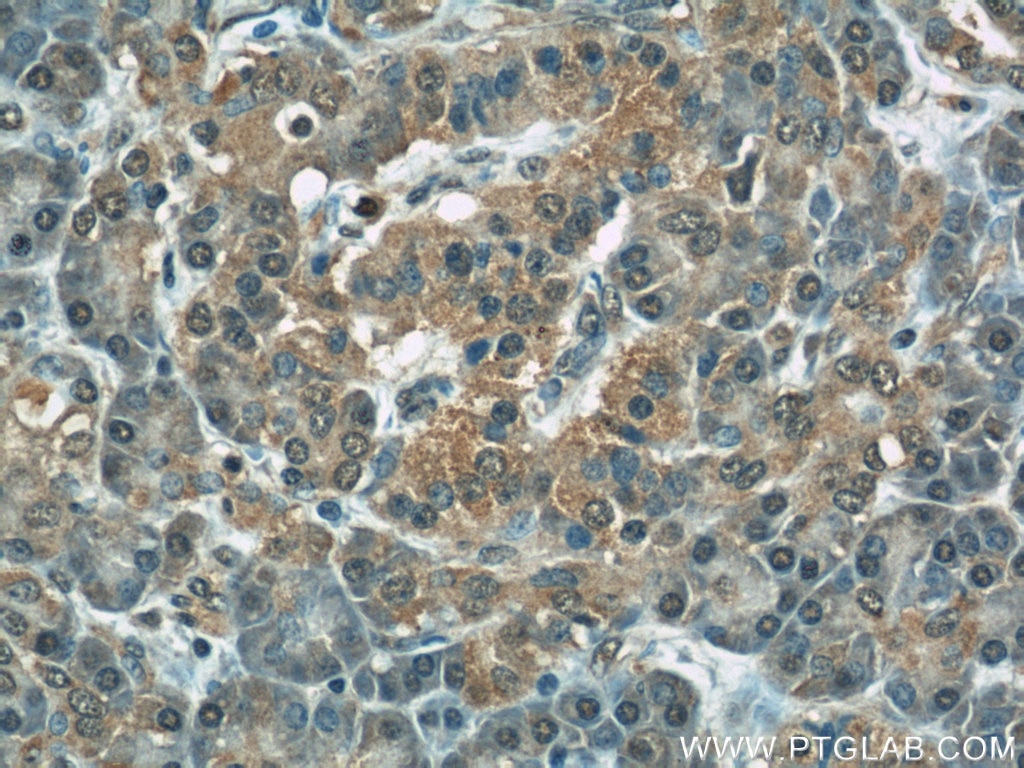
  • Immunohistochemistry (IHC) staining of human pancreas tissue using COXIV Monoclonal antibody (66110-1-Ig)

    IHC staining of human pancreas using 66110-1-Ig

    Immunohistochemical analysis of paraffin-embedded human pancreas using 66110-1-Ig(COXIV antibody) at dilution of 1:100 (under 40x lens).

  • Immunohistochemistry (IHC) staining of human pancreas tissue using COXIV Monoclonal antibody (66110-1-Ig)

    IHC staining of human pancreas using 66110-1-Ig

    Immunohistochemical analysis of paraffin-embedded human pancreas using 66110-1-Ig(COXIV antibody) at dilution of 1:200 (under 10x lens).

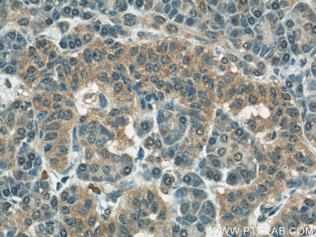
  • Immunohistochemistry (IHC) staining of human pancreas tissue using COXIV Monoclonal antibody (66110-1-Ig)

    IHC staining of human pancreas using 66110-1-Ig

    Immunohistochemical analysis of paraffin-embedded human pancreas using 66110-1-Ig(COXIV antibody) at dilution of 1:200 (under 40x lens).

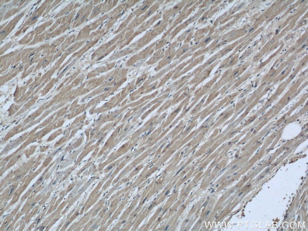
  • Immunohistochemistry (IHC) staining of human heart tissue using COXIV Monoclonal antibody (66110-1-Ig)

    IHC staining of human heart using 66110-1-Ig

    Immunohistochemical analysis of paraffin-embedded human heart using 66110-1-Ig(COXIV antibody) at dilution of 1:100 (under 10x lens).

  • Immunohistochemistry (IHC) staining of human heart tissue using COXIV Monoclonal antibody (66110-1-Ig)

    IHC staining of human heart using 66110-1-Ig

    Immunohistochemical analysis of paraffin-embedded human heart using 66110-1-Ig(COXIV antibody) at dilution of 1:100 (under 40x lens).

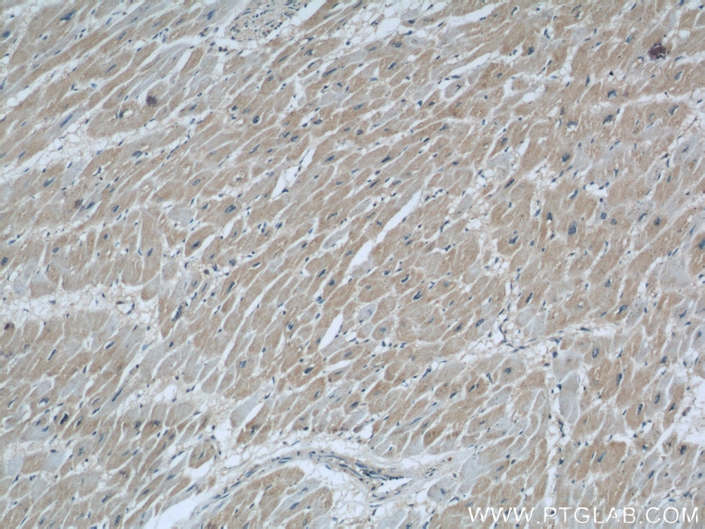
  • Immunohistochemistry (IHC) staining of human heart tissue using COXIV Monoclonal antibody (66110-1-Ig)

    IHC staining of human heart using 66110-1-Ig

    Immunohistochemical analysis of paraffin-embedded human heart using 66110-1-Ig(COXIV antibody) at dilution of 1:200 (under 10x lens).

  • Immunofluorescence (IF) / fluorescent staining of HepG2 cells using COXIV Monoclonal antibody (66110-1-Ig)

    IF Staining of HepG2 using 66110-1-Ig

    Immunofluorescent analysis of (4% PFA) fixed HepG2 cells using 66110-1-Ig (COXIV antibody, 1:100) at dilution of 1:100 and Alexa Fluor 488-Conjugated AffiniPure Goat Anti-Mouse IgG(H+L); Red is the staining with alpha Tubulin antibody (11224-1-AP, 1:100).

  • Flow cytometry (FC) experiment of HeLa cells using COXIV Monoclonal antibody (66110-1-Ig)

    FC experiment of HeLa using 66110-1-Ig

    1X10^6 HeLa cells were stained with 0.2ug COXIV antibody (66110-1-Ig, red) and control antibody (blue). Fixed with 90% MeOH blocked with 3% BSA (30 min). Alexa Fluor 488-conjugated AffiniPure Goat Anti-Mouse IgG(H+L) with dilution 1:1000.

"COXIV Antibodies" Comparison

View side-by-side comparison of COXIV antibodies from other vendors to find the one that best suits your research needs.

Applications testées

Résultats positifs en WBcellules A431, cellules HEK-293, cellules HeLa, cellules HepG2, cellules Jurkat, cellules K-562, cellules LNCaP
Résultats positifs en IHCtissu de cancer de la prostate humain, tissu cardiaque humain, tissu pancréatique humain
il est suggéré de démasquer l'antigène avec un tampon de TE buffer pH 9.0; (*) À défaut, 'le démasquage de l'antigène peut être 'effectué avec un tampon citrate pH 6,0.
Résultats positifs en IFcellules HepG2,
Résultats positifs en cytométriecellules HeLa

Dilution recommandée

ApplicationDilution
Western Blot (WB)WB : 1:5000-1:50000
Immunohistochimie (IHC)IHC : 1:250-1:1000
Immunofluorescence (IF)IF : 1:50-1:500
Flow Cytometry (FC)FC : 0.20 ug per 10^6 cells in a 100 µl suspension
It is recommended that this reagent should be titrated in each testing system to obtain optimal results.
Sample-dependent, check data in validation data gallery

Informations sur le produit

66110-1-Ig cible COXIV dans les applications de WB, IHC, IF, FC, ELISA et montre une réactivité avec des échantillons Humain

Réactivité Humain
Réactivité citéerat, Humain, souris
Hôte / Isotype Mouse / IgG1
Clonalité Monoclonal
Type Anticorps
Immunogène COXIV Protéine recombinante Ag20551
Nom complet cytochrome c oxidase subunit IV isoform 1
Masse moléculaire calculée19.6 kDa
Poids moléculaire observé 17-18 kDa
Numéro d’acquisition GenBankBC021236
Symbole du gène COX4I1
Identification du gène (NCBI) 1327
Conjugaison Non conjugué
Forme Liquide
Méthode de purification Purification par protéine G
Tampon de stockage PBS avec azoture de sodium à 0,02 % et glycérol à 50 % pH 7,3
Conditions de stockageStocker à -20°C. Stable pendant un an après l'expédition. L'aliquotage n'est pas nécessaire pour le stockage à -20oC Les 20ul contiennent 0,1% de BSA.

Informations générales

COX4I1, also named as COX4 and COXIV-1, belongs to the cytochrome c oxidase IV family. It is one of the nuclear-coded polypeptide chains of cytochrome c oxidase, the terminal oxidase in mitochondrial electron transport. COX4I1 is a marker for mitochondria. It has two isoforms (isoform 1 and 2). Isoform 1(COX4I1) is ubiquitously expressed and isoform 2 is highly expressed in lung tissues. COX4I1 is commonly used as a loading control. This antibody is specific to COX4I1 and do not cross reacts with COX4I2.

Protocole

Product Specific Protocols
WB protocol for COXIV antibody 66110-1-IgDownload protocol
IHC protocol for COXIV antibody 66110-1-IgDownload protocol
IF protocol for COXIV antibody 66110-1-IgDownload protocol
FC protocol for COXIV antibody 66110-1-IgDownload protocol
Standard Protocols
Click here to view our Standard Protocols

Publications

SpeciesApplicationTitle
humanWB

Science

Structural insight into the SAM-mediated assembly of the mitochondrial TOM core complex.

Authors - Qiang Wang
humanWB

ACS Nano

Mitochondria-Targeting Polymer Micelle of Dichloroacetate Induced Pyroptosis to Enhance Osteosarcoma Immunotherapy.

Authors - Jiakang Jin
humanWB

Nat Cell Biol

Mitochondria-localised ZNFX1 functions as a dsRNA sensor to initiate antiviral responses through MAVS.

Authors - Yao Wang
humanWB

Mol Cell

Global mitochondrial protein import proteomics reveal distinct regulation by translation and translocation machinery

Authors - Jasmin Adriana Schafer
humanIF

Nat Commun

PINCH-1 regulates mitochondrial dynamics to promote proline synthesis and tumor growth.

Authors - Ling Guo
humanWB

Elife

Genome-wide CRISPRi screening identifies OCIAD1 as a prohibitin client and regulatory determinant of mitochondrial Complex III assembly in human cells.

Authors - Maxence Le Vasseur

Avis

The reviews below have been submitted by verified Proteintech customers who received an incentive forproviding their feedback.


FH

Bryce (Verified Customer) (02-18-2022)

Decent loading control for mitochondria. I cut the blot too low, but you get the point.

  • Applications: Western Blot
  • Primary Antibody Dilution: 1:1000
  • Cell Tissue Type: Mouse Kidney
COXIV Antibody Western Blot validation (1:1000 dilution) in Mouse Kidney (Cat no:66110-1-Ig)