"COXIV Antibodies" Comparison
View side-by-side comparison of COXIV antibodies from other vendors to find the one that best suits your research needs.
Tested Applications
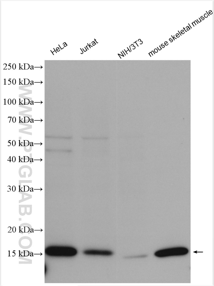
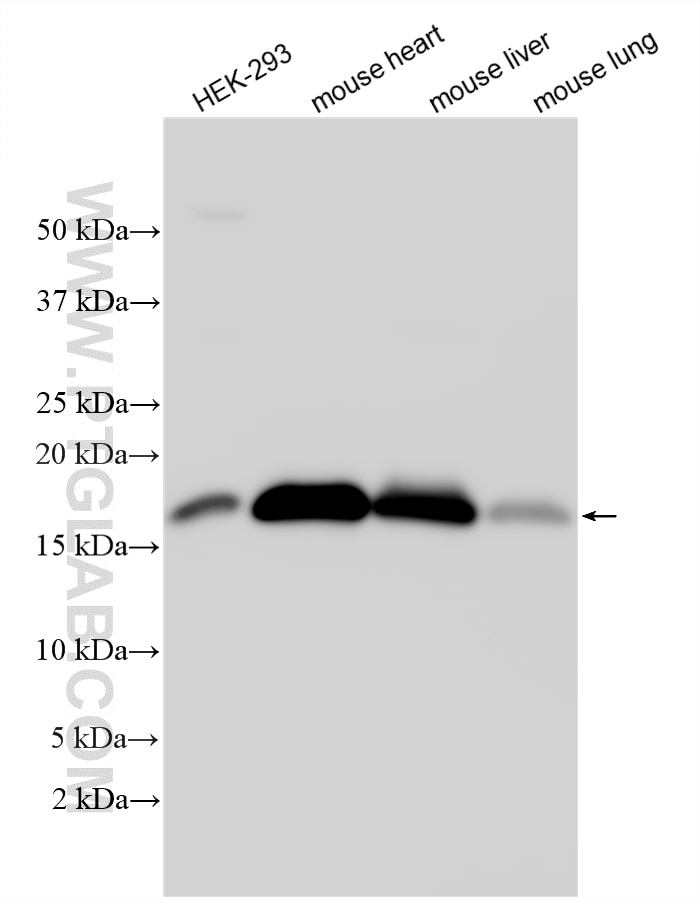
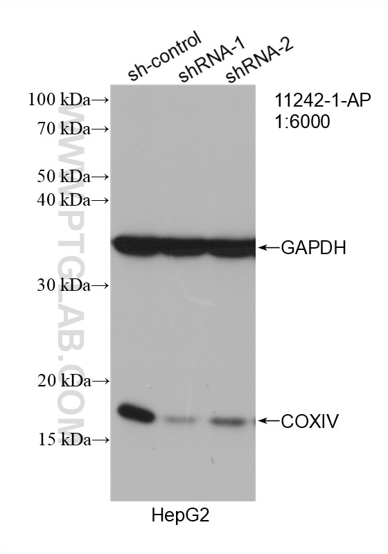
| Positive WB detected in | HeLa cells, HEK-293 cells, HepG2 cells, rat brain tissue, rat liver tissue, mouse heart tissue, mouse liver tissue, mouse lung tissue, Jurkat cells, NIH/3T3 cells, mouse skeletal muscle tissue, MCF-7 cells |
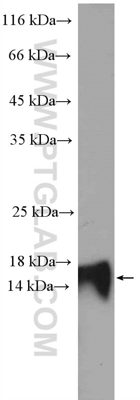
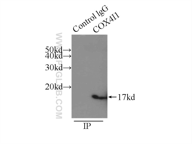
| Positive IP detected in | mouse skeletal muscle tissue |
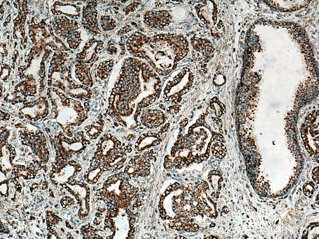
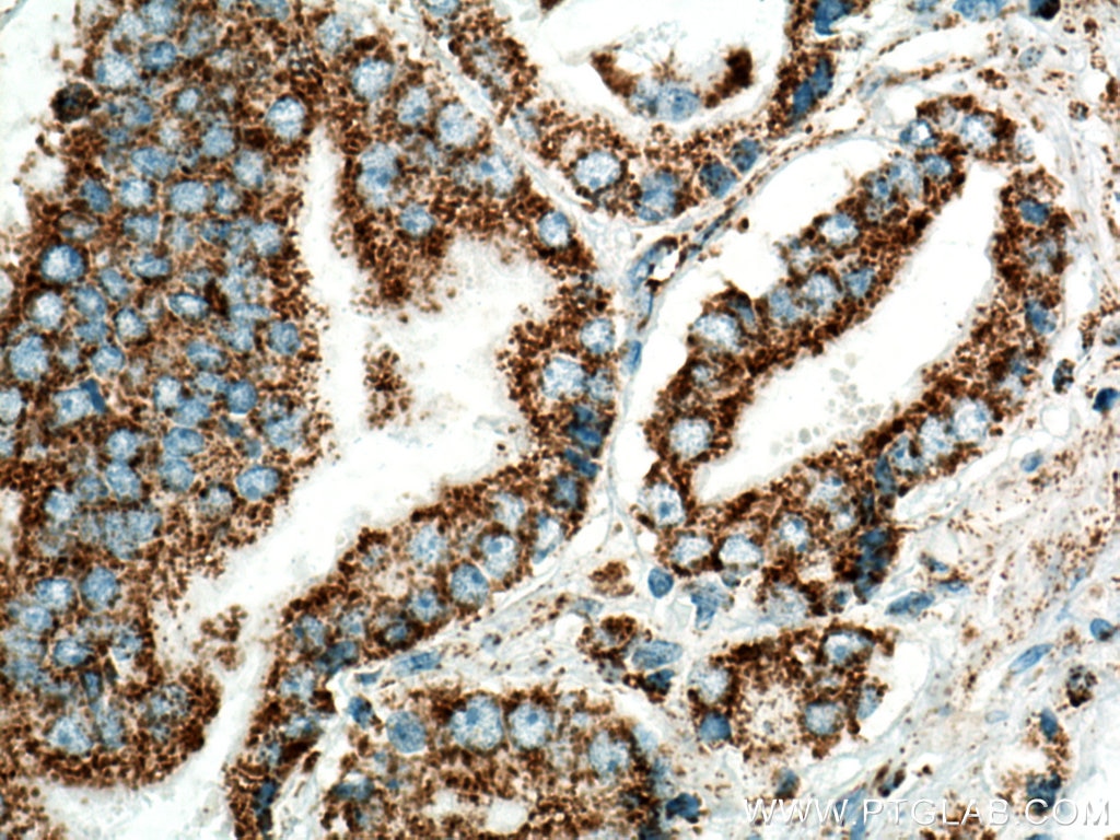
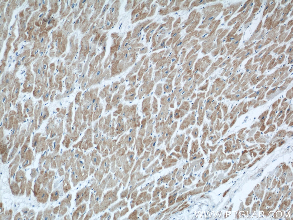
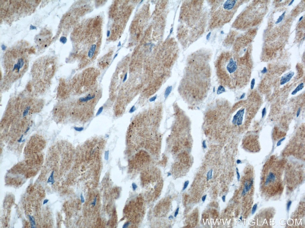
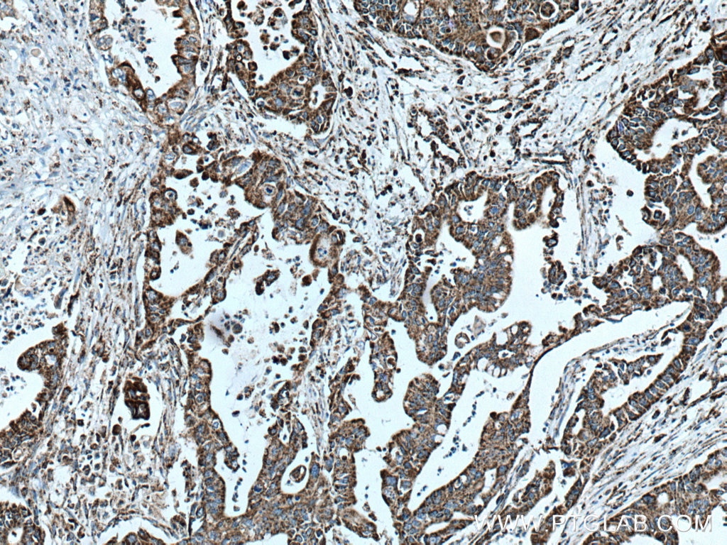
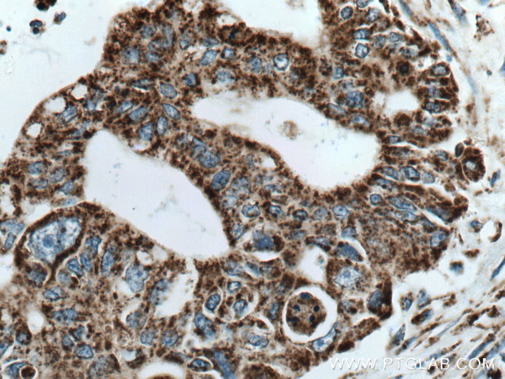
| Positive IHC detected in | human prostate cancer tissue, human pancreas cancer tissue, human heart tissue Note: suggested antigen retrieval with TE buffer pH 9.0; (*) Alternatively, antigen retrieval may be performed with citrate buffer pH 6.0 |
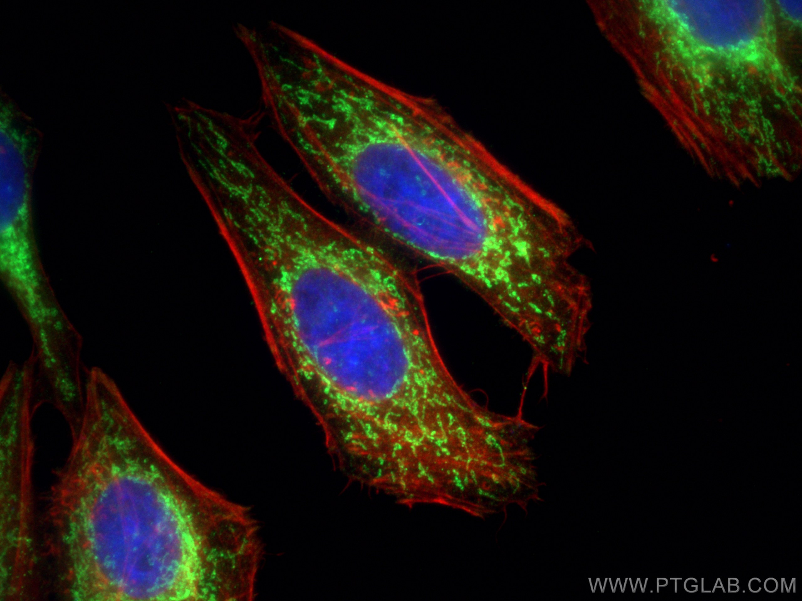
| Positive IF/ICC detected in | HepG2 cells |
Recommended dilution
| Application | Dilution |
|---|---|
| Western Blot (WB) | WB : 1:5000-1:20000 |
| Immunoprecipitation (IP) | IP : 0.5-4.0 ug for 1.0-3.0 mg of total protein lysate |
| Immunohistochemistry (IHC) | IHC : 1:200-1:1000 |
| Immunofluorescence (IF)/ICC | IF/ICC : 1:50-1:500 |
| It is recommended that this reagent should be titrated in each testing system to obtain optimal results. | |
| Sample-dependent, Check data in validation data gallery. | |
Published Applications
| KD/KO | See 4 publications below |
| WB | See 543 publications below |
| IHC | See 12 publications below |
| IF | See 75 publications below |
| IP | See 3 publications below |
| ChIP | See 1 publications below |
Product Information
11242-1-AP targets COXIV in WB, IHC, IF/ICC, IP, ChIP, ELISA applications and shows reactivity with human, mouse, rat samples.
| Tested Reactivity | human, mouse, rat |
| Cited Reactivity | human, mouse, rat, pig, canine, bovine, goat |
| Host / Isotype | Rabbit / IgG |
| Class | Polyclonal |
| Type | Antibody |
| Immunogen |
CatNo: Ag1640 Product name: Recombinant human COXIV protein Source: e coli.-derived, PGEX-4T Tag: GST Domain: 1-169 aa of BC021236 Sequence: MLATRVFSLVGKRAISTSVCVRAHESVVKSEDFSLPAYMDRRDHPLPEVAHVKHLSASQKALKEKEKASWSSLSMDEKVELYRIKFKESFAEMNRGSNEWKTVVGGAMFFIGFTALVIMWQKHYVYGPLPQSFDKEWVAKQTKRMLDMKVNPIQGLASKWDYEKNEWKK Predict reactive species |
| Full Name | cytochrome c oxidase subunit IV isoform 1 |
| Calculated Molecular Weight | 19.6 kDa |
| Observed Molecular Weight | 17-18 kDa |
| GenBank Accession Number | BC021236 |
| Gene Symbol | COX IV |
| Gene ID (NCBI) | 1327 |
| RRID | AB_2085278 |
| Conjugate | Unconjugated |
| Form | Liquid |
| Purification Method | Antigen affinity purification |
| UNIPROT ID | P13073 |
| Storage Buffer | PBS with 0.02% sodium azide and 50% glycerol, pH 7.3. |
| Storage Conditions | Store at -20°C. Stable for one year after shipment. Aliquoting is unnecessary for -20oC storage. 20ul sizes contain 0.1% BSA. |
Background Information
COX4I1, also named as COX4 and COXIV-1, belongs to the cytochrome c oxidase IV family. It is one of the nuclear-coded polypeptide chains of cytochrome c oxidase, the terminal oxidase in mitochondrial electron transport. COX4I1 is a marker for mitochondria. It has two isoforms (isoform 1 and 2). Isoform 1(COX4I1) is ubiquitously expressed and isoform 2 is highly expressed in lung tissues. COX4I1 is commonly used as a loading control. This antibody was generated against full length COX4I1 protein and cross reacts with COX4I2.
Protocols
| Product Specific Protocols | |
|---|---|
| IF protocol for COXIV antibody 11242-1-AP | Download protocol |
| IHC protocol for COXIV antibody 11242-1-AP | Download protocol |
| IP protocol for COXIV antibody 11242-1-AP | Download protocol |
| WB protocol for COXIV antibody 11242-1-AP | Download protocol |
| Standard Protocols | |
|---|---|
| Click here to view our Standard Protocols |
Publications
| Species | Application | Title |
|---|---|---|
Cell Res Mitochondria-localized cGAS suppresses ferroptosis to promote cancer progression | ||
Cell Res Nonenzymatic lysine D-lactylation induced by glyoxalase II substrate SLG dampens inflammatory immune responses | ||
Immunity Excessive Polyamine Generation in Keratinocytes Promotes Self-RNA Sensing by Dendritic Cells in Psoriasis. | ||
Cell Metab Tyrosine Phosphorylation of Mitochondrial Creatine Kinase 1 Enhances a Druggable Tumor Energy Shuttle Pathway. | ||
Nat Cell Biol AIDA directly connects sympathetic innervation to adaptive thermogenesis by UCP1. |
Reviews
The reviews below have been submitted by verified Proteintech customers who received an incentive for providing their feedback.
FH Matthieu (Verified Customer) (09-24-2025) | Slightly diffuse band at the expected size
|
FH Vignesh (Verified Customer) (09-03-2025) | Good product with good specificity.
|
FH Jimmy (Verified Customer) (02-27-2024) | Strong signal in primary human cell fractions.
|
FH Maria (Verified Customer) (08-17-2021) | Good ab, works well 1:1000 in BSA 3% O/N 4ºC incubation.
 |
FH Mohammed (Verified Customer) (08-19-2020) | Very strong staining in the midpiece of the sperm tail.
|
FH SITING (Verified Customer) (07-13-2020) | THIS IS A GOOD ANTIBODY, CAN SEE CLEAR BAND
 |