- Phare
- Validé par KD/KO
Anticorps Polyclonal de lapin anti-Lamin B1
Lamin B1 Polyclonal Antibody for WB, IHC, IF/ICC, IF-P, FC (Intra), IP, ELISA
Hôte / Isotype
Lapin / IgG
Réactivité testée
Humain, rat, souris et plus (6)
Applications
WB, IHC, IF/ICC, IF-P, FC (Intra), IP, CoIP, ChIP, ELISA
Conjugaison
Non conjugué
N° de cat : 12987-1-AP
Synonymes
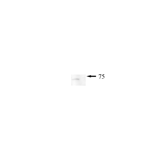
 at dilution of 1:20000 incubated at room temperature for 1.5 hours.")
 at dilution of 1:20000 incubated at room temperature for 1.5 hours.")
 at dilution of 1:10000 incubated at room temperature for 1.5 hours.")
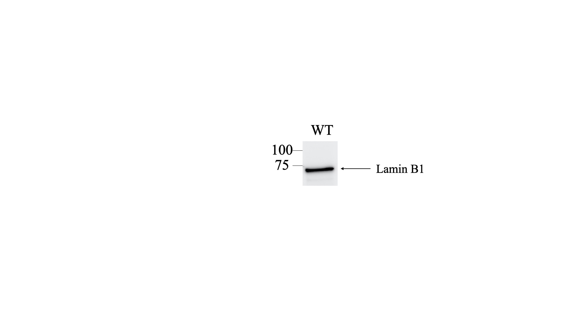
 with HeLa cells lysate 1320 ug.")
 at dilution of 1:100 (under 10x lens). Heat mediated antigen retrieval with Tris-EDTA buffer (pH 9.0).")
 at dilution of 1:1000 (under 10x lens). Heat mediated antigen retrieval with Tris-EDTA buffer (pH 9.0).")
 at dilution of 1:2000 (under 10x lens). Heat mediated antigen retrieval with Tris-EDTA buffer (pH 9.0).")
 at dilution of 1:2000 (under 40x lens). Heat mediated antigen retrieval with Tris-EDTA buffer (pH 9.0).")
 at dilution of 1:4000 (under 20x lens). Heat mediated antigen retrieval with Tris-EDTA buffer (pH 9.0).")
 at dilution of 1:4000 (under 20x lens). Heat mediated antigen retrieval with Tris-EDTA buffer (pH 9.0).")
 at dilution of 1:1200 (under 20x lens). Heat mediated antigen retrieval with Tris-EDTA buffer (pH 9.0).")
 at dilution of 1:50.")
 at dilution of 1:50.")
 fixed human skin cancer tissue using 12987-1-AP (Lamin B1 antibody) at dilution of 1:50 and Alexa Fluor 488-conjugated AffiniPure Goat Anti-Rabbit IgG(H+L).")
 fixed HepG2 cells using 12987-1-AP (Lamin B1 antibody) at dilution of 1:200 and Alexa Fluor 488-conjugated AffiniPure Goat Anti-Rabbit IgG(H+L).")
 fixed HepG2 cells using Lamin B1 antibody (12987-1-AP) at dilution of 1:400 and CoraLite®488-Conjugated AffiniPure Goat Anti-Rabbit IgG(H+L), CL594-Phalloidin (red).")
 fixed NIH/3T3 cells using 12987-1-AP (Lamin B1 antibody) at dilution of 1:100 and Alexa Fluor 488-conjugated AffiniPure Goat Anti-Rabbit IgG(H+L).")
.")
 and CoraLite®488-Conjugated AffiniPure Goat Anti-Rabbit IgG(H+L) at dilution 1:1000 (red), or 0.4 ug Isotype Control. Cells were fixed with 4% PFA and permeabilized with Flow Cytometry Perm Buffer (PF00011-C).")
"Lamin B1 Antibodies" Comparison
View side-by-side comparison of Lamin B1 antibodies from other vendors to find the one that best suits your research needs.
Applications testées
| Résultats positifs en WB | cellules HeLa, cellules HepG2, cellules Jurkat, cellules K-562, cellules MCF-7, cellules NIH/3T3, cellules RAW 264.7 |
| Résultats positifs en IP | cellules HeLa, |
| Résultats positifs en IHC | tissu cardiaque de souris, tissu de cancer du côlon humain, tissu de cancer du foie humain, tissu de côlon humain, tissu rénal de souris, tissu rénal humain il est suggéré de démasquer l'antigène avec un tampon de TE buffer pH 9.0; (*) À défaut, 'le démasquage de l'antigène peut être 'effectué avec un tampon citrate pH 6,0. |
| Résultats positifs en IF-P | tissu de cancer de la peau humain, |
| Résultats positifs en IF/ICC | cellules HepG2, cellules NIH/3T3, tissu de cancer de la peau humain |
| Résultats positifs en FC (Intra) | cellules HEK-293 |
Dilution recommandée
| Application | Dilution |
|---|---|
| Western Blot (WB) | WB : 1:5000-1:50000 |
| Immunoprécipitation (IP) | IP : 0.5-4.0 ug for 1.0-3.0 mg of total protein lysate |
| Immunohistochimie (IHC) | IHC : 1:1000-1:4000 |
| Immunofluorescence (IF)-P | IF-P : 1:50-1:500 |
| Immunofluorescence (IF)/ICC | IF/ICC : 1:200-1:800 |
| Flow Cytometry (FC) (INTRA) | FC (INTRA) : 0.40 ug per 10^6 cells in a 100 µl suspension |
| It is recommended that this reagent should be titrated in each testing system to obtain optimal results. | |
| Sample-dependent, check data in validation data gallery | |
Informations sur le produit
12987-1-AP cible Lamin B1 dans les applications de WB, IHC, IF/ICC, IF-P, FC (Intra), IP, CoIP, ChIP, ELISA et montre une réactivité avec des échantillons Humain, rat, souris
| Réactivité | Humain, rat, souris |
| Réactivité citée | rat, canin, Humain, porc, poulet, singe, souris, Hamster, Bombyx Mori |
| Hôte / Isotype | Lapin / IgG |
| Clonalité | Polyclonal |
| Type | Anticorps |
| Immunogène | Lamin B1 Protéine recombinante Ag3631 |
| Nom complet | lamin B1 |
| Masse moléculaire calculée | 66 kDa |
| Poids moléculaire observé | 66-70 kDa |
| Numéro d’acquisition GenBank | BC012295 |
| Symbole du gène | Lamin B1 |
| Identification du gène (NCBI) | 4001 |
| Conjugaison | Non conjugué |
| Forme | Liquide |
| Méthode de purification | Purification par affinité contre l'antigène |
| Tampon de stockage | PBS with 0.02% sodium azide and 50% glycerol |
| Conditions de stockage | Stocker à -20°C. Stable pendant un an après l'expédition. L'aliquotage n'est pas nécessaire pour le stockage à -20oC Les 20ul contiennent 0,1% de BSA. |
Informations générales
Lamins are components of the nuclear lamina, a fibrous layer on the nucleoplasmic side of the inner nuclear membrane, which is thought to provide a framework for the nuclear envelope and may also interact with chromatin. The nuclear lamina consists of a two-dimensional matrix of proteins located next to the inner nuclear membrane. The lamin family of proteins make up the matrix and are highly conserved in evolution. During mitosis, the lamina matrix is reversibly disassembled as the lamin proteins are phosphorylated. Vertebrate lamins consist of two types, A and B. This gene encodes one of the two B type proteins, B1. Expression of uncleavable mutant lamin A or B caused significant delays in the onset of chromatin condensation and nuclear shrinkage during apoptosis (PMID:11953316). This protein is not suitable for samples where the nuclear envelope has been removed.
Protocole
| Product Specific Protocols | |
|---|---|
| WB protocol for Lamin B1 antibody 12987-1-AP | Download protocol |
| IHC protocol for Lamin B1 antibody 12987-1-AP | Download protocol |
| IF protocol for Lamin B1 antibody 12987-1-AP | Download protocol |
| IP protocol for Lamin B1 antibody 12987-1-AP | Download protocol |
| FC protocol for Lamin B1 antibody 12987-1-AP | Download protocol |
| Standard Protocols | |
|---|---|
| Click here to view our Standard Protocols |
Publications
| Species | Application | Title |
|---|---|---|
Nat Biotechnol Drag-and-drop genome insertion of large sequences without double-strand DNA cleavage using CRISPR-directed integrases | ||
Cell Res Disruption of ER ion homeostasis maintained by an ER anion channel CLCC1 contributes to ALS-like pathologies | ||
Nature Stella safeguards the oocyte methylome by preventing de novo methylation mediated by DNMT1. | ||
Mol Cancer Diversifying the anthracycline class of anti-cancer drugs identifies aclarubicin for superior survival of acute myeloid leukemia patients | ||
Immunity 25-Hydroxycholesterol regulates lysosome AMP kinase activation and metabolic reprogramming to educate immunosuppressive macrophages |
Avis
The reviews below have been submitted by verified Proteintech customers who received an incentive for providing their feedback.
FH Rajkumar (Verified Customer) (04-18-2025) | I used this antibody for HRP-based detection and it worked very well at the suggested dilution. The signal was strong. Highly recommend for similar applications.
|
FH Mi (Verified Customer) (02-21-2023) | Works great in human adipocytes.
|
FH Ning (Verified Customer) (01-28-2023) | Excellent antibody to probe lamin B1.
 |
FH Tsimafei (Verified Customer) (12-04-2022) | 20µg of total protein were loaded. Incubation for 2h at room temperature. Excellent antibody
 |
FH Alejandro (Verified Customer) (08-07-2022) | Works fine for flow cytometry
|
FH Iram (Verified Customer) (09-04-2020) | Very sharp bands for nuclear protein
|
FH Chun (Verified Customer) (12-05-2019) | Excellent antibody
|
FH Marco (Verified Customer) (09-02-2019) | good staining in conjunction with Alexa Fluor Plus 488 secondary
|

