- Phare
- Validé par KD/KO
Anticorps Polyclonal de lapin anti-LC3
LC3 Polyclonal Antibody for WB, IHC, IF/ICC, FC (Intra), ELISA
Hôte / Isotype
Lapin / IgG
Réactivité testée
Humain, rat, souris et plus (6)
Applications
WB, IHC, IF/ICC, FC (Intra), IP, CoIP, ELISA
Conjugaison
Non conjugué
N° de cat : 14600-1-AP
Synonymes
Galerie de données de validation
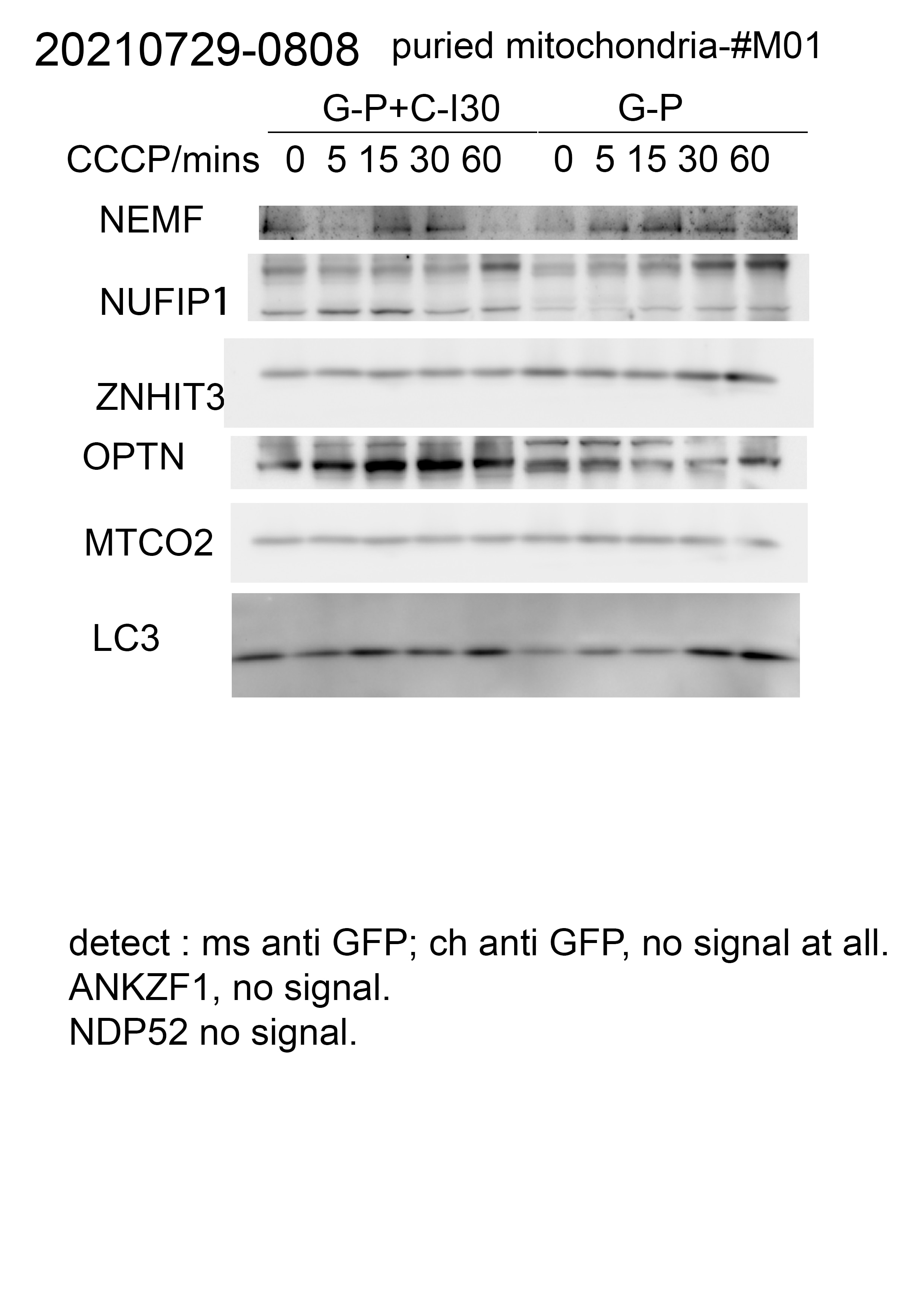
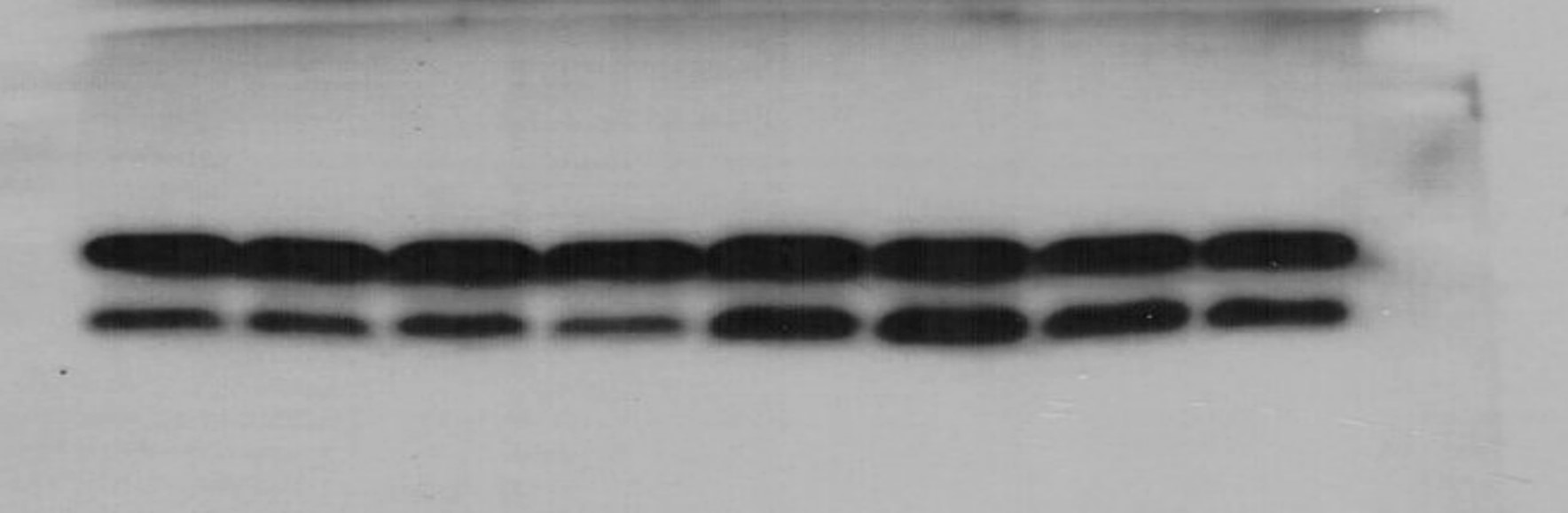
 with negative Control and LC3 knockout HeLa cells.")
 with sh-Control and sh-LC3 transfected HeLa cells.")
 at dilution of 1:8000 incubated at room temperature for 1.5 hours.")
 at dilution of 1:6000 incubated at room temperature for 1.5 hours.")
 at dilution of 1:200 (under 40x lens). Heat mediated antigen retrieval with Tris-EDTA buffer (pH 9.0).")
 at dilution of 1:600 (under 40x lens). Heat mediated antigen retrieval with Tris-EDTA buffer (pH 9.0).")
 at dilution of 1:200 (under 10x lens). Heat mediated antigen retrieval with Tris-EDTA buffer (pH 9.0).")
 at dilution of 1:200 (under 40x lens). Heat mediated antigen retrieval with Tris-EDTA buffer (pH 9.0).")
 at dilution of 1:200 (under 10x lens). Heat mediated antigen retrieval with Tris-EDTA buffer (pH 9.0).")
 at dilution of 1:600 (under 10x lens). Heat mediated antigen retrieval with Tris-EDTA buffer (pH 9.0).")
 at dilution of 1:200 (under 40x lens). Heat mediated antigen retrieval with Tris-EDTA buffer (pH 9.0).")
 at dilution of 1:200 (under 10x lens). Heat mediated antigen retrieval with Tris-EDTA buffer (pH 9.0).")
 fixed Chloroquine treated HeLa cells using LC3 antibody (14600-1-AP) at dilution of 1:500 and CoraLite®488-Conjugated AffiniPure Goat Anti-Rabbit IgG(H+L).")
 fixed CQ treated HepG2 cells using 14600-1-AP (LC3 antibody) at dilution of 1:200 and CoraLite488-Conjugated AffiniPure Goat Anti-Rabbit IgG(H+L).")
 fixed CQ treated HepG2 cells using 14600-1-AP (LC3 antibody) at dilution of 1:100 and CoraLite488-Conjugated AffiniPure Goat Anti-Rabbit IgG(H+L).")
 and CoraLite®488-Conjugated AffiniPure Goat Anti-Rabbit IgG(H+L) at dilution 1:1000 (red), or 0.5 ug rabbit IgG isotype control (blue). Cells were fixed with 4% PFA and permeabilized with Flow Cytometry Perm Buffer (PF00011-C).")
Applications testées
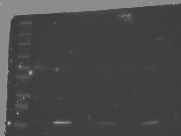
| Résultats positifs en WB | cellules HeLa, cellules HeLa traitées à la chloroquine, tissu cérébral de rat, tissu cérébral de souris |
| Résultats positifs en IHC | tissu hépatique humain, tissu cérébral de souris, tissu de gliome humain il est suggéré de démasquer l'antigène avec un tampon de TE buffer pH 9.0; (*) À défaut, 'le démasquage de l'antigène peut être 'effectué avec un tampon citrate pH 6,0. |
| Résultats positifs en IF/ICC | cellules HeLa traitées à la chloroquine, cellules HepG2 traitées à la chloroquine |
| Résultats positifs en FC (Intra) | cellules HeLa, |
Dilution recommandée
| Application | Dilution |
|---|---|
| Western Blot (WB) | WB : 1:2000-1:8000 |
| Immunohistochimie (IHC) | IHC : 1:50-1:500 |
| Immunofluorescence (IF)/ICC | IF/ICC : 1:250-1:1000 |
| Flow Cytometry (FC) (INTRA) | FC (INTRA) : 0.50 ug per 10^6 cells in a 100 µl suspension |
| It is recommended that this reagent should be titrated in each testing system to obtain optimal results. | |
| Sample-dependent, check data in validation data gallery | |
Applications publiées
| KD/KO | See 1 publications below |
| WB | See 1475 publications below |
| IHC | See 168 publications below |
| IF | See 456 publications below |
| IP | See 6 publications below |
| ELISA | See 1 publications below |
| CoIP | See 5 publications below |
Informations sur le produit
14600-1-AP cible LC3 dans les applications de WB, IHC, IF/ICC, FC (Intra), IP, CoIP, ELISA et montre une réactivité avec des échantillons Humain, rat, souris
| Réactivité | Humain, rat, souris |
| Réactivité citée | rat, Chèvre, Humain, Lapin, poisson-zèbre, poulet, singe, souris, Hamster |
| Hôte / Isotype | Lapin / IgG |
| Clonalité | Polyclonal |
| Type | Anticorps |
| Immunogène | LC3 Protéine recombinante Ag6144 |
| Nom complet | microtubule-associated protein 1 light chain 3 beta |
| Masse moléculaire calculée | 15 kDa |
| Poids moléculaire observé | 14-18 kDa |
| Numéro d’acquisition GenBank | BC067797 |
| Symbole du gène | LC3B |
| Identification du gène (NCBI) | 81631 |
| Conjugaison | Non conjugué |
| Forme | Liquide |
| Méthode de purification | Purification par affinité contre l'antigène |
| Tampon de stockage | PBS with 0.02% sodium azide and 50% glycerol |
| Conditions de stockage | Stocker à -20°C. Stable pendant un an après l'expédition. L'aliquotage n'est pas nécessaire pour le stockage à -20oC Les 20ul contiennent 0,1% de BSA. |
Informations générales
Map1LC3, also known as LC3, is the human homolog of yeast Atg8 and is involved in the formation of autophagosomal vacuoles, called autophagosomes. Three human Map1LC3 isoforms, MAP1LC3A, MAP1LC3B, and MAP1LC3C, undergo post-translational modifications during autophagy. And they differ in their post-translation modifications during autophagy. Map1LC3 also exists in two modified forms, an 18 kDa cytoplasmic form that was originally identified as a subunit of the microtubule-associated protein 1, and a 14-16 kDa form that is associated with the autophagosome membrane. This antibody can cross react with MAP1LC3A, MAP1LC3B, and MAP1LC3C.
Protocole
| Product Specific Protocols | |
|---|---|
| WB protocol for LC3 antibody 14600-1-AP | Download protocol |
| IHC protocol for LC3 antibody 14600-1-AP | Download protocol |
| IF protocol for LC3 antibody 14600-1-AP | Download protocol |
| FC protocol for LC3 antibody 14600-1-AP | Download protocol |
| Standard Protocols | |
|---|---|
| Click here to view our Standard Protocols |
Publications
| Species | Application | Title |
|---|---|---|
Signal Transduct Target Ther Targeting CRL4 suppresses chemoresistant ovarian cancer growth by inducing mitophagy | ||
Nat Methods Visualizing the native cellular organization by coupling cryofixation with expansion microscopy (Cryo-ExM). | ||
Gastroenterology Pancreatic acinar cells-derived sphingosine-1-phosphate contributes to fibrosis of chronic pancreatitis via inducing autophagy and activation of pancreatic stellate cells | ||
Nat Cell Biol Ammonia-induced lysosomal and mitochondrial damage causes cell death of effector CD8+ T cells | ||
Bioact Mater Local delivery of EGFR+NSCs-derived exosomes promotes neural regeneration post spinal cord injury via miR-34a-5p/HDAC6 pathway | ||
Acta Neuropathol C9orf72 intermediate repeats are associated with corticobasal degeneration, increased C9orf72 expression and disruption of autophagy. |
Avis
The reviews below have been submitted by verified Proteintech customers who received an incentive for providing their feedback.
FH A (Verified Customer) (07-15-2025) | Used for caco2 cells and animal colon tissue
|
FH Aditya (Verified Customer) (11-01-2024) | great with ecl
|
FH David (Verified Customer) (01-02-2024) | Good antibody, single band at the predicted molecular weight, no background.
|
FH Hala (Verified Customer) (05-09-2023) | works very good in milk5%
 |
FH Hala (Verified Customer) (04-28-2023) | works very good with milk blocking
|
FH Zhihao (Verified Customer) (07-25-2022) | We could detect one form of LC3 in our cell samples, which should be two.
 |
FH S (Verified Customer) (12-31-2021) | I have tried three different LC3B antibodies from three different companies so far. This antibody is by far the best (cost efficient as well)
 |
FH YING (Verified Customer) (09-25-2021) | The antibody works very well. Distinguished LC3-I and LC3-II bands. An increase of LC3-II level in response to treatment with bafilomycin A.
 |
FH Eric (Verified Customer) (03-13-2021) | Worked very well. I was able to get the LC3A/B bands on western blot using a 12.5% gel and prominent bands on a 8% gel. IF imaging worked fairly well, I used 1:100 which was likely too low so I will repeat at higher but got signal nonetheless. This performed better than some of the other LC3 antibodies we tested.
 |
FH Elzbieta (Verified Customer) (09-23-2020) | The antibody works very nicely in WB. Clear signal, no aspecific bands. Our lab discarded other products and switched to this antibody.
|
FH An (Verified Customer) (09-17-2020) | Used for IHC on zebrafish cryosections of eyes. Worked well.
|
FH David (Verified Customer) (03-30-2020) | Very nice visualisation of LC3-I and LC3-II. Run on 20% gel and significant changes in response to treatment with bafilomycin A and concanamycin A.
|

