Anticorps Polyclonal de lapin anti-S100B
S100B Polyclonal Antibody for WB, IHC, IF/ICC, IF-P, FC (Intra), ELISA
Hôte / Isotype
Lapin / IgG
Réactivité testée
Humain, rat, souris
Applications
WB, IHC, IF/ICC, IF-P, FC (Intra), ELISA
Conjugaison
Non conjugué
N° de cat : 15146-1-AP
Synonymes
Galerie de données de validation
 at dilution of 1:15000 incubated at room temperature for 1.5 hours.")
 at dilution of 1:500 incubated at room temperature for 1.5 hours.")
 at dilution of 1:1000 incubated at room temperature for 1.5 hours.")
 at dilution of 1:400 incubated at room temperature for 1.5 hours.")
 at dilution of 1:300 incubated at room temperature for 1.5 hours.")
 at dilution of 1:6000 (under 10x lens). Heat mediated antigen retrieval with Tris-EDTA buffer (pH 9.0).")
 at dilution of 1:6000 (under 40x lens). Heat mediated antigen retrieval with Tris-EDTA buffer (pH 9.0).")
 at dilution of 1:6000 (under 10x lens). Heat mediated antigen retrieval with Tris-EDTA buffer (pH 9.0).")
 at dilution of 1:6000 (under 40x lens). Heat mediated antigen retrieval with Tris-EDTA buffer (pH 9.0).")
 at dilution of 1:6000 (under 40x lens). Heat mediated antigen retrieval with Tris-EDTA buffer (pH 9.0).")
 at dilution of 1:6000 (under 10x lens). Heat mediated antigen retrieval with Tris-EDTA buffer (pH 9.0).")
 at dilution of 1:6000 (under 40x lens). Heat mediated antigen retrieval with Tris-EDTA buffer (pH 9.0).")
 at dilution of 1:6000 (under 10x lens). Heat mediated antigen retrieval with Tris-EDTA buffer (pH 9.0).")
 at dilution of 1:6000 (under 40x lens). Heat mediated antigen retrieval with Tris-EDTA buffer (pH 9.0).")
 at dilution of 1:6000 (under 10x lens). Heat mediated antigen retrieval with Tris-EDTA buffer (pH 9.0).")
 at dilution of 1:6000 (under 40x lens). Heat mediated antigen retrieval with Tris-EDTA buffer (pH 9.0).")
 fixed mouse brain tissue using S100 Beta antibody (15146-1-AP) at dilution of 1:200 and CoraLite®488-Conjugated AffiniPure Goat Anti-Rabbit IgG(H+L).")
 fixed mouse brain tissue using S100 Beta antibody (15146-1-AP) at dilution of 1:200 and CoraLite®488-Conjugated AffiniPure Goat Anti-Rabbit IgG(H+L).")
 fixed mouse cerebellum tissue using S100 Beta antibody (15146-1-AP) at dilution of 1:200 and CoraLite®488-Conjugated AffiniPure Goat Anti-Rabbit IgG(H+L).")
 fixed mouse cerebellum tissue using S100 Beta antibody (15146-1-AP) at dilution of 1:200 and CoraLite®488-Conjugated AffiniPure Goat Anti-Rabbit IgG(H+L).")
 fixed paraffin-embedded rat brain tissue using S100B antibody (15146-1-AP) at dilution of 1:200 and CoraLite®488-Conjugated Goat Anti-Rabbit IgG(H+L) (SA00013-2). Heat mediated antigen retrieval with Tris-EDTA buffer (pH 9.0).")
 fixed paraffin-embedded rat brain tissue using S100B antibody (15146-1-AP) at dilution of 1:200 and CoraLite®488-Conjugated Goat Anti-Rabbit IgG(H+L) (SA00013-2). Heat mediated antigen retrieval with Tris-EDTA buffer (pH 9.0).")
 fixed paraffin-embedded mouse brain tissue using S100B antibody (15146-1-AP) at dilution of 1:200 and CoraLite®488-Conjugated Goat Anti-Rabbit IgG(H+L) (SA00013-2). Heat mediated antigen retrieval with Tris-EDTA buffer (pH 9.0).")
 fixed human malignant melanoma tissue using S100 Beta antibody (15146-1-AP) at dilution of 1:100 and CoraLite®488-Conjugated AffiniPure Goat Anti-Rabbit IgG(H+L).")
 fixed human malignant melanoma tissue using S100 Beta antibody (15146-1-AP) at dilution of 1:100 and CoraLite®488-Conjugated AffiniPure Goat Anti-Rabbit IgG(H+L).")
 at 1/200 (Magenta) and neurons with TUJ1 (66375-1-Ig) at 1:500 (Green). The sample was fixed with 4% Paraformaldehyde and permeabilized with 0.3% Triton X-100. Alexa Fluor 488-conjugated goat anti-mouse IgG (1/500) and Alexa Fluor 594-conjugated goat anti-rabbit IgG (1/500) were used as the secondary antibodies. Nuclei were counterstained with DAPI (blue).")
 fixed A375 cells using 15146-1-AP (S100 beta antibody) at dilution of 1:50 and Alexa Fluor 488-conjugated AffiniPure Goat Anti-Rabbit IgG(H+L).")
 and CoraLite®488-Conjugated AffiniPure Goat Anti-Rabbit IgG(H+L) at dilution 1:1000 (red), or 0.5 ug Control Antibody. Cells were fixed with 4% PFA and permeabilized with Flow Cytometry Perm Buffer (PF00011-C).")
Applications testées
| Résultats positifs en WB | tissu cérébral de souris, cellules A375, cellules C6, cellules U-251, tissu cérébral de rat, tissu testiculaire humain |
| Résultats positifs en IHC | tissu de mélanome malin humain, tissu cérébral de rat, tissu cérébral de souris, tissu d'appendicite humain, tissu de gliome humain il est suggéré de démasquer l'antigène avec un tampon de TE buffer pH 9.0; (*) À défaut, 'le démasquage de l'antigène peut être 'effectué avec un tampon citrate pH 6,0. |
| Résultats positifs en IF-P | tissu cérébral de souris, tissu cérébral de rat, tissu de cervelet de souris, tissu de mélanome malin humain |
| Résultats positifs en IF/ICC | astrocytes humains, cellules A375 |
| Résultats positifs en FC (Intra) | cellules A375 |
Dilution recommandée
| Application | Dilution |
|---|---|
| Western Blot (WB) | WB : 1:4000-1:20000 |
| Immunohistochimie (IHC) | IHC : 1:1000-1:6000 |
| Immunofluorescence (IF)-P | IF-P : 1:50-1:500 |
| Immunofluorescence (IF)/ICC | IF/ICC : 1:50-1:500 |
| Flow Cytometry (FC) (INTRA) | FC (INTRA) : 0.50 ug per 10^6 cells in a 100 µl suspension |
| It is recommended that this reagent should be titrated in each testing system to obtain optimal results. | |
| Sample-dependent, check data in validation data gallery | |
Applications publiées
| WB | See 26 publications below |
| IHC | See 28 publications below |
| IF | See 80 publications below |
| ELISA | See 1 publications below |
Informations sur le produit
15146-1-AP cible S100B dans les applications de WB, IHC, IF/ICC, IF-P, FC (Intra), ELISA et montre une réactivité avec des échantillons Humain, rat, souris
| Réactivité | Humain, rat, souris |
| Réactivité citée | rat, Humain, souris |
| Hôte / Isotype | Lapin / IgG |
| Clonalité | Polyclonal |
| Type | Anticorps |
| Immunogène | S100B Protéine recombinante Ag7440 |
| Nom complet | S100 calcium binding protein B |
| Masse moléculaire calculée | 11 kDa |
| Poids moléculaire observé | 11 kDa |
| Numéro d’acquisition GenBank | BC001766 |
| Symbole du gène | S100 Beta |
| Identification du gène (NCBI) | 6285 |
| Conjugaison | Non conjugué |
| Forme | Liquide |
| Méthode de purification | Purification par affinité contre l'antigène |
| Tampon de stockage | PBS with 0.02% sodium azide and 50% glycerol |
| Conditions de stockage | Stocker à -20°C. Stable pendant un an après l'expédition. L'aliquotage n'est pas nécessaire pour le stockage à -20oC Les 20ul contiennent 0,1% de BSA. |
Informations générales
S100B belongs to the EF-hand calcium binding proteins and is found primarily in astrocytes in the central nervous system (CNS). S100B has a variety of functions, including calcium homeostasis, cell proliferation, differentiation, migration, and survival, as well as neurite outgrowth and regeneration.
1. What is the molecular weight of S100B?
S100B protein is composed of non-covalently linked homodimers of 11 kDa size.
2. What is the subcellular localization of S100B?
S100B localizes to the nucleus and cytoplasm, associating with intracellular membranes, the centrosomes, microtubules, and type III intermediate filaments (PMID: 19110011). Additionally, it can be released from astrocytes into the extracellular space and can enter the bloodstream.
3. What is the expression pattern of S100B?
S100B is predominantly expressed in astrocytes and maturing oligodendrocytes but is also present in other cell types such as kidney epithelial cells, neural progenitor cells, pituicytes, ependymocytes, chondrocytes, adipocytes, melanocytes, Langerhans cells, dendritic cells, certain lymphocyte subpopulations, skeletal myofibers, myoblasts, and muscle satellite cells (PMID: 19110011). S100B is a commonly used marker of Schwann cells and reactive astrocytes in ICC, IHC, and WB applications.
4. What is the diagnostic use of S100B in the clinic?
S100B is naturally secreted by astrocytes into the extracellular space and low amounts of S100B can pass through the brain-blood barrier and enter the bloodstream. Elevated levels of S100B in the serum are observed in patients with traumatic head injuries, as well as in patients suffering from neurodegenerative diseases (PMID: 30144068). This increase in S100B levels is attributed to the elevated secretion of S100B protein from astrocytes as part of the physiological response to the injury, as well as to the physical damage of astrocytes and increased blood-brain barrier permeability.
Protocole
| Product Specific Protocols | |
|---|---|
| WB protocol for S100B antibody 15146-1-AP | Download protocol |
| IHC protocol for S100B antibody 15146-1-AP | Download protocol |
| IF protocol for S100B antibody 15146-1-AP | Download protocol |
| FC protocol for S100B antibody 15146-1-AP | Download protocol |
| Standard Protocols | |
|---|---|
| Click here to view our Standard Protocols |
Publications
| Species | Application | Title |
|---|---|---|
Cell Mechanoreceptor synapses in the brainstem shape the central representation of touch. | ||
Sci Bull (Beijing) Restoring sweat gland function in mice using regenerative sweat gland cells derived from chemically reprogrammed human epidermal keratinocytes | ||
Nat Commun Alk1 acts in non-endothelial VE-cadherin+ perineurial cells to maintain nerve branching during hair homeostasis | ||
Neuron γ-Protocadherins control synapse formation and peripheral branching of touch sensory neurons |
Avis
The reviews below have been submitted by verified Proteintech customers who received an incentive for providing their feedback.
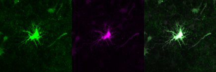
FH Clarisse (Verified Customer) (08-08-2022) | This antibody works very well as an astrocyte marker. In the image: in green mouse anti-S100B (1:500 - 66616-1-Ig) and in magenta rabbit anti-S100B (1:500 - 15146-1-AP). The rabbit antibody has a better performance.
 |
FH Tongcheng (Verified Customer) (09-08-2021) | This antibody works perfect on human astrocytes.
|
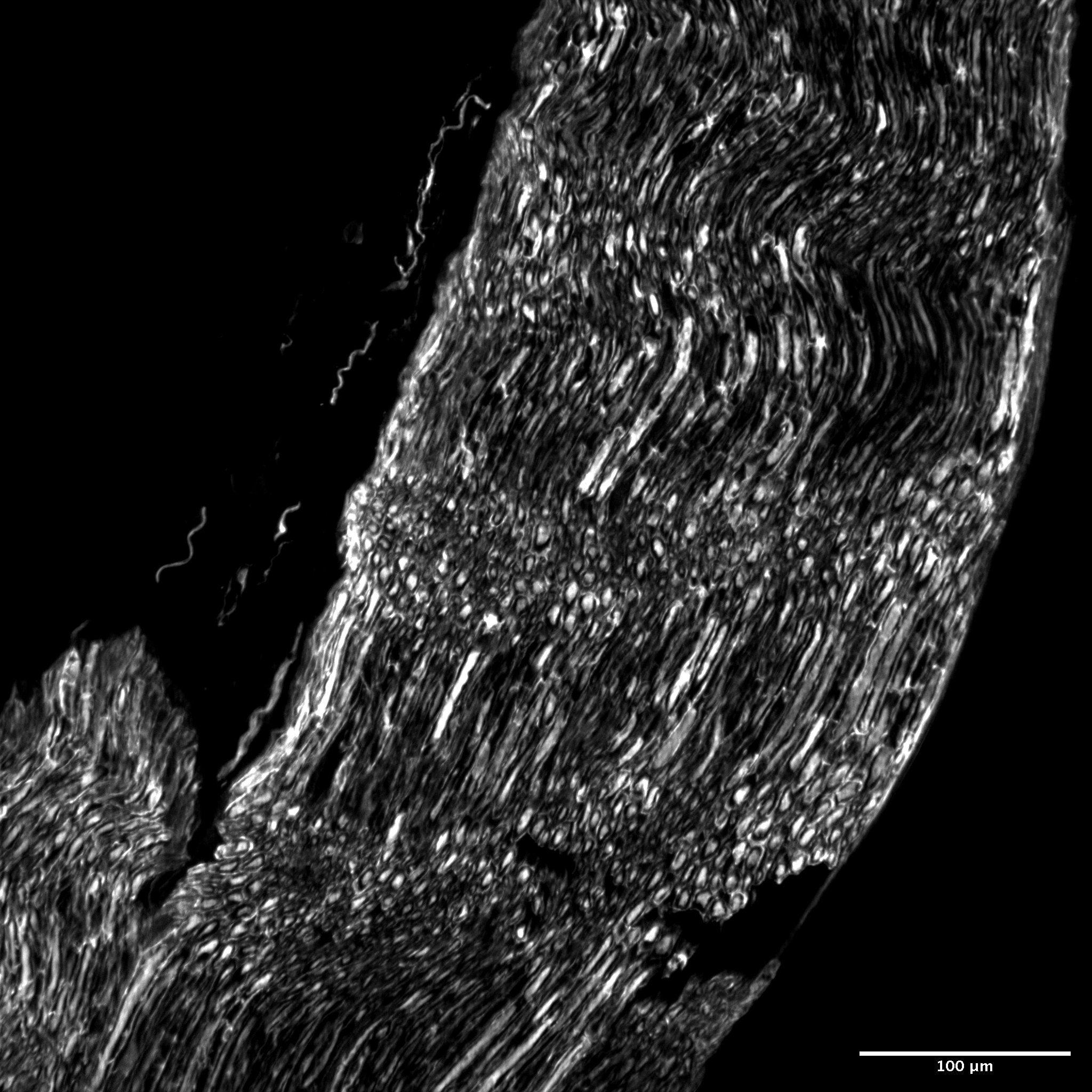
FH Sonia (Verified Customer) (10-21-2019) | Works really well on mouse sciatic nerve cross section (cryostat)
 |

