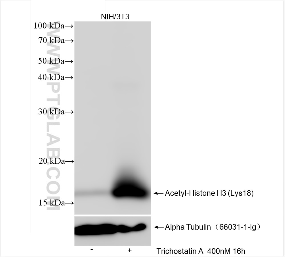
Trichostatin A treated NIH/3T3 cells were subjected to SDS PAGE followed by western blot with 82832-1-RR (Acetyl-Histone H3 (Lys18) antibody) at dilution of 1:10000 incubated at room temperature for 1.5 hours. The membrane was stripped and reblotted with Alpha Tubulin Monoclonal antibody (66031-1-Ig) as loading control.
Trichostatin A treated NIH/3T3 cells were subjected to SDS PAGE followed by western blot with 82832-1-RR (Acetyl-Histone H3 (Lys18) antibody) at dilution of 1:10000 incubated at room temperature for 1.5 hours. The membrane was stripped and reblotted with Alpha Tubulin Monoclonal antibody (66031-1-Ig) as loading control.
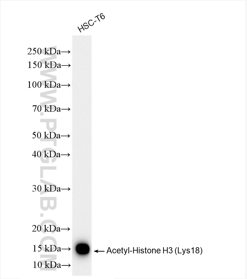
WB analysis of HSC-T6 using 82832-1-RR
HSC-T6 cells were subjected to SDS PAGE followed by western blot with 82832-1-RR (Acetyl-Histone H3 (Lys18) antibody) at dilution of 1:20000 incubated at room temperature for 1.5 hours.
HSC-T6 cells were subjected to SDS PAGE followed by western blot with 82832-1-RR (Acetyl-Histone H3 (Lys18) antibody) at dilution of 1:20000 incubated at room temperature for 1.5 hours.
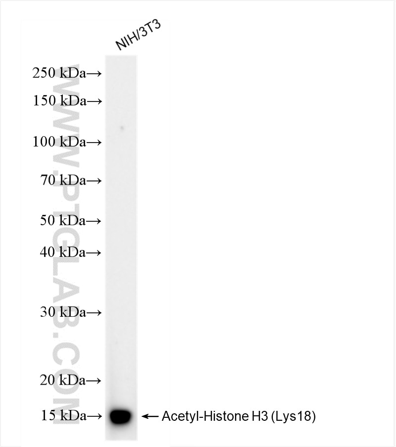
WB analysis of NIH/3T3 using 82832-1-RR
NIH/3T3 cells were subjected to SDS PAGE followed by western blot with 82832-1-RR (Acetyl-Histone H3 (Lys18) antibody) at dilution of 1:20000 incubated at room temperature for 1.5 hours.
NIH/3T3 cells were subjected to SDS PAGE followed by western blot with 82832-1-RR (Acetyl-Histone H3 (Lys18) antibody) at dilution of 1:20000 incubated at room temperature for 1.5 hours.
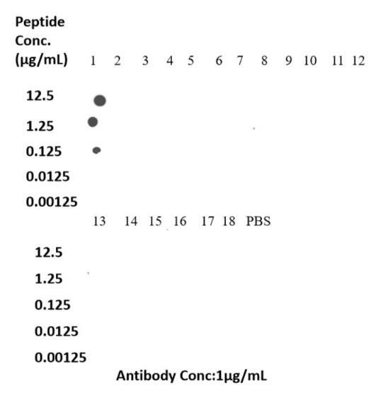
Dot Blot experiment of peptide using 82832-1-RR
Dot blot analysis was used to confirm the specificity of 82832-1-RR Acetyl-Histone H3 (Lys18) antibody. Acetylated peptides were spotted onto NC and probed with antibody at 1 µg/ml.The amount of peptide (μg/mL) spotted is indicated next to each row.
Column 1: H3AK18Ac. Column 2: Unmodified H3AK18. Column 3: H3AK36Ac. Column 4: Unmodified H3AK36. Column 5: H3AK23Ac. Column 6: Unmodified H3AK23. Column 7: H3AK122Ac. Column 8:Unmodified H3AK122. Column 9: H3AK27Ac. Column 10: Unmodified H3AK27. Column 11: H3AK14Ac. Column 12: Unmodified H3AK14. Column 13: H3AK56Ac. Column 14:Unmodified H3AK56. Column 15:H3AK9Ac. Column 16:Unmodified H3AK9. Column 17:H3AK4Ac. Column 18:Unmodified H3AK4. Column 19:Blank(PBS).
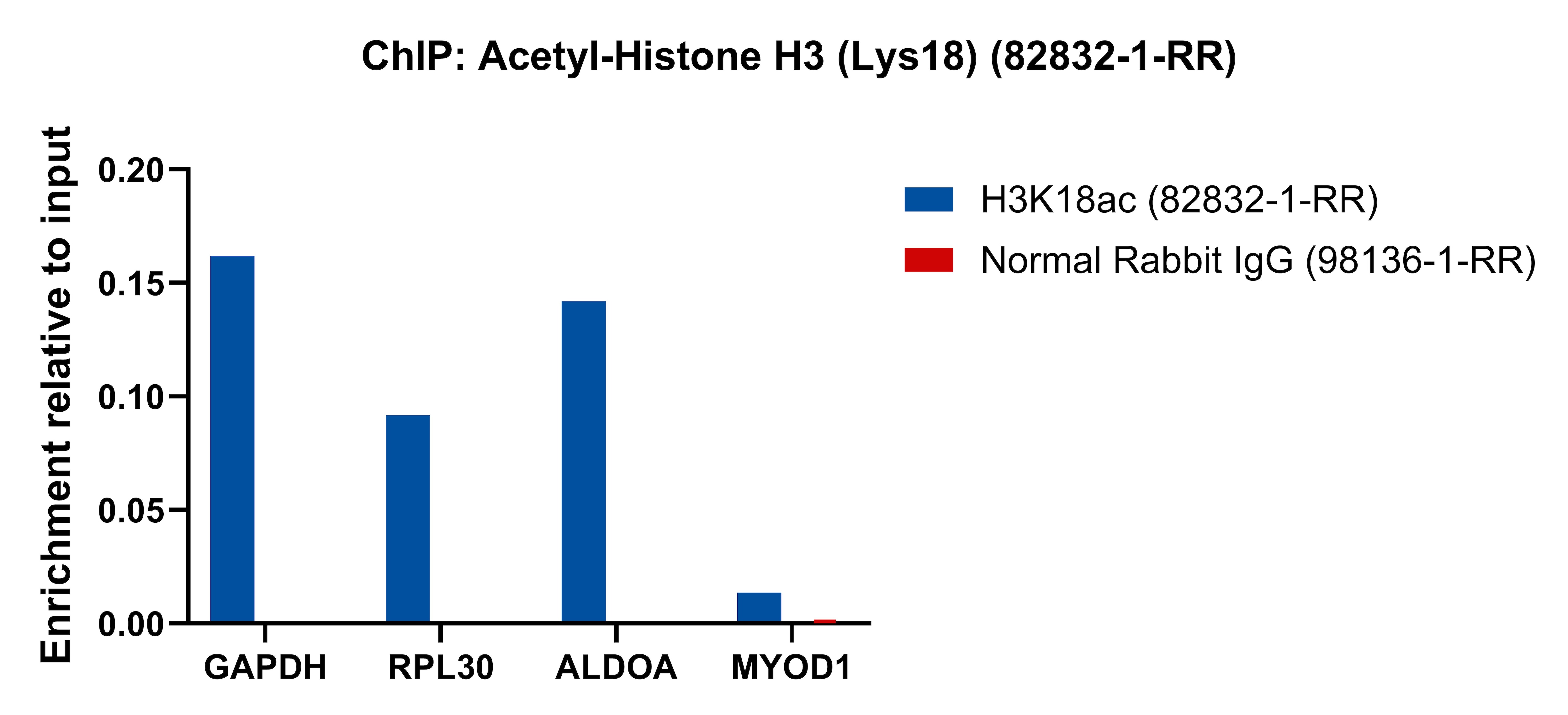
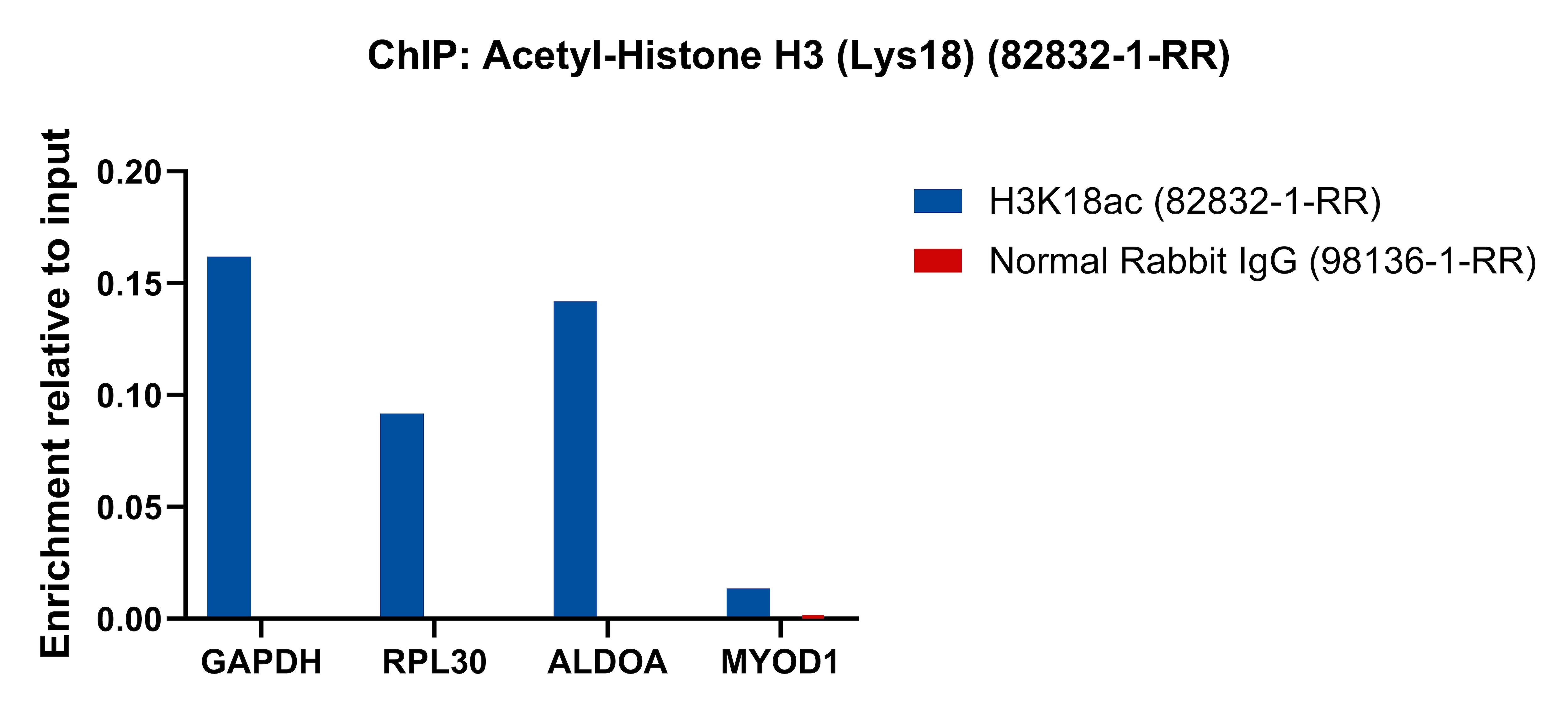
ChIP experiment of HeLa using 82832-1-RR
Chromatin was prepared from HeLa cells. Cells were fixed with formaldehyde for 10 minutes. The ChIP was performed with 15 µg of cross-linked chromatin, 5 µg of Acetyl-Histone H3 (Lys18) (82832-1-RR) or 5 ug of Normal Rabbit IgG (98136-1-RR), and 20 µl of Protein A Magarose Beads. The immunoprecipitated DNA was quantified by real-time PCR.
Chromatin was prepared from HeLa cells. Cells were fixed with formaldehyde for 10 minutes. The ChIP was performed with 15 µg of cross-linked chromatin, 5 µg of Acetyl-Histone H3 (Lys18) (82832-1-RR) or 5 ug of Normal Rabbit IgG (98136-1-RR), and 20 µl of Protein A Magarose Beads. The immunoprecipitated DNA was quantified by real-time PCR.
ChIP experiment of HeLa using 82832-1-RR
Chromatin was prepared from HeLa cells, cells were fixed with formaldehyde for 10 minutes. The ChIP was performed with 15 µg of cross-linked chromatin, 5 µg of Acetyl-Histone H3 (Lys18) (82832-1-RR) or 5 ug of Normal Rabbit IgG (98136-1-RR), and 20 µl of Protein A Magarose Beads. The immunoprecipitated DNA was quantified by real-time PCR.
Chromatin was prepared from HeLa cells, cells were fixed with formaldehyde for 10 minutes. The ChIP was performed with 15 µg of cross-linked chromatin, 5 µg of Acetyl-Histone H3 (Lys18) (82832-1-RR) or 5 ug of Normal Rabbit IgG (98136-1-RR), and 20 µl of Protein A Magarose Beads. The immunoprecipitated DNA was quantified by real-time PCR.
The Proteintech guarantee covers Proteintech antibodies in any species and any application, including those not listed on the datasheet. If the antibody doesn’t perform, you can receive a hassle-free refund or credit note.
PBS with 0.02% sodium azide and 50% glycerol, pH 7.3.
Storage Conditions
Store at -20°C. Stable for one year after shipment. Aliquoting is unnecessary for -20oC storage. 20ul sizes contain 0.1% BSA.
Background Information
Histones, including H1/H5 (linker histones), H2, H3, and H4 (core histones), are nucleic proteins which interact with DNA to form the nucleosomes and play important roles in gene regulation and DNA replication. Histone proteins are highly post-translationally modified while Histone H3 is the most extensively modified.
Protocols
Product Specific Protocols
WB protocol for Acetyl-Histone H3 (Lys18) antibody 82832-1-RR
Chromatin was prepared from HeLa cells, cells were fixed with formaldehyde for 10 minutes. The ChIP was performed with 15 µg of cross-linked chromatin, 5 µg of Acetyl-Histone H3 (Lys18) (82832-1-RR) or 5 ug of Normal Rabbit IgG (98136-1-RR), and 20 µl of Protein A Magarose Beads. The immunoprecipitated DNA was quantified by real-time PCR.
WB Figures
WB analysis of NIH/3T3 using 82832-1-RR
Trichostatin A treated NIH/3T3 cells were subjected to SDS PAGE followed by western blot with 82832-1-RR (Acetyl-Histone H3 (Lys18) antibody) at dilution of 1:10000 incubated at room temperature for 1.5 hours. The membrane was stripped and reblotted with Alpha Tubulin Monoclonal antibody (66031-1-Ig) as loading control.
WB analysis of HSC-T6 using 82832-1-RR
HSC-T6 cells were subjected to SDS PAGE followed by western blot with 82832-1-RR (Acetyl-Histone H3 (Lys18) antibody) at dilution of 1:20000 incubated at room temperature for 1.5 hours.
WB analysis of NIH/3T3 using 82832-1-RR
NIH/3T3 cells were subjected to SDS PAGE followed by western blot with 82832-1-RR (Acetyl-Histone H3 (Lys18) antibody) at dilution of 1:20000 incubated at room temperature for 1.5 hours.
DOT BLOT Figures
Dot Blot experiment of peptide using 82832-1-RR
Dot blot analysis was used to confirm the specificity of 82832-1-RR Acetyl-Histone H3 (Lys18) antibody. Acetylated peptides were spotted onto NC and probed with antibody at 1 µg/ml.The amount of peptide (μg/mL) spotted is indicated next to each row.
Column 1: H3AK18Ac. Column 2: Unmodified H3AK18. Column 3: H3AK36Ac. Column 4: Unmodified H3AK36. Column 5: H3AK23Ac. Column 6: Unmodified H3AK23. Column 7: H3AK122Ac. Column 8:Unmodified H3AK122. Column 9: H3AK27Ac. Column 10: Unmodified H3AK27. Column 11: H3AK14Ac. Column 12: Unmodified H3AK14. Column 13: H3AK56Ac. Column 14:Unmodified H3AK56. Column 15:H3AK9Ac. Column 16:Unmodified H3AK9. Column 17:H3AK4Ac. Column 18:Unmodified H3AK4. Column 19:Blank(PBS).
CHIP-QPCR Figures
ChIP experiment of HeLa using 82832-1-RR
Chromatin was prepared from HeLa cells. Cells were fixed with formaldehyde for 10 minutes. The ChIP was performed with 15 µg of cross-linked chromatin, 5 µg of Acetyl-Histone H3 (Lys18) (82832-1-RR) or 5 ug of Normal Rabbit IgG (98136-1-RR), and 20 µl of Protein A Magarose Beads. The immunoprecipitated DNA was quantified by real-time PCR.
The species listed in Tested Reactivity are in-house verified and applicable species. For unlisted species, please refer to the homology analysis of the immunogen sequence and related species. For rabbit polyclonal antibodies, homology >70% is recommended. For mouse monoclonal antibodies and rabbit recombinant antibodies, homology >90% is recommended. Generally, the higher the homology, the greater the applicability. However, there will be certain differences in protein expression in different species, tissues or cells. Therefore, the homology analysis results are for reference only and do not serve as a guarantee.
At Proteintech, we pride ourselves on our antibody quality, customer service and transparency. As such, we are comparing our antibodies with other vendors, enabling easy identification and comparisons of key data to help you choose the suitable antibody for your needs.
We have selected the top cited antibodies from these vendors for you to compare.
 antibody) at dilution of 1:10000 incubated at room temperature for 1.5 hours. The membrane was stripped and reblotted with Alpha Tubulin Monoclonal antibody (66031-1-Ig) as loading control.")
 antibody) at dilution of 1:20000 incubated at room temperature for 1.5 hours.")
 antibody) at dilution of 1:20000 incubated at room temperature for 1.5 hours.")
 antibody. Acetylated peptides were spotted onto NC and probed with antibody at 1 µg/ml.The amount of peptide (μg/mL) spotted is indicated next to each row.
Column 1: H3AK18Ac. Column 2: Unmodified H3AK18. Column 3: H3AK36Ac. Column 4: Unmodified H3AK36. Column 5: H3AK23Ac. Column 6: Unmodified H3AK23. Column 7: H3AK122Ac. Column 8:Unmodified H3AK122. Column 9: H3AK27Ac. Column 10: Unmodified H3AK27. Column 11: H3AK14Ac. Column 12: Unmodified H3AK14. Column 13: H3AK56Ac. Column 14:Unmodified H3AK56. Column 15:H3AK9Ac. Column 16:Unmodified H3AK9. Column 17:H3AK4Ac. Column 18:Unmodified H3AK4. Column 19:Blank(PBS).")
 (82832-1-RR) or 5 ug of Normal Rabbit IgG (98136-1-RR), and 20 µl of Protein A Magarose Beads. The immunoprecipitated DNA was quantified by real-time PCR.")
 (82832-1-RR) or 5 ug of Normal Rabbit IgG (98136-1-RR), and 20 µl of Protein A Magarose Beads. The immunoprecipitated DNA was quantified by real-time PCR.")