
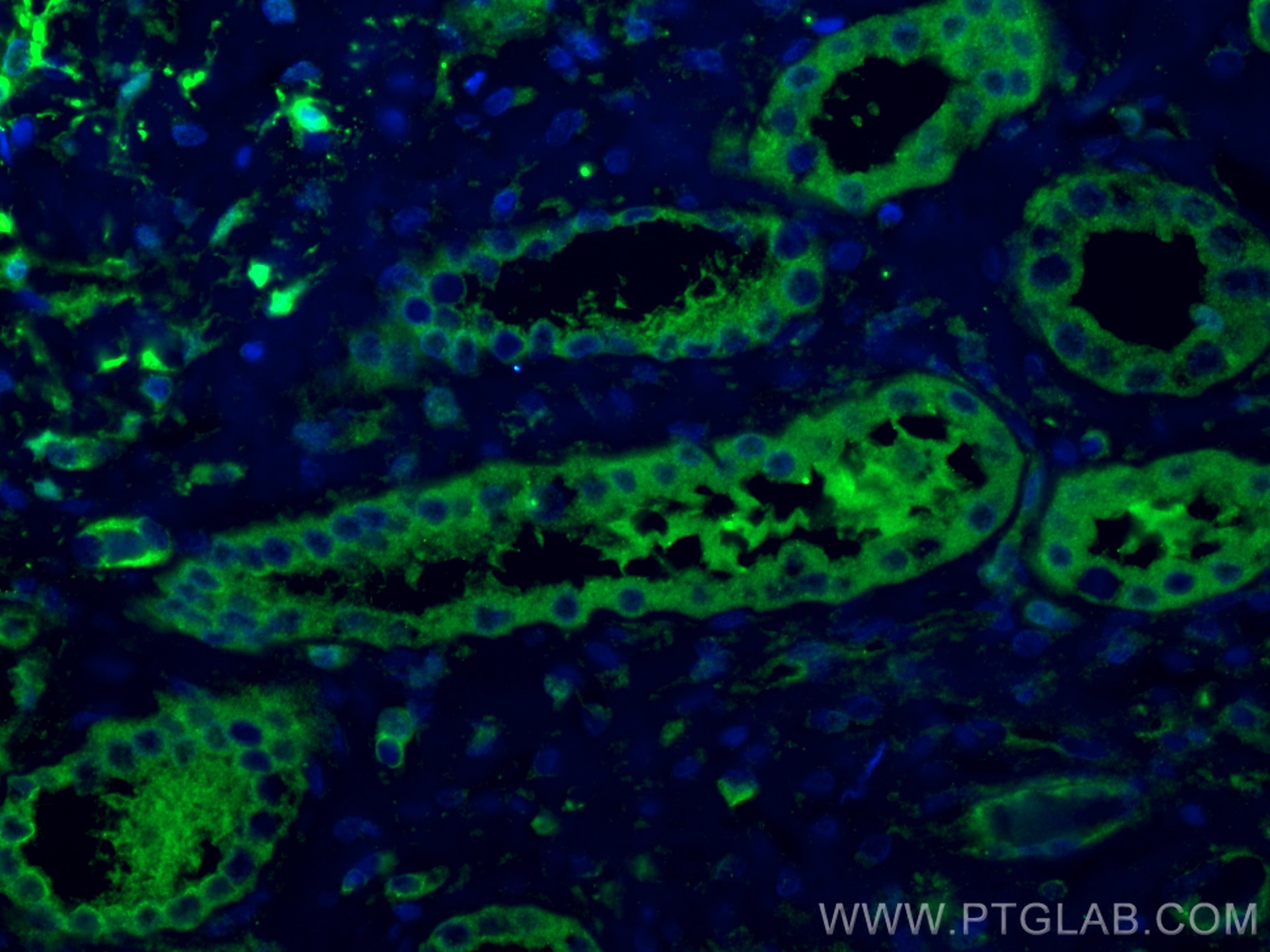

Tested Applications
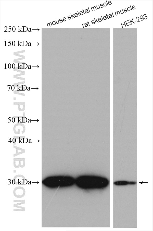
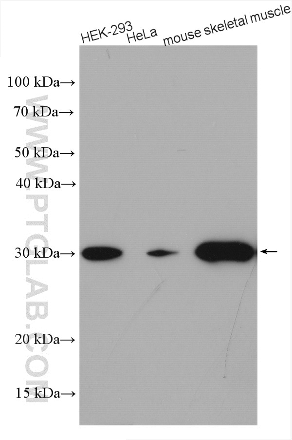
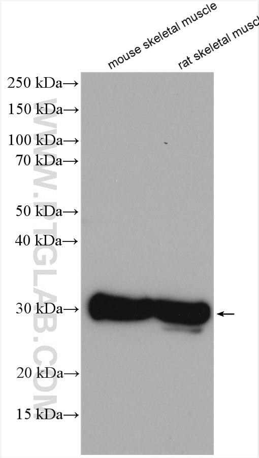
| Positive WB detected in | HEK-293 cells, mouse skeletal muscle tissue, rat skeletal muscle tissue, HeLa cells |
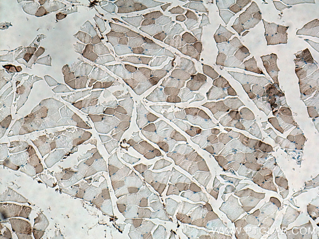
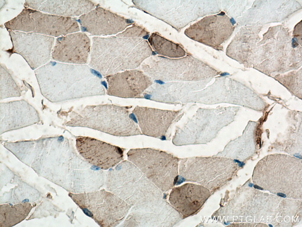
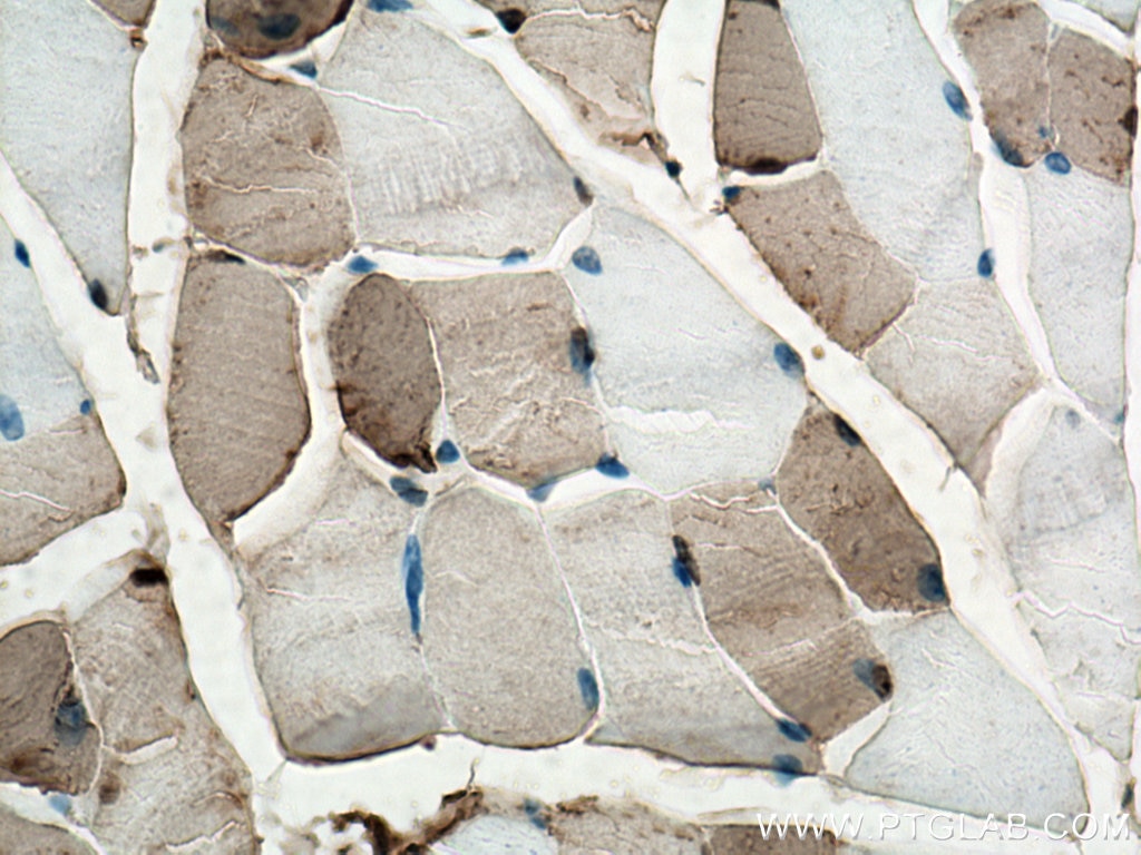
| Positive IHC detected in | mouse skeletal muscle tissue Note: suggested antigen retrieval with TE buffer pH 9.0; (*) Alternatively, antigen retrieval may be performed with citrate buffer pH 6.0 |
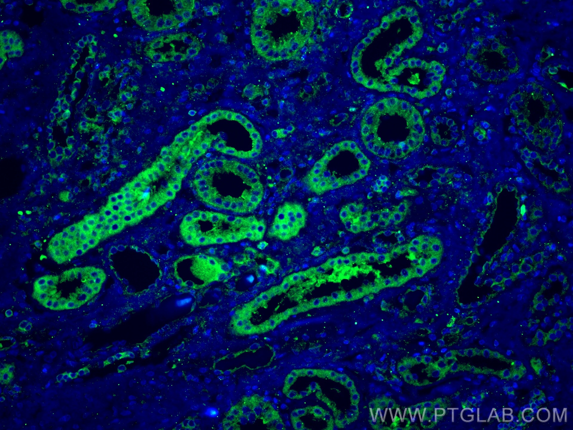
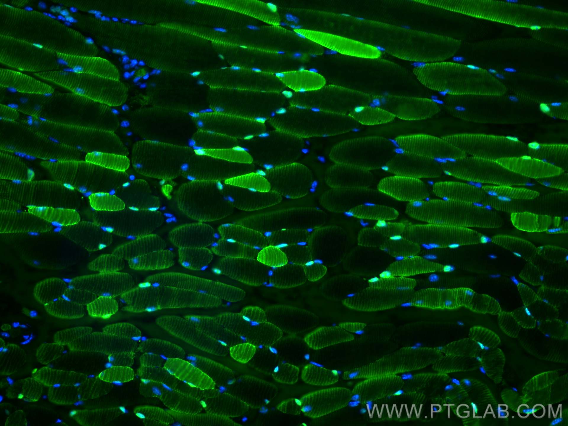
| Positive IF-P detected in | mouse skeletal muscle tissue, human kidney tissue |
Recommended dilution
| Application | Dilution |
|---|---|
| Western Blot (WB) | WB : 1:2000-1:16000 |
| Immunohistochemistry (IHC) | IHC : 1:200-1:1000 |
| Immunofluorescence (IF)-P | IF-P : 1:50-1:500 |
| It is recommended that this reagent should be titrated in each testing system to obtain optimal results. | |
| Sample-dependent, Check data in validation data gallery. | |
Product Information
15197-1-AP targets CA3 in WB, IHC, IF-P, ELISA applications and shows reactivity with human, mouse, rat samples.
| Tested Reactivity | human, mouse, rat |
| Cited Reactivity | human, mouse, rat |
| Host / Isotype | Rabbit / IgG |
| Class | Polyclonal |
| Type | Antibody |
| Immunogen |
CatNo: Ag7344 Product name: Recombinant human CA3 protein Source: e coli.-derived, PGEX-4T Tag: GST Domain: 1-260 aa of BC004897 Sequence: MAKEWGYASHNGPDHWHELFPNAKGENQSPIELHTKDIRHDPSLQPWSVSYDGGSAKTILNNGKTCRVVFDDTYDRSMLRGGPLPGPYRLRQFHLHWGSSDDHGSEHTVDGVKYAAELHLVHWNPKYNTFKEALKQRDGIAVIGIFLKIGHENGEFQIFLDALDKIKTKGKEAPFTKFDPSCLFPACRDYWTYQGSFTTPPCEECIVWLLLKEPMTVSSDQMAKLRSLLSSAENEPPVPLVSNWRPPQPINNRVVRASFK Predict reactive species |
| Full Name | carbonic anhydrase III, muscle specific |
| Calculated Molecular Weight | 29 kDa |
| Observed Molecular Weight | 30 kDa |
| GenBank Accession Number | BC004897 |
| Gene Symbol | CA3 |
| Gene ID (NCBI) | 761 |
| RRID | AB_2243603 |
| Conjugate | Unconjugated |
| Form | Liquid |
| Purification Method | Antigen affinity purification |
| UNIPROT ID | P07451 |
| Storage Buffer | PBS with 0.02% sodium azide and 50% glycerol, pH 7.3. |
| Storage Conditions | Store at -20°C. Stable for one year after shipment. Aliquoting is unnecessary for -20oC storage. 20ul sizes contain 0.1% BSA. |
Background Information
Carbonic anhydrase III (CA3), which belongs to the alpha-carbonic anhydrase family, is a cytoplasmic enzyme that exhibits a relatively low carbon dioxide hydratase activity. It is expressed at a very high level in skeletal muscle, where physical exercise has been shown to increase free radical production. In addition to its carbon dioxide hydratase activity, CA3 has been demonstrated to have a carboxyl esterase activity and phosphatase activity, which suggests that it is a tyrosine phosphatase (PMID: 10064618). CA3 was found to be localized in Type-I muscle fibers and could be used as a marker for abnormal Type-I muscle fibers in several neuromuscular diseases (PMID: 6221502).
Protocols
| Product Specific Protocols | |
|---|---|
| IF protocol for CA3 antibody 15197-1-AP | Download protocol |
| IHC protocol for CA3 antibody 15197-1-AP | Download protocol |
| WB protocol for CA3 antibody 15197-1-AP | Download protocol |
| Standard Protocols | |
|---|---|
| Click here to view our Standard Protocols |
Publications
What published studies show
The antibody is Carbonic Anhydrase 3 (CA3), catalog number 15197-1-AP, with a total of 13 citations. These appear in journals including Nature Communications, International Journal of Oral Science, Genome Medicine, Food Chemistry, Journal of Nutritional Biochemistry, iScience, Clinical Proteomics, American Journal of Physiology–Renal Physiology, PLoS ONE, FEBS Open Bio, and Aging. Research fields span intervertebral disc repair, bone formation, cornea aging, muscle proteomics, hypertension, otic mesenchyme, muscular dystrophy biomarkers, cystitis, bladder fibroblasts, tooth development, and liver adipogenesis.
| Species | Application | Title |
|---|---|---|
Nat Commun Targeting the ROS-ferroptosis-inflammation cycle with a nanozyme-functionalized hydrogel for intervertebral disc repair. | ||
Int J Oral Sci Osteoblast-derived CAR3 synergizing with collagen and bone sialoprotein enhances bone formation.
| ||
Genome Med Spatiotemporal single-cell analysis elucidates the cellular and molecular dynamics of human cornea aging | ||
Food Chem Integrative transcriptomic, proteomic and metabolomic analyses yields insights into muscle fiber type in cattle | ||
J Nutr Biochem Postnatal high-fat diet sex-specifically exacerbates prenatal dexamethasone-induced hypertension: Mass spectrometry-based quantitative proteomic approach. | ||
iScience Spatially distinct otic mesenchyme cells show molecular and functional heterogeneity patterns before hearing onset |
Reviews
What customers say
The single review for this product reports a negative experience. The reviewer tested it for immunofluorescence on kidney tissue at a 1:500 dilution, using 5% blocking serum and 1% BSA, and found it did not work. They noted plans to retest at a 1:150 dilution and to evaluate it for IHC and WB. Overall, the feedback is critical, with a low rating reflecting the failed IF result.
The reviews below have been submitted by verified Proteintech customers who received an incentive for providing their feedback.
Elise (Verified Customer) (06-21-2022) | Do not Work for IF with blocking serum 5% and antibody in BSA 1%. Will be tested on IHC and WB and Tested again in IF with 1: 150 dilution
|










