Tested Applications
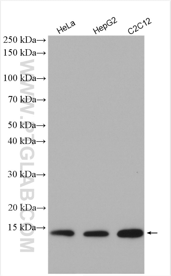
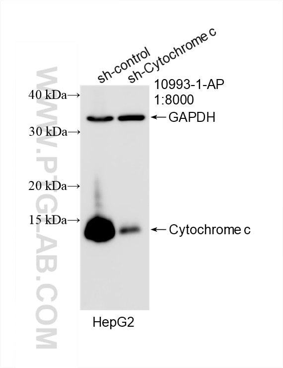
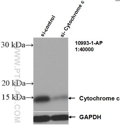
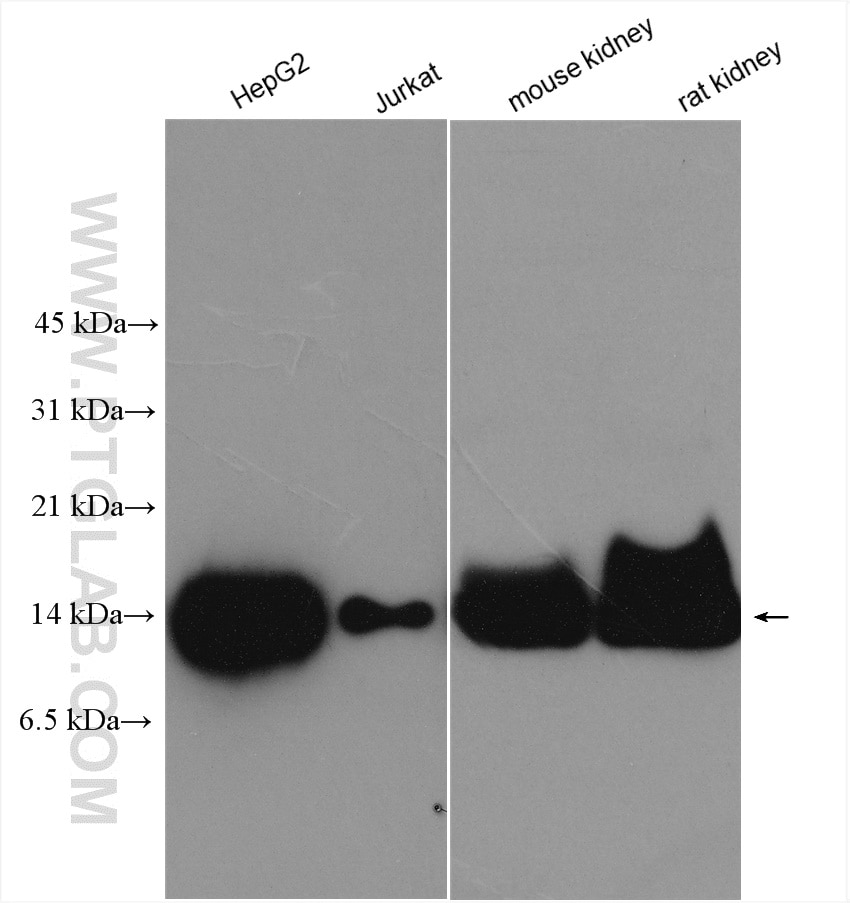
| Positive WB detected in | HeLa cells, mouse skeletal muscle tissue, rat skeletal muscle tissue, NIH/3T3 cells, rat liver tissue, HEK-293 cells, HepG2 cells, C2C12 cells, Jurkat cells, mouse kidney tissue, rat kidney tissue |
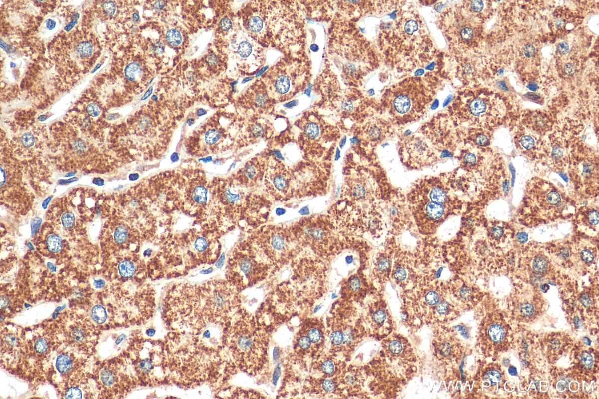
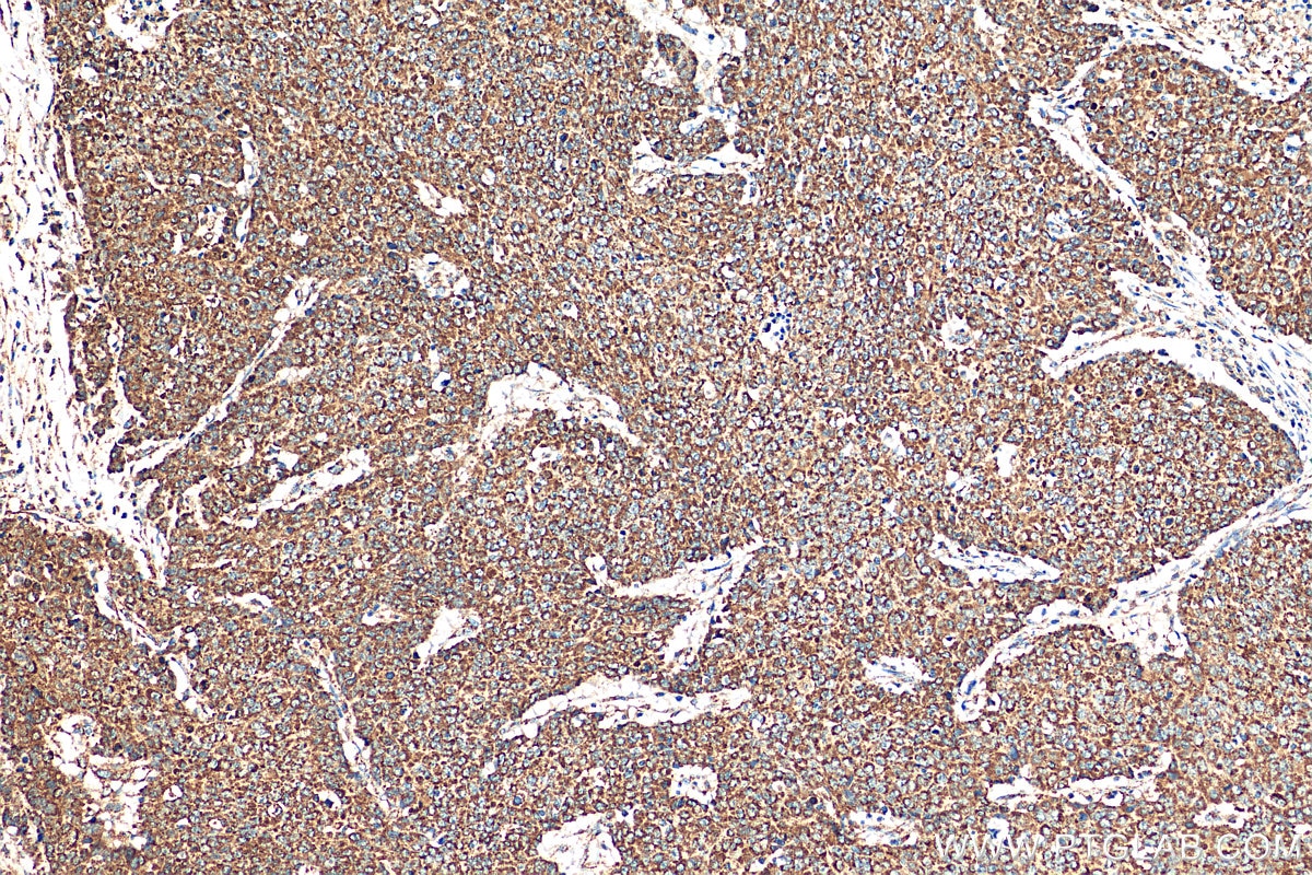
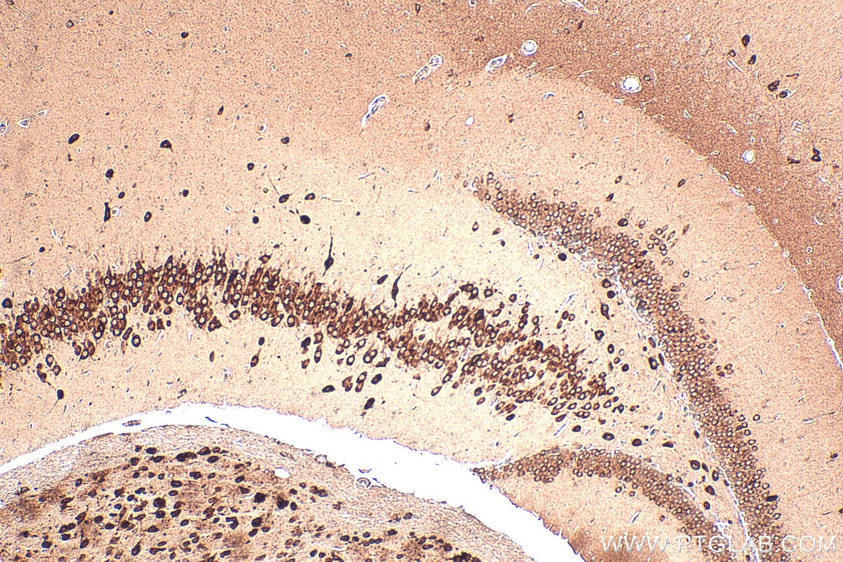
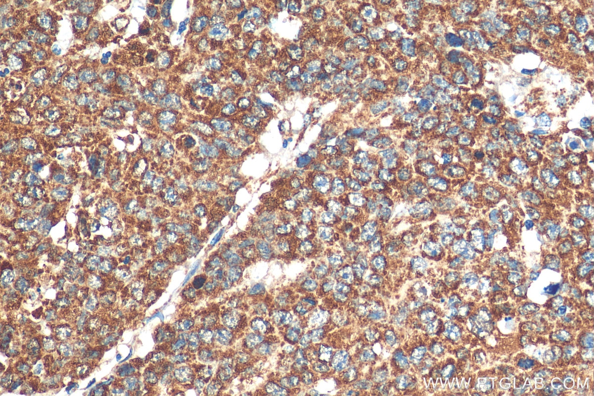
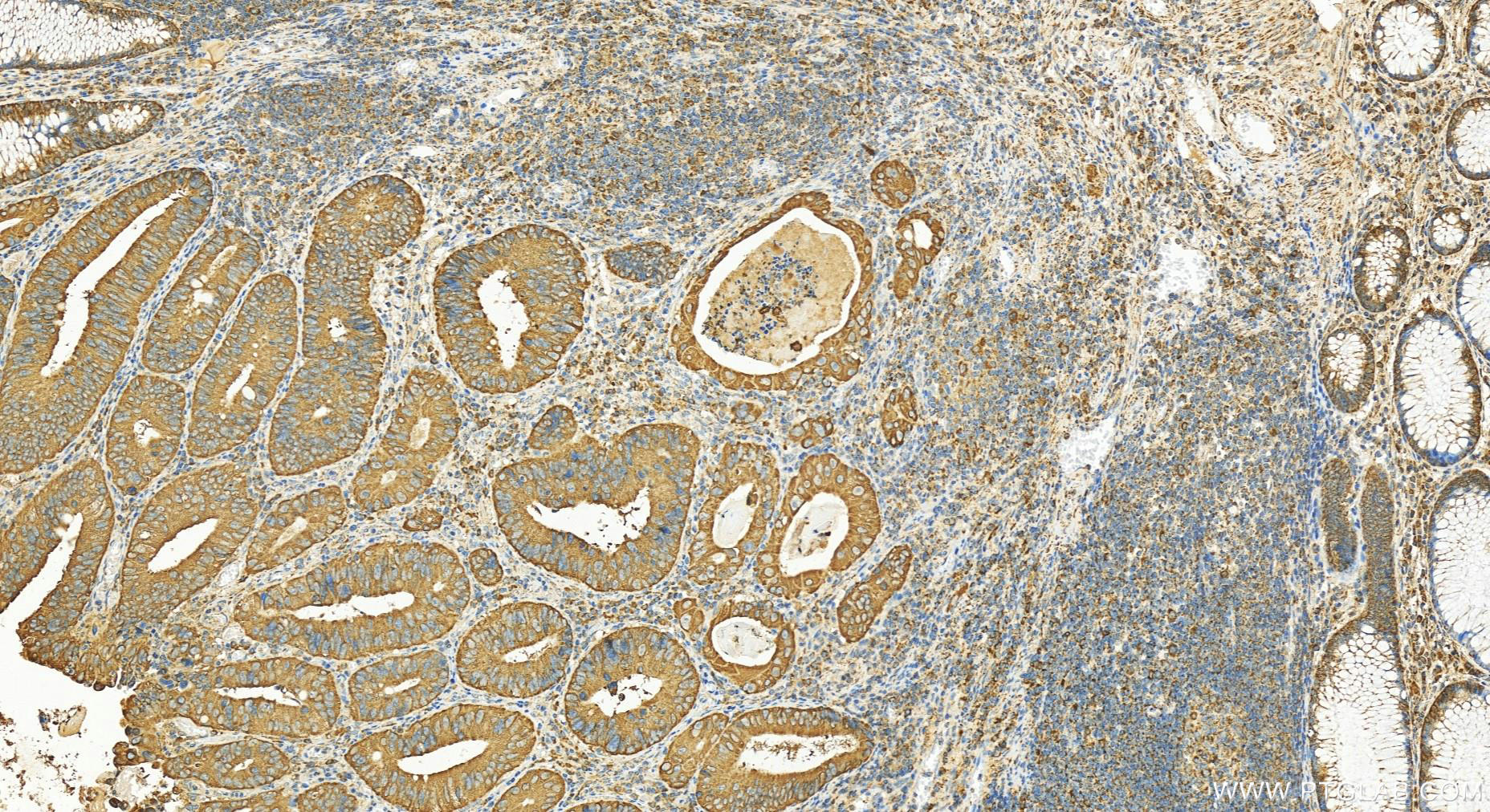
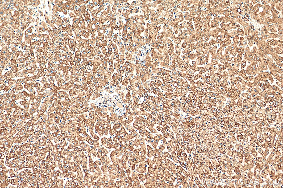
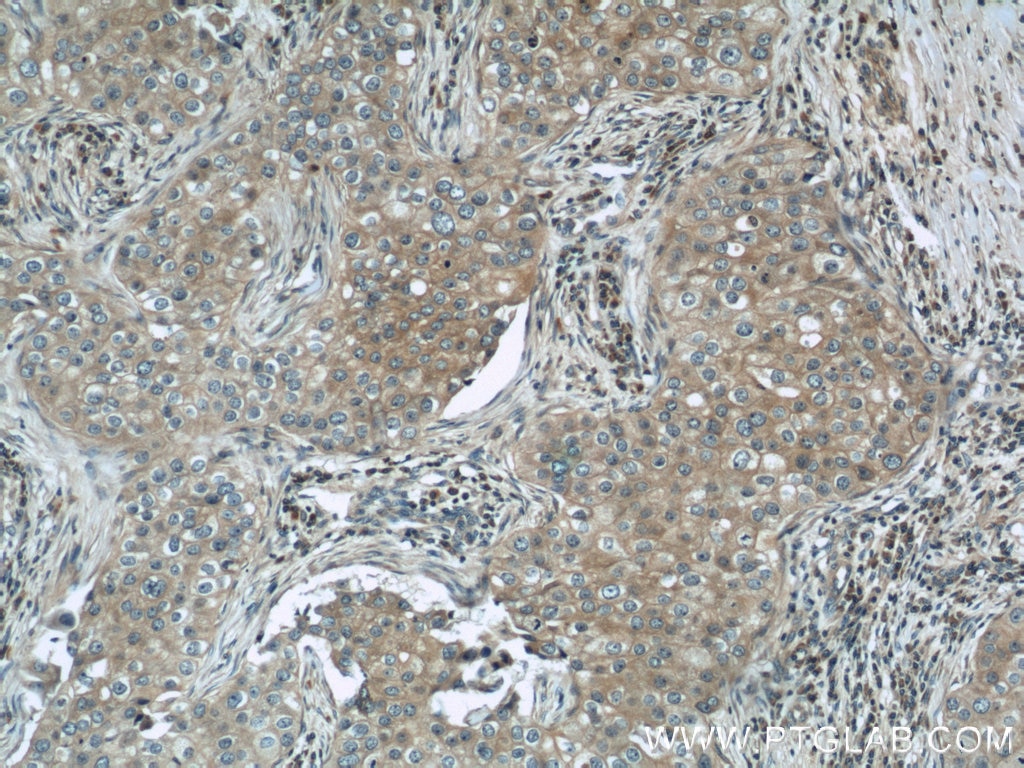
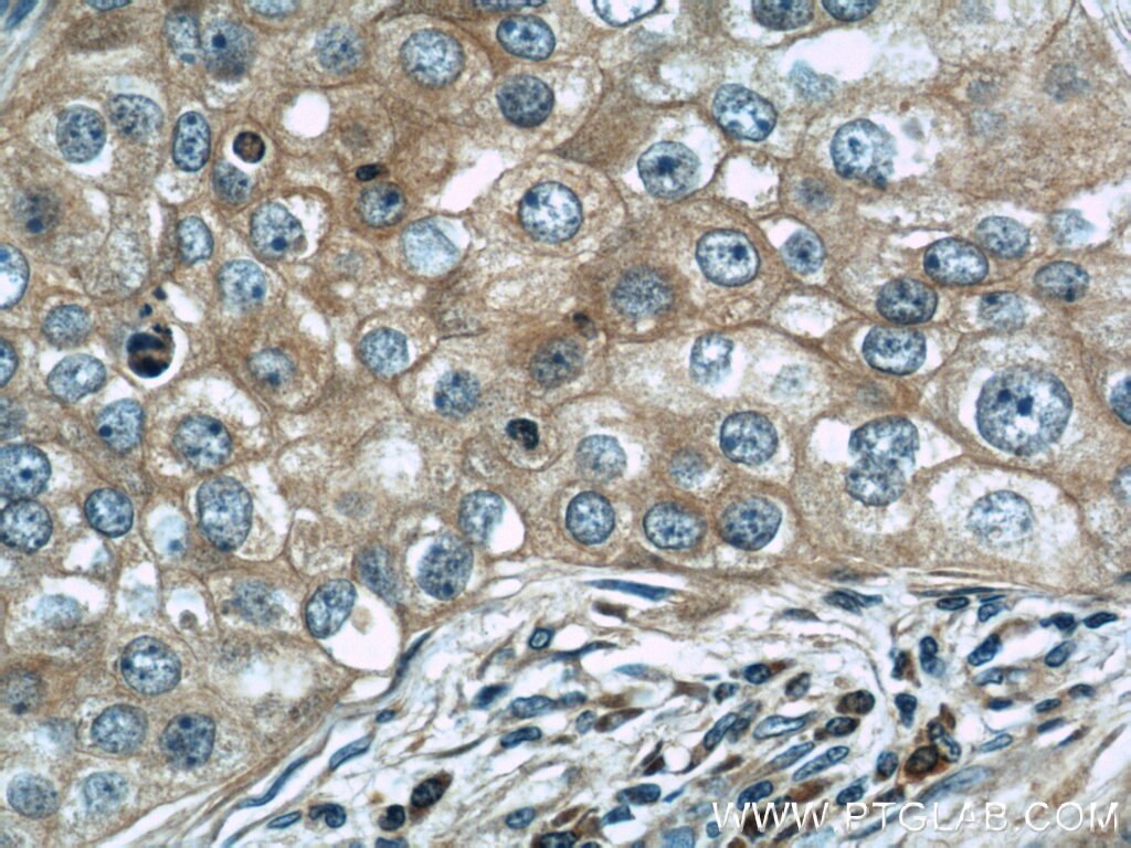
| Positive IHC detected in | human liver tissue, human breast cancer tissue, human colon cancer tissue, mouse brain tissue Note: suggested antigen retrieval with TE buffer pH 9.0; (*) Alternatively, antigen retrieval may be performed with citrate buffer pH 6.0 |
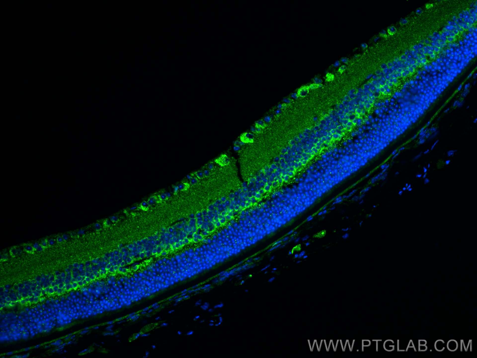
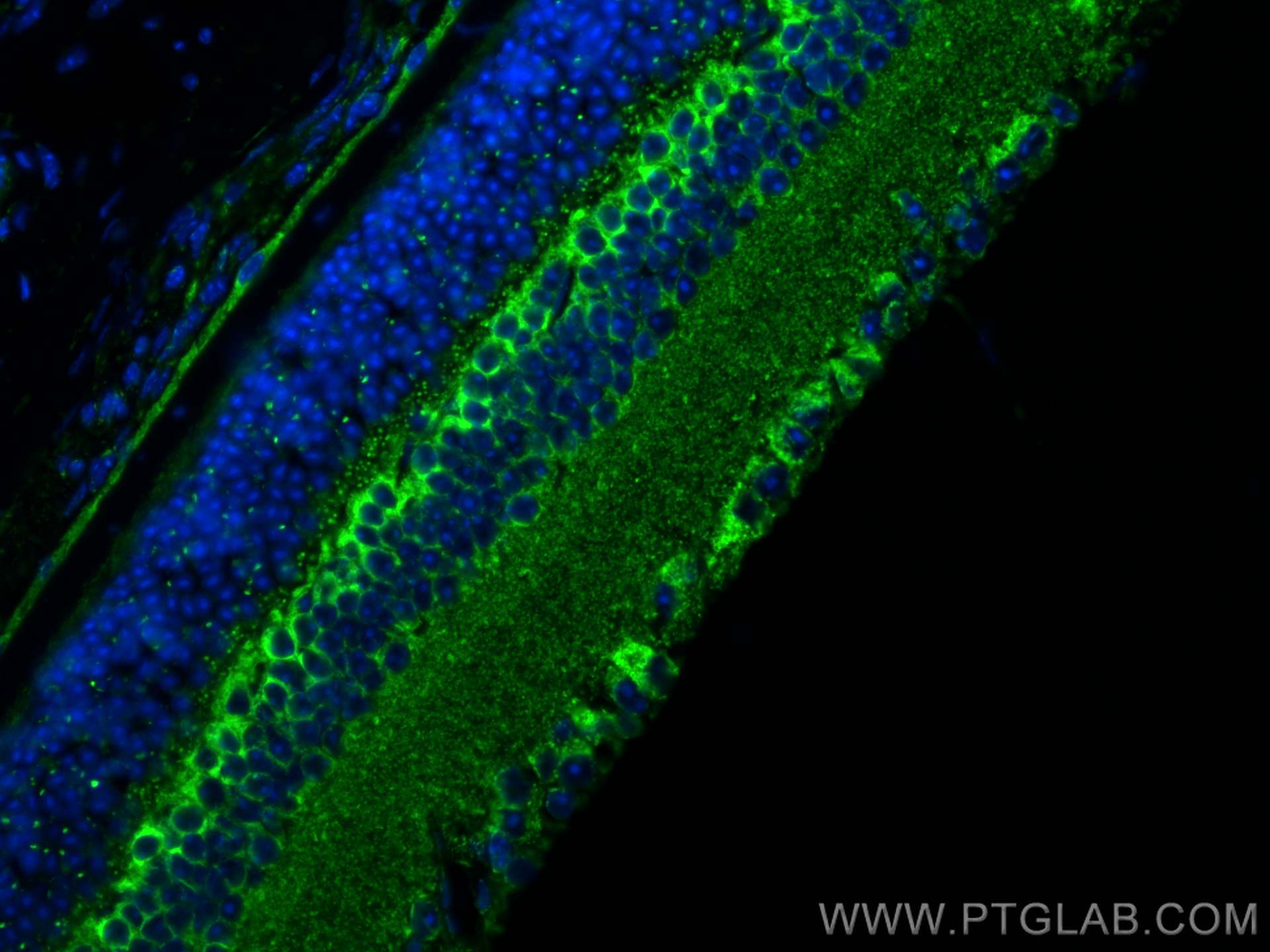
| Positive IF-P detected in | mouse eye tissue |
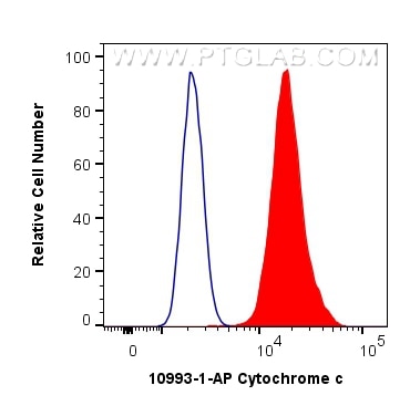
| Positive FC (Intra) detected in | HepG2 cells |
This antibody is not recommended for immunocytofluorescent assays.
Recommended dilution
| Application | Dilution |
|---|---|
| Western Blot (WB) | WB : 1:1000-1:8000 |
| Immunohistochemistry (IHC) | IHC : 1:500-1:2000 |
| Immunofluorescence (IF)-P | IF-P : 1:50-1:500 |
| Flow Cytometry (FC) (INTRA) | FC (INTRA) : 0.40 ug per 10^6 cells in a 100 µl suspension |
| It is recommended that this reagent should be titrated in each testing system to obtain optimal results. | |
| Sample-dependent, Check data in validation data gallery. | |
Published Applications
| WB | See 650 publications below |
| IHC | See 37 publications below |
| IF | See 47 publications below |
| IP | See 1 publications below |
Product Information
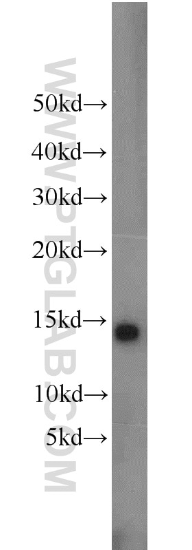
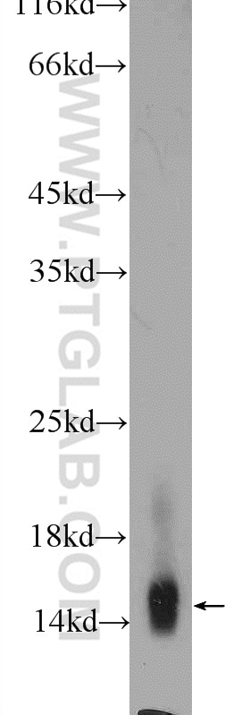
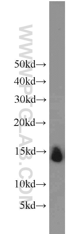
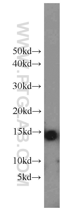
10993-1-AP targets Cytochrome c in WB, IHC, IF-P, FC (Intra), IP, ELISA applications and shows reactivity with human, mouse, rat samples.
| Tested Reactivity | human, mouse, rat |
| Cited Reactivity | human, mouse, rat, monkey, chicken, hamster, sheep, goat, hippospongia |
| Host / Isotype | Rabbit / IgG |
| Class | Polyclonal |
| Type | Antibody |
| Immunogen |
CatNo: Ag1455 Product name: Recombinant human Cytochrome c protein Source: e coli.-derived, PGEX-4T Tag: GST Domain: 1-105 aa of BC009578 Sequence: MGDVEKGKKIFIMKCSQCHTVEKGGKHKTGPNLHGLFGRKTGQAPGYSYTAANKNKGIIWGEDTLMEYLENPKKYIPGTKMIFVGIKKKEERADLIAYLKKATNE Predict reactive species |
| Full Name | cytochrome c, somatic |
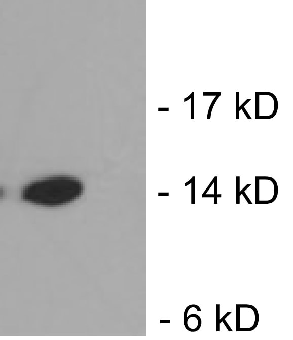
| Calculated Molecular Weight | 12 kDa |
| Observed Molecular Weight | 12-15 kDa |
| GenBank Accession Number | BC009578 |
| Gene Symbol | Cytochrome c |
| Gene ID (NCBI) | 54205 |
| RRID | AB_2090467 |
| Conjugate | Unconjugated |
| Form | Liquid |
| Purification Method | Antigen affinity purification |
| UNIPROT ID | P99999 |
| Storage Buffer | PBS with 0.02% sodium azide and 50% glycerol, pH 7.3. |
| Storage Conditions | Store at -20°C. Stable for one year after shipment. Aliquoting is unnecessary for -20oC storage. 20ul sizes contain 0.1% BSA. |
Background Information
Cytochrome c is a 12-15 kDa electron transporting protein located in the inner mitochondrial membrane. As a part of respiratory chain, cytochrome c plays a critical role in the process of oxidative phosphorylation and ATP producing. Besides, cytochrome c also gets implicated in apoptosis process. Upon apoptotic stimulation, cytochrome c ca99n be released from mitochondria into cytoplasm, which is required for caspase-3 activation and the occurrence of apoptosis.
Protocols
| Product Specific Protocols | |
|---|---|
| FC protocol for Cytochrome c antibody 10993-1-AP | Download protocol |
| IF protocol for Cytochrome c antibody 10993-1-AP | Download protocol |
| IHC protocol for Cytochrome c antibody 10993-1-AP | Download protocol |
| WB protocol for Cytochrome c antibody 10993-1-AP | Download protocol |
| Standard Protocols | |
|---|---|
| Click here to view our Standard Protocols |
Publications
| Species | Application | Title |
|---|---|---|
Cell Tau interactome maps synaptic and mitochondrial processes associated with neurodegeneration. | ||
Nat Commun MYG1 drives glycolysis and colorectal cancer development through nuclear-mitochondrial collaboration | ||
Mol Cell Filamentous GLS1 promotes ROS-induced apoptosis upon glutamine deprivation via insufficient asparagine synthesis. | ||
Cell Death Differ TRIM32 promotes anoikis resistance and metastasis in NSCLC by degrading CHEK2 to enhance IL-6 secretion | ||
Adv Sci (Weinh) Hierarchical Targeting Nanodrug with Holistic DNA Protection for Effective Treatment of Acute Kidney Injury | ||
Adv Sci (Weinh) Mitochondrial tRNAGlu 14693A>G Mutation, an "Enhancer" to the Phenotypic Expression of Leber's Hereditary Optic Neuropathy |
Reviews
The reviews below have been submitted by verified Proteintech customers who received an incentive for providing their feedback.
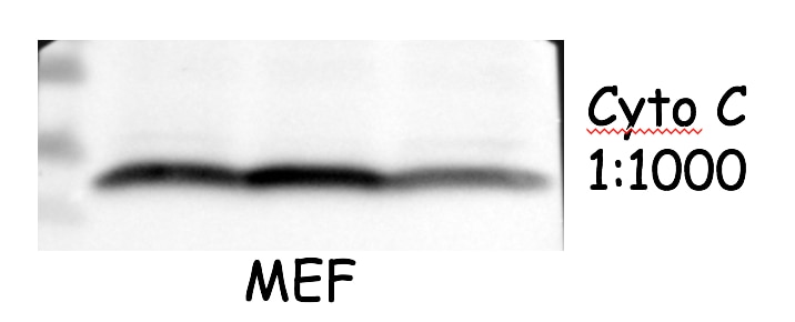
FH Lucas (Verified Customer) (10-31-2025) | Successfully used in Western blot on mitochondrial fractions from mouse muscle. Produced strong and specific bands at a 1:1000 dilution.
|
FH Kazu (Verified Customer) (12-14-2022) | Worked well for 4% PFA fixed mouse optic nerve using this antibody at dilution of 1:200. Little background observed.
|
FH Azita (Verified Customer) (06-02-2021) | Western blot analysis using Cytochrome c polyclonal antibody in NSC34 cell line at dilution of 1:1000.
|
FH Ying (Verified Customer) (04-22-2021) | good antibody. work well with mouse cells
 |
FH Chi (Verified Customer) (09-26-2019) | The antibody works very well with good signal and low background.
 |
FH Xiaoping (Verified Customer) (10-22-2018) | The signal is good and the size is between 10-15kDa.
 |