Tested Applications
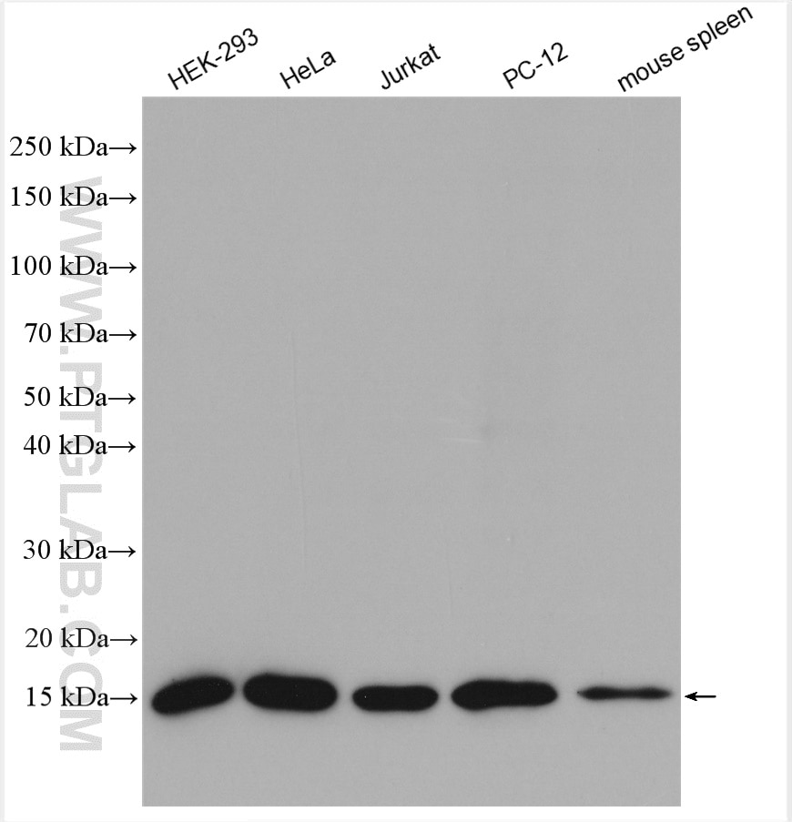
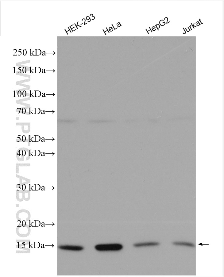
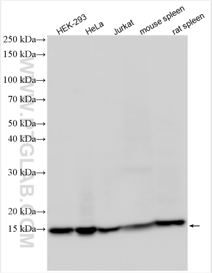
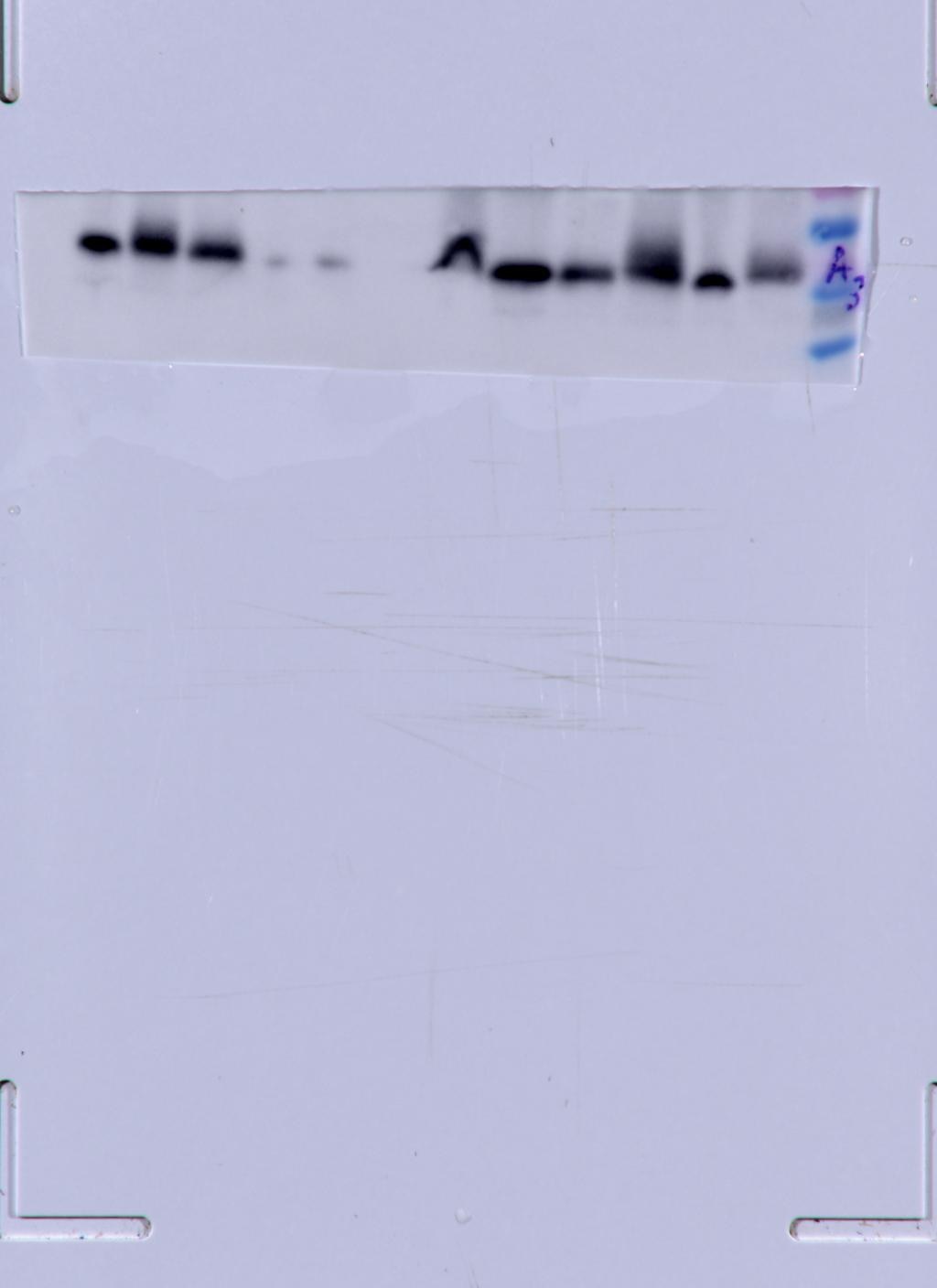
| Positive WB detected in | HEK-293 cells, HeLa cells, mouse brain tissue, mouse heart tissue, pig brain tissue, rat brain tissue, rat heart tissue, HepG2 cells, Jurkat cells, PC-12 cells, mouse spleen tissue, rat spleen tissue |
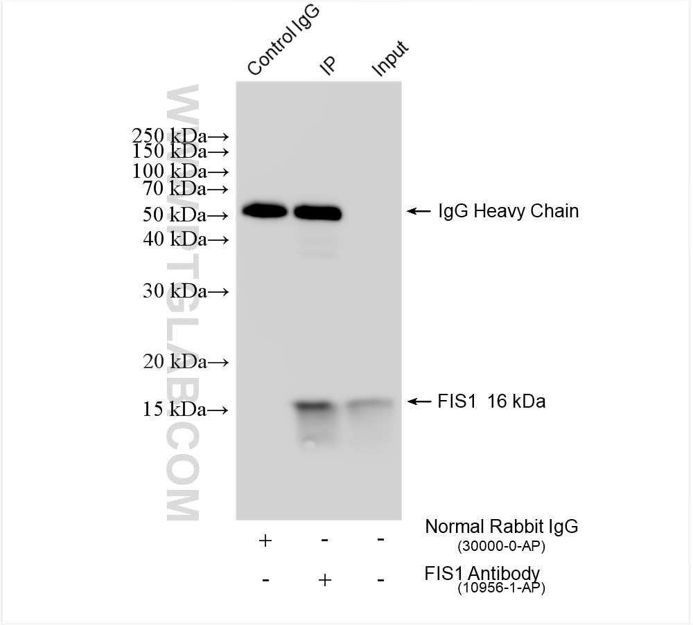
| Positive IP detected in | HeLa cells |
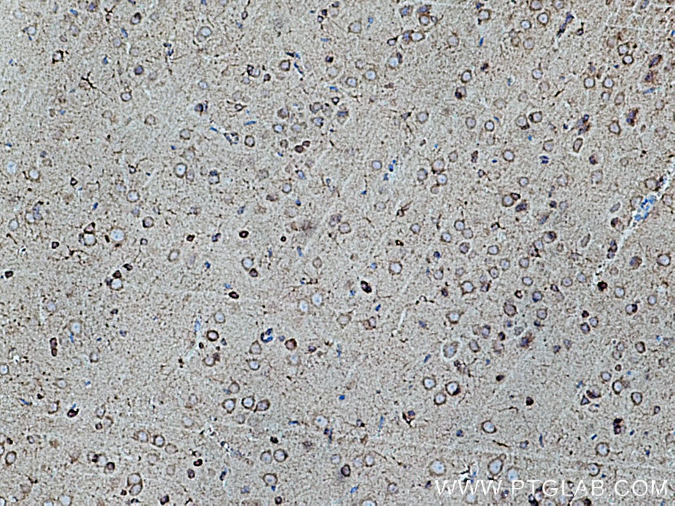
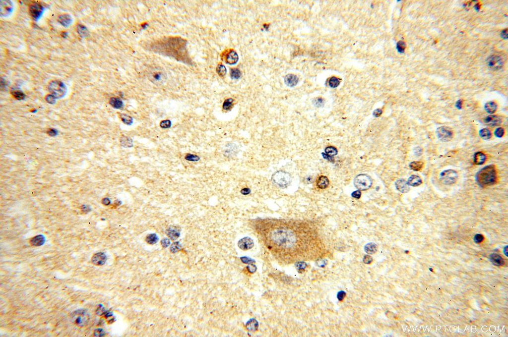
| Positive IHC detected in | rat brain tissue, human brain tissue Note: suggested antigen retrieval with TE buffer pH 9.0; (*) Alternatively, antigen retrieval may be performed with citrate buffer pH 6.0 |
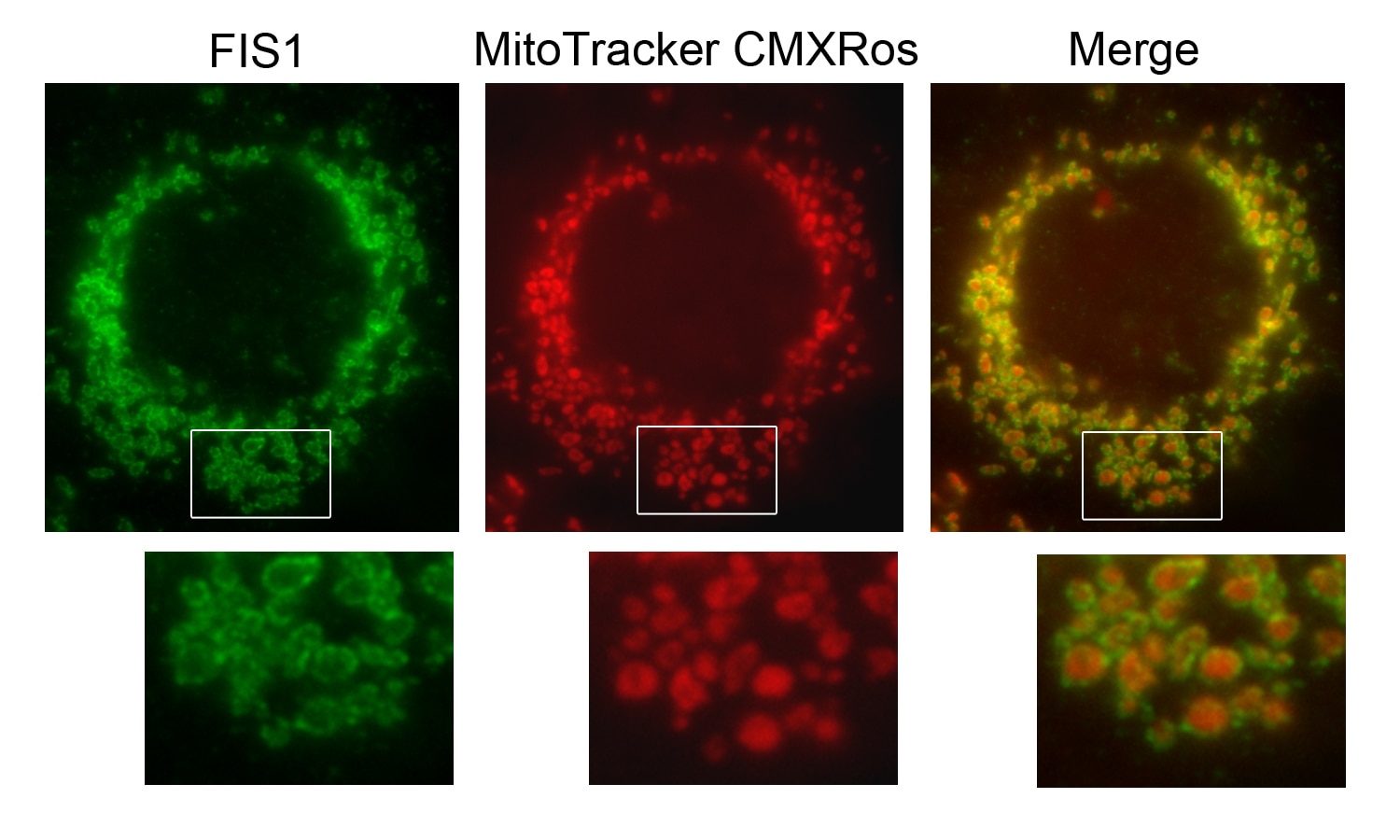
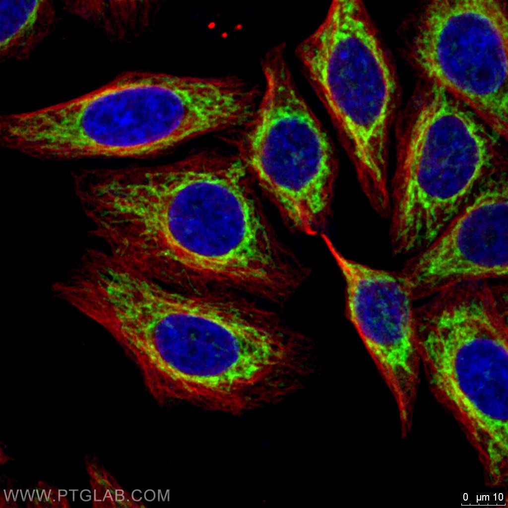
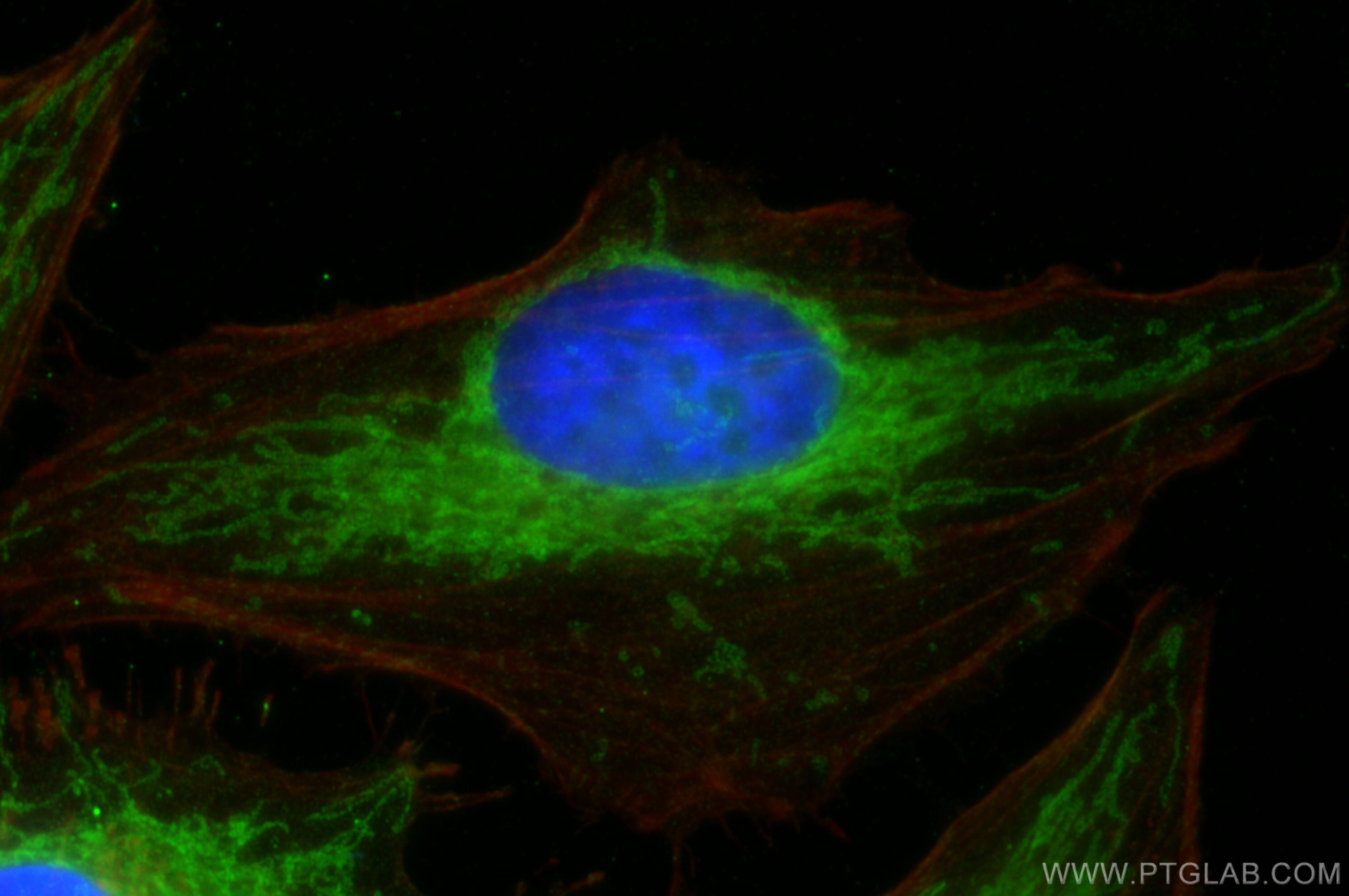
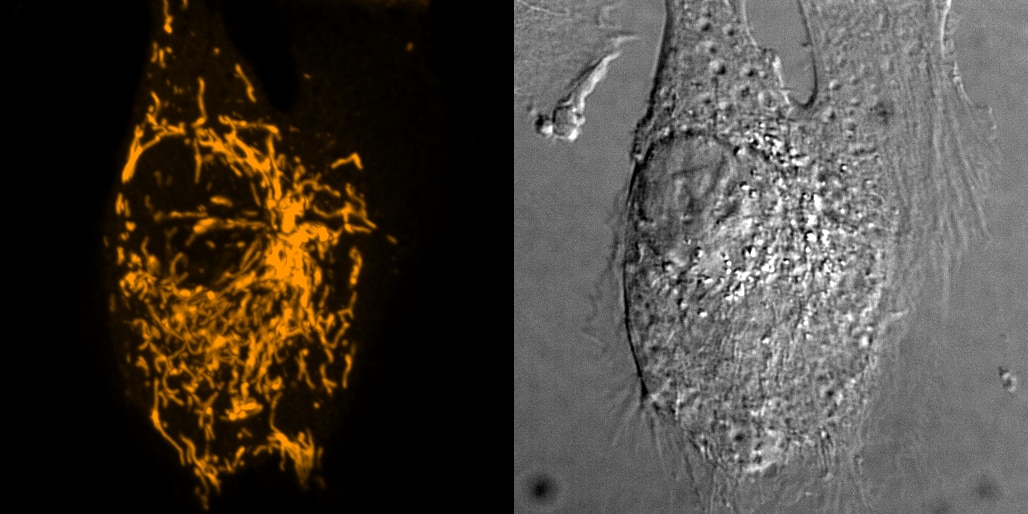
| Positive IF/ICC detected in | HeLa cells, Hepa1-6 cells, HepG2 cells |
Recommended dilution
| Application | Dilution |
|---|---|
| Western Blot (WB) | WB : 1:2000-1:14000 |
| Immunoprecipitation (IP) | IP : 0.5-4.0 ug for 1.0-3.0 mg of total protein lysate |
| Immunohistochemistry (IHC) | IHC : 1:100-1:800 |
| Immunofluorescence (IF)/ICC | IF/ICC : 1:200-1:800 |
| It is recommended that this reagent should be titrated in each testing system to obtain optimal results. | |
| Sample-dependent, Check data in validation data gallery. | |
Published Applications
| KD/KO | See 18 publications below |
| WB | See 444 publications below |
| IHC | See 20 publications below |
| IF | See 51 publications below |
| IP | See 6 publications below |
| CoIP | See 3 publications below |
Product Information
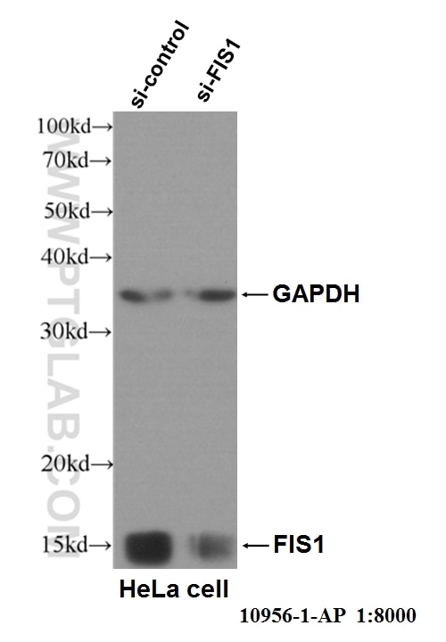
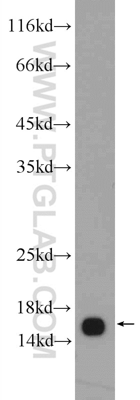
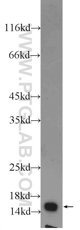
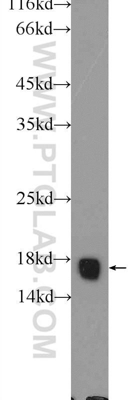
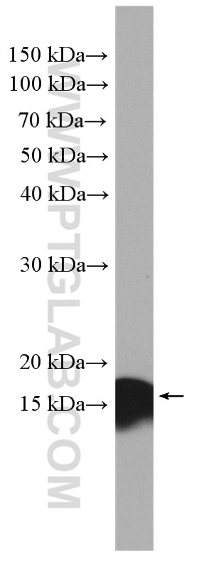
10956-1-AP targets FIS1 in WB, IHC, IF/ICC, IP, CoIP, ELISA applications and shows reactivity with human, mouse, rat, pig samples.
| Tested Reactivity | human, mouse, rat, pig |
| Cited Reactivity | human, mouse, rat, pig, monkey, chicken, zebrafish, hamster, goat, duck |
| Host / Isotype | Rabbit / IgG |
| Class | Polyclonal |
| Type | Antibody |
| Immunogen |
CatNo: Ag1409 Product name: Recombinant human FIS1 protein Source: e coli.-derived, PGEX-4T Tag: GST Domain: 1-152 aa of BC009428 Sequence: MEAVLNELVSVEDLLKFEKKFQSEKAAGSVSKSTQFEYAWCLVRSKYNDDIRKGIVLLEELLPKGSKEEQRDYVFYLAVGNYRLKEYEKALKYVRGLLQTEPQNNQAKELERLIDKAMKKDGLVGMAIVGGMALGVAGLAGLIGLAVSKSKS Predict reactive species |
| Full Name | fission 1 (mitochondrial outer membrane) homolog (S. cerevisiae) |
| Calculated Molecular Weight | 17 kDa |
| Observed Molecular Weight | 17 kDa |
| GenBank Accession Number | BC009428 |
| Gene Symbol | FIS1 |
| Gene ID (NCBI) | 51024 |
| RRID | AB_2102532 |
| Conjugate | Unconjugated |
| Form | Liquid |
| Purification Method | Antigen affinity purification |
| UNIPROT ID | Q9Y3D6 |
| Storage Buffer | PBS with 0.02% sodium azide and 50% glycerol, pH 7.3. |
| Storage Conditions | Store at -20°C. Stable for one year after shipment. Aliquoting is unnecessary for -20oC storage. 20ul sizes contain 0.1% BSA. |
Background Information
Fis1 (fission 1) is an integral mitochondrial outer membrane protein that participates in mitochondrial fission by interacting with dynamin-related protein 1 (Drp1). Excessive mitochondrial fission is associated with the pathology of a number of neurodegenerative or neurodevelopmental diseases. Increased expression of Fis1 has been found in Huntington's disease (HD)-affected brain, Alzheimer's disease (AD) patients, and autism spectrum disorder. This antibody was raised against the full-length of human Fis1 protein, and recognizes endogenous Fis1 protein in various lysates. (PMID: 21257639, 21459773, 23333625)
Protocols
| Product Specific Protocols | |
|---|---|
| IF protocol for FIS1 antibody 10956-1-AP | Download protocol |
| IHC protocol for FIS1 antibody 10956-1-AP | Download protocol |
| IP protocol for FIS1 antibody 10956-1-AP | Download protocol |
| WB protocol for FIS1 antibody 10956-1-AP | Download protocol |
| Standard Protocols | |
|---|---|
| Click here to view our Standard Protocols |
Publications
| Species | Application | Title |
|---|---|---|
Nature Distinct fission signatures predict mitochondrial degradation or biogenesis.
| ||
Nat Immunol RNA viruses promote activation of the NLRP3 inflammasome through a RIP1-RIP3-DRP1 signaling pathway.
| ||
Cell Stem Cell AMPK/FIS1-Mediated Mitophagy Is Required for Self-Renewal of Human AML Stem Cells.
| ||
Nat Metab Mitochondrial fission drives neuronal metabolic burden to promote stress susceptibility in male mice |
Reviews
The reviews below have been submitted by verified Proteintech customers who received an incentive for providing their feedback.
FH Megan (Verified Customer) (06-22-2021) | RPE cell over-expressing pcDNA hFis1. Note 1:500 was used here to detect over-expressed Fis1, but we also use 1:100 when detecting endogenous Fis1. Also successfully used to detect endogenous Fis1 levels via Western Blot (used @ 1:1000).
 |
FH Siting (Verified Customer) (01-28-2020) | This antibody worked perfectly for western blot.
|
FH Chun (Verified Customer) (07-03-2019) | This antibody is excellent/
|
FH Kishor (Verified Customer) (12-13-2018) | I got good results in both rat liver protein and H69 cells.
 |