Tested Applications
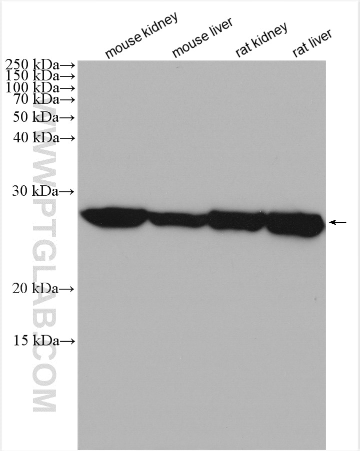
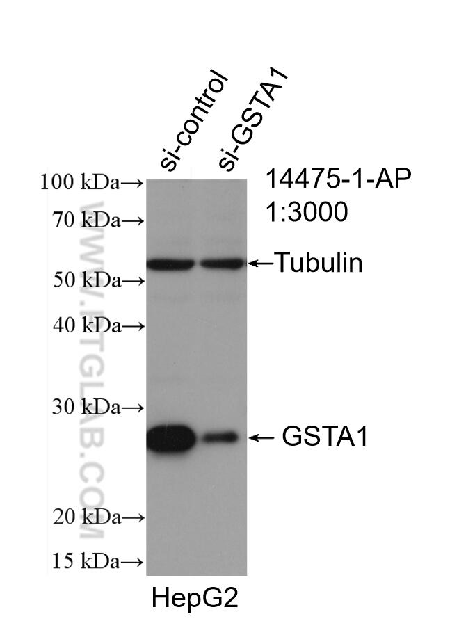
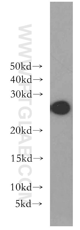
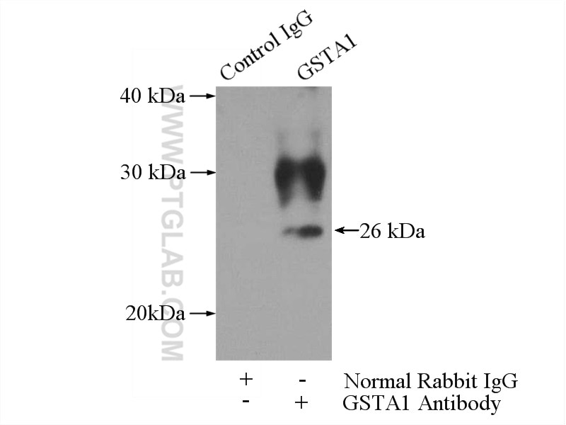
| Positive WB detected in | mouse kidney tissue, human kidney tissue, HepG2 cells, mouse liver tissue, rat kidney tissue, rat liver tissue |
| Positive IP detected in | mouse liver tissue |
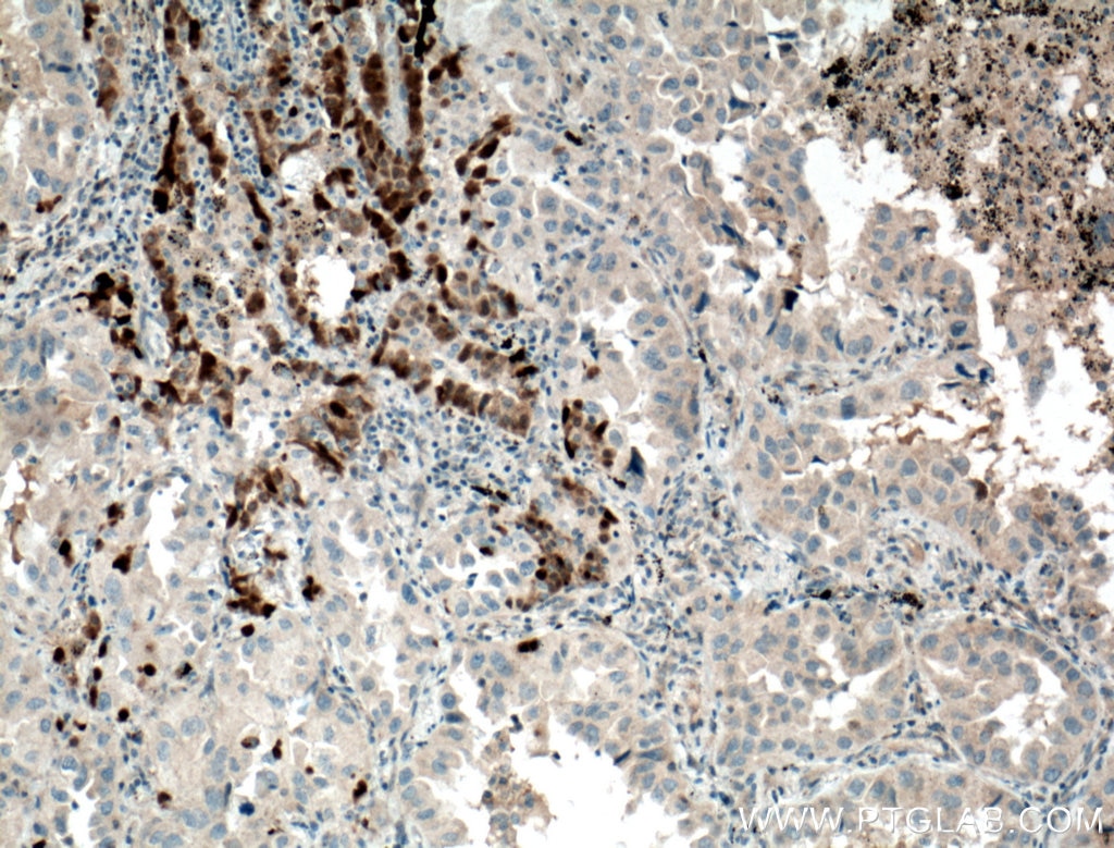
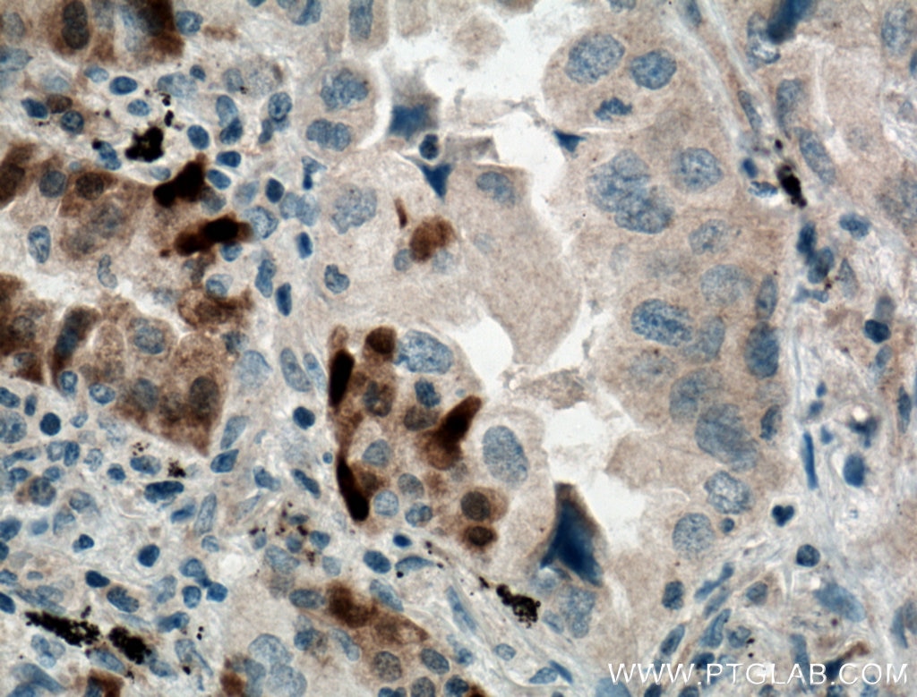
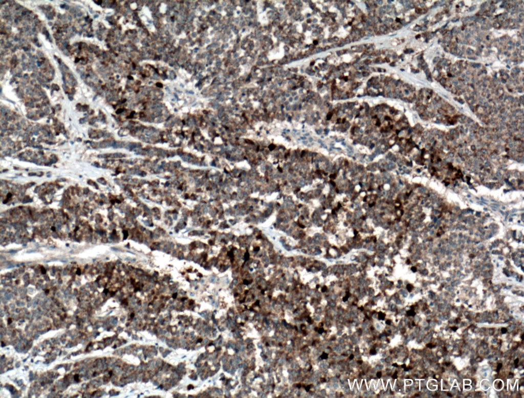
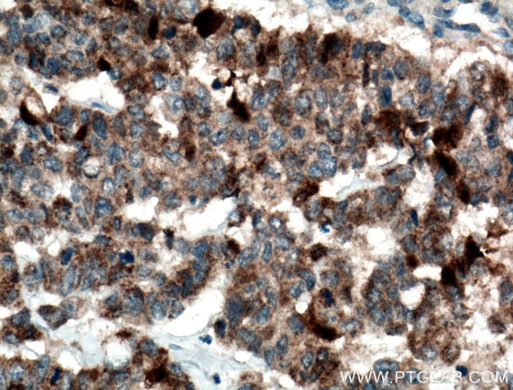
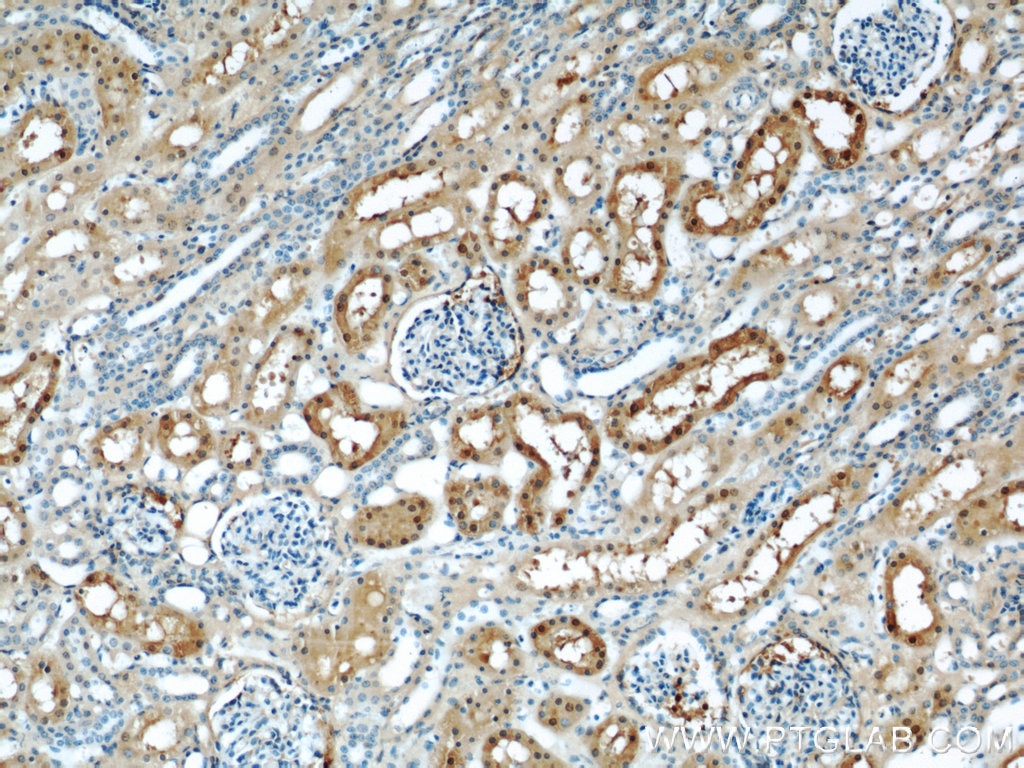
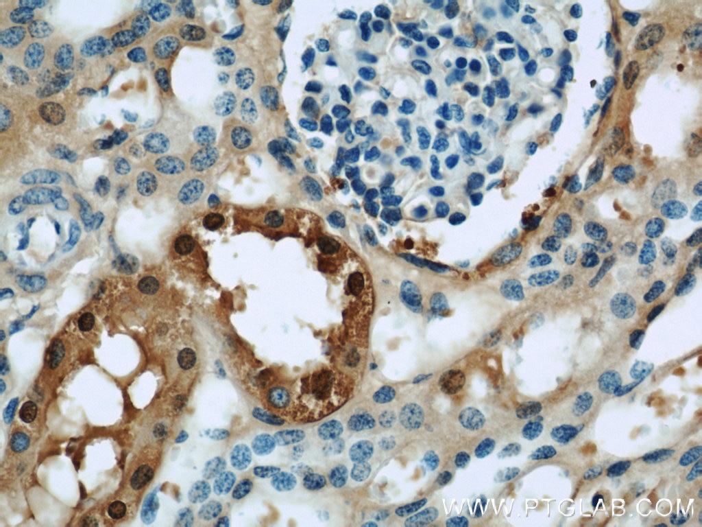
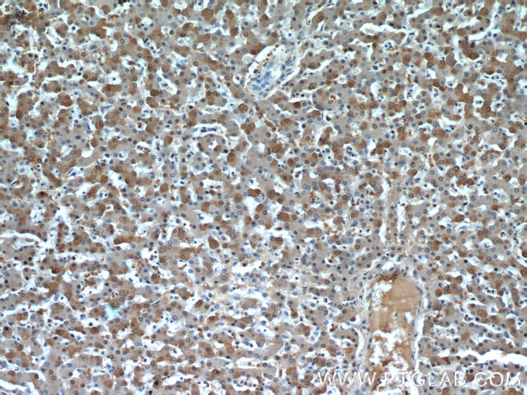
| Positive IHC detected in | human lung cancer tissue, human liver tissue, human colon cancer tissue, human kidney tissue Note: suggested antigen retrieval with TE buffer pH 9.0; (*) Alternatively, antigen retrieval may be performed with citrate buffer pH 6.0 |
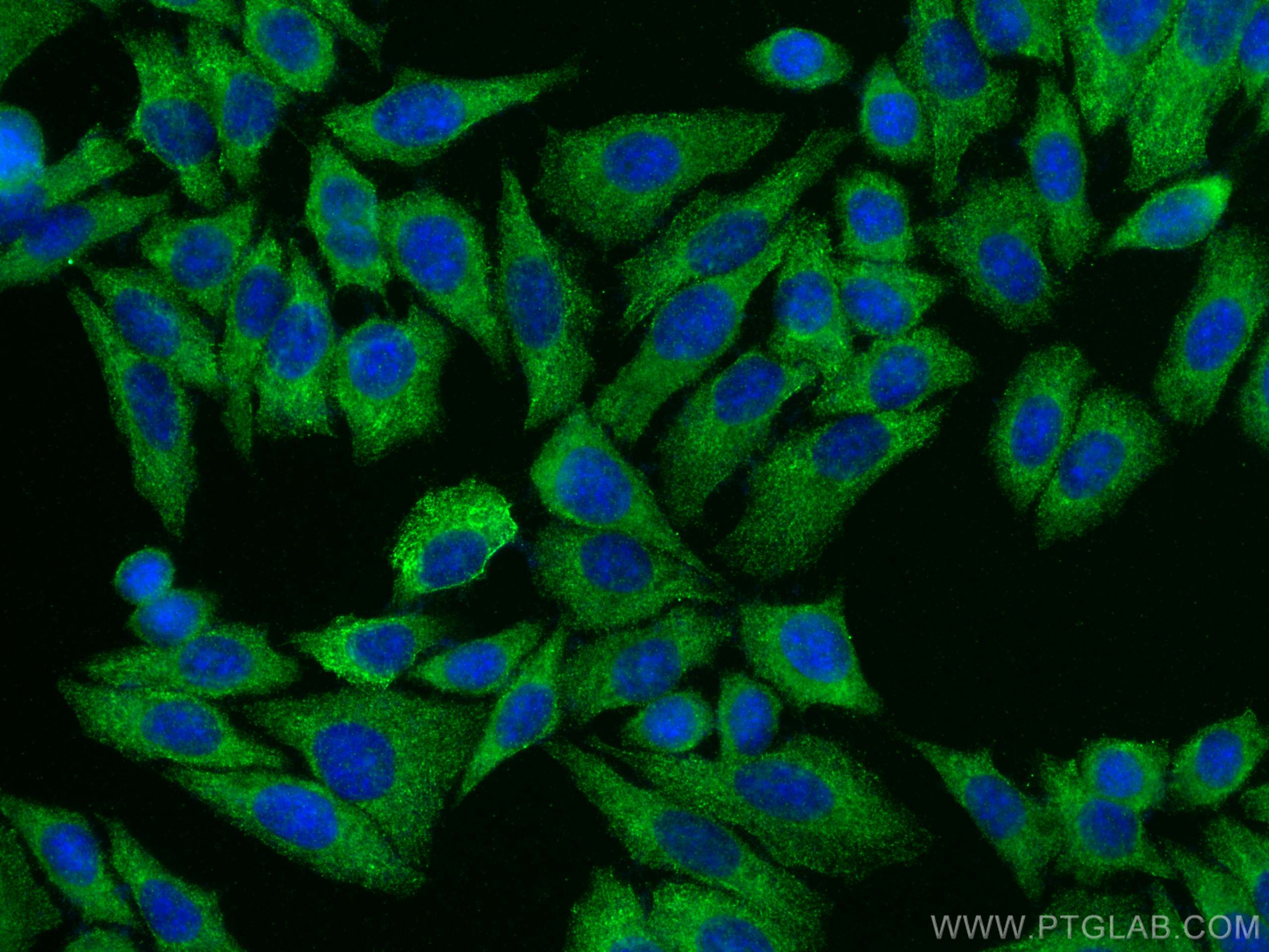
| Positive IF/ICC detected in | HepG2 cells |
Recommended dilution
| Application | Dilution |
|---|---|
| Western Blot (WB) | WB : 1:1000-1:6000 |
| Immunoprecipitation (IP) | IP : 0.5-4.0 ug for 1.0-3.0 mg of total protein lysate |
| Immunohistochemistry (IHC) | IHC : 1:200-1:800 |
| Immunofluorescence (IF)/ICC | IF/ICC : 1:50-1:500 |
| It is recommended that this reagent should be titrated in each testing system to obtain optimal results. | |
| Sample-dependent, Check data in validation data gallery. | |
Published Applications
| KD/KO | See 2 publications below |
| WB | See 23 publications below |
| IHC | See 6 publications below |
| IF | See 1 publications below |
Product Information
14475-1-AP targets GSTA1 in WB, IHC, IF/ICC, IP, ELISA applications and shows reactivity with human, mouse, rat samples.
| Tested Reactivity | human, mouse, rat |
| Cited Reactivity | human, mouse, rat |
| Host / Isotype | Rabbit / IgG |
| Class | Polyclonal |
| Type | Antibody |
| Immunogen |
CatNo: Ag5849 Product name: Recombinant human GSTA1 protein Source: e coli.-derived, PGEX-4T Tag: GST Domain: 1-222 aa of BC053578 Sequence: MAEKPKLHYFNARGRMESTRWLLAAAGVEFEEKFIKSAEDLDKLRNDGYLMFQQVPMVEIDGMKLVQTRAILNYIASKYNLYGKDIKERALIDMYIEGIADLGEMILLLPVCPPEEKDAKLALIKEKIKNRYFPAFEKVLKSHGQDYLVGNKLSRADIHLVELLYYVEELDSSLISSFPLLKALKTRISNLPTVKKFLQPGSPRKPPMDEKSLEEARKIFRF Predict reactive species |
| Full Name | glutathione S-transferase alpha 1 |
| Calculated Molecular Weight | 26 kDa |
| Observed Molecular Weight | 26 kDa |
| GenBank Accession Number | BC053578 |
| Gene Symbol | GSTA1 |
| Gene ID (NCBI) | 2938 |
| RRID | AB_2232810 |
| Conjugate | Unconjugated |
| Form | Liquid |
| Purification Method | Antigen affinity purification |
| UNIPROT ID | P08263 |
| Storage Buffer | PBS with 0.02% sodium azide and 50% glycerol, pH 7.3. |
| Storage Conditions | Store at -20°C. Stable for one year after shipment. Aliquoting is unnecessary for -20oC storage. 20ul sizes contain 0.1% BSA. |
Background Information
GSTA1 encodes one of the GST alpha isozymes, belongs to GST family, which is mainly expressed in the liver. GST can improve the detoxification of organism by enzymatic and non-enzymatic methods, and has anti-mutagenic and anti-tumor effects. Studies have shown that overexpression of GSTA1 can promote the development of lung cancer cells, so the expression level of GSTA1 can be used to predict the degree of malignancy of lung cancer tissues. To some extent, GSTA1 is regarded as a potential lung cancer candidate targets.
Protocols
| Product Specific Protocols | |
|---|---|
| IF protocol for GSTA1 antibody 14475-1-AP | Download protocol |
| IHC protocol for GSTA1 antibody 14475-1-AP | Download protocol |
| IP protocol for GSTA1 antibody 14475-1-AP | Download protocol |
| WB protocol for GSTA1 antibody 14475-1-AP | Download protocol |
| Standard Protocols | |
|---|---|
| Click here to view our Standard Protocols |
Publications
| Species | Application | Title |
|---|---|---|
Nat Commun Pregnane X receptor agonist nomilin extends lifespan and healthspan in preclinical models through detoxification functions | ||
Sci Adv Treatment with siRNAs is commonly associated with GPX4 up-regulation and target knockdown-independent sensitization to ferroptosis | ||
Cell Death Dis Poly(ADP-ribosyl)ated PXR is a critical regulator of acetaminophen-induced hepatotoxicity. | ||