Recombinant Human CD302 protein (rFc Tag)
Species
Human
Purity
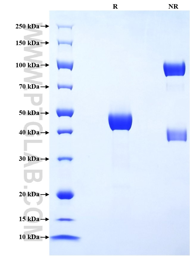
>90 %, SDS-PAGE
Tag
rFc Tag
Activity
not tested
Cat no : Eg2671
Validation Data Gallery
Product Information
| Purity | >90 %, SDS-PAGE |
| Endotoxin | <0.1 EU/μg protein, LAL method |
| Activity |
Not tested |
| Expression | HEK293-derived Human CD302 protein Asp23-His168 (Accession# Q8IX05-1) with a rabbit IgG Fc tag at the C-terminus. |
| GeneID | 9936 |
| Accession | Q8IX05-1 |
| PredictedSize | 43.0 kDa |
| SDS-PAGE | 40-50 kDa, reducing (R) conditions |
| Formulation | Lyophilized from 0.22 μm filtered solution in PBS, pH 7.4. Normally 5% trehalose and 5% mannitol are added as protectants before lyophilization. |
| Reconstitution | Briefly centrifuge the tube before opening. Reconstitute at 0.1-0.5 mg/mL in sterile water. |
| Storage Conditions |
It is recommended that the protein be aliquoted for optimal storage. Avoid repeated freeze-thaw cycles.
|
| Shipping | The product is shipped at ambient temperature. Upon receipt, store it immediately at the recommended temperature. |
Background
CD302, also known as CD302 molecule, is a C-type lectin receptor (CLR) that plays a significant role in the immune system, particularly in cell adhesion, migration, endocytosis, and phagocytosis. CD302 is expressed mainly on myeloid phagocytes in human blood and has been implicated in the functional regulation of immune cells. As a CLR, CD302 recognizes molecular patterns expressed by exogenous and endogenous threats, capturing and internalizing antigens and mediating other important immune cell functions. In the liver, CD302 is expressed by hepatocytes, liver sinusoidal endothelial cells, and Kupffer cells. Among immune cells, myeloid cells such as macrophages, granulocytes, and myeloid dendritic cells (mDC) express the highest levels of CD302. CD302 is also considered a prognostic marker in various types of cancer, including Glioblastoma multiforme, Kidney renal clear cell carcinoma, and Lung adenocarcinoma. It has been detected in blood by mass spectrometry and proximity extension assays, suggesting its potential as a biomarker for certain diseases.
References:
1. Masato Kato. et al. (2007). J Immunol. 179(9):6052-6063.
2. Tsun-Ho Lo. et al.(2016). J Immunol.197(3):885-898.
3. Birthe Reinecke. et al. (2022). J Virol. 96(7):e0199521.

