Recombinant Human Pentraxin 3 protein (His Tag)
Species
Human
Purity
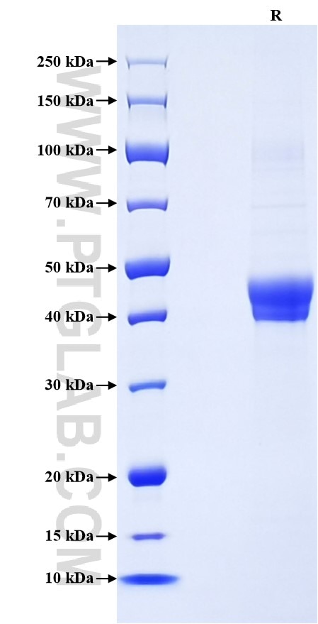
>90 %, SDS-PAGE
Tag
His Tag
Activity
not tested
Cat no : Eg1303
Validation Data Gallery
Product Information
| Purity | >90 %, SDS-PAGE |
| Endotoxin | <0.1 EU/μg protein, LAL method |
| Activity |
Not tested |
| Expression | HEK293-derived Human Pentraxin 3 protein Glu18-Ser381 (Accession# P26022) with a His tag at the C-terminus. |
| GeneID | 5806 |
| Accession | P26022 |
| PredictedSize | 41.2 kDa |
| SDS-PAGE | 40-48 kDa, reducing (R) conditions |
| Formulation | Lyophilized from 0.22 μm filtered solution in PBS, pH 7.4. Normally 5% trehalose and 5% mannitol are added as protectants before lyophilization. |
| Reconstitution | Briefly centrifuge the tube before opening. Reconstitute at 0.1-0.5 mg/mL in sterile water. |
| Storage Conditions |
It is recommended that the protein be aliquoted for optimal storage. Avoid repeated freeze-thaw cycles.
|
| Shipping | The product is shipped at ambient temperature. Upon receipt, store it immediately at the recommended temperature. |
Background
Pentraxin 3 (PTX3), also known as TNF-inducible gene 14 protein (TSG-14), is a member of the pentraxin superfamily which consists of evolutionarily conserved proteins characterized by a structural motif99, the pentraxin domain. Pentraxin 3 can be produced by a variety of cell types including endothelial cells, smooth muscle cells, adipocytes, fibroblasts, mononuclear phagocytes, and dendritic cells, and its expression is induced by primary inflammatory signals, such as IL-1, TNF, and microbial moieties. Pentraxin 3 is an acute-phase glycoprotein that plays a role in regulating innate resistance to pathogens, inflammation, tissue remodeling and repair, female fertility, and cancer. Pentraxin 3 is a biomarker for cardiovascular disease. Increased plasma Pentraxin 3 levels have been found in patients with cardiovascular disorders.
References:
1. Garlanda C. et al. (2005). Annu Rev Immunol. 23:337-66.
2. Garlanda C. et al. (2018). Physiol Rev. 98(2):623-639.
3. Inoue K. et al. (2012). Int J Vasc Med. 2012:657025.
4. Ristagno G. et al. (2019). Front Immunol. 10:823.

