WB Figures
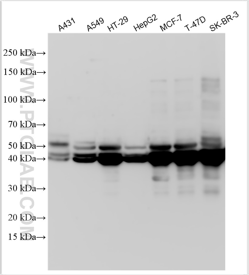
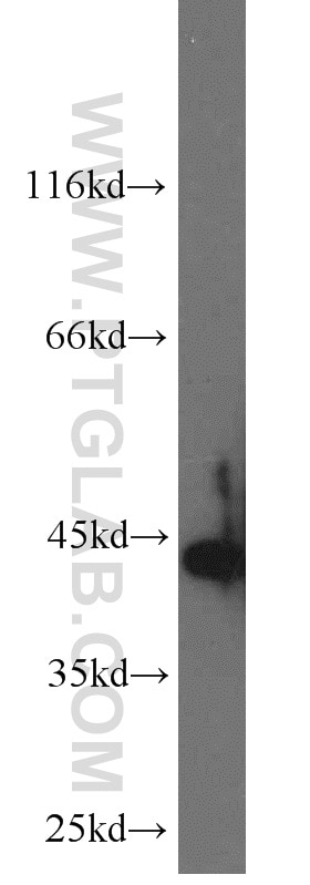
WB analysis using 10712-1-AP
Various lysates were subjected to SDS PAGE followed by western blot with 10712-1-AP (Cytokeratin 19 antibody) at dilution of 1:80000 incubated at room temperature for 1.5 hours.
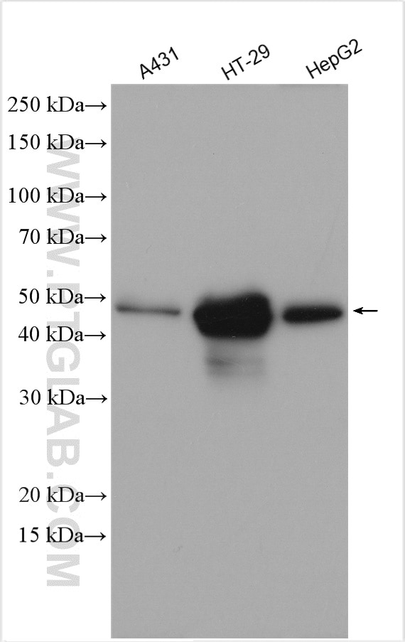
WB analysis using 10712-1-AP
Various lysates were subjected to SDS PAGE followed by western blot with 10712-1-AP (Cytokeratin 19 antibody) at dilution of 1:5000 incubated at room temperature for 1.5 hours.
WB analysis of mouse brain using 10712-1-AP
mouse brain tissue were subjected to SDS PAGE followed by western blot with 10712-1-AP (KRT19 antibody) at dilution of 1:3000 incubated at room temperature for 1.5 hours.
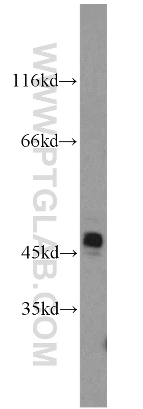
WB analysis of A549 using 10712-1-AP
A549 cells were subjected to SDS PAGE followed by western blot with 10712-1-AP (KRT19 antibody) at dilution of 1:5000 incubated at room temperature for 1.5 hours.
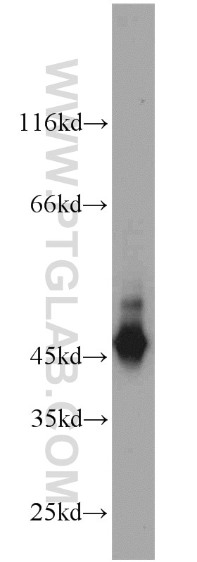
WB analysis of mouse placenta using 10712-1-AP
mouse placenta tissue were subjected to SDS PAGE followed by western blot with 10712-1-AP (KRT19 antibody) at dilution of 1:1000 incubated at room temperature for 1.5 hours.
IHC Figures
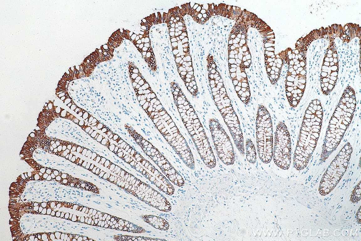
IHC staining of human colon using 10712-1-AP
Immunohistochemical analysis of paraffin-embedded human colon tissue slide using 10712-1-AP (Cytokeratin 19 antibody) at dilution of 1:6000 (under 10x lens). Heat mediated antigen retrieval with Tris-EDTA buffer (pH 9.0).
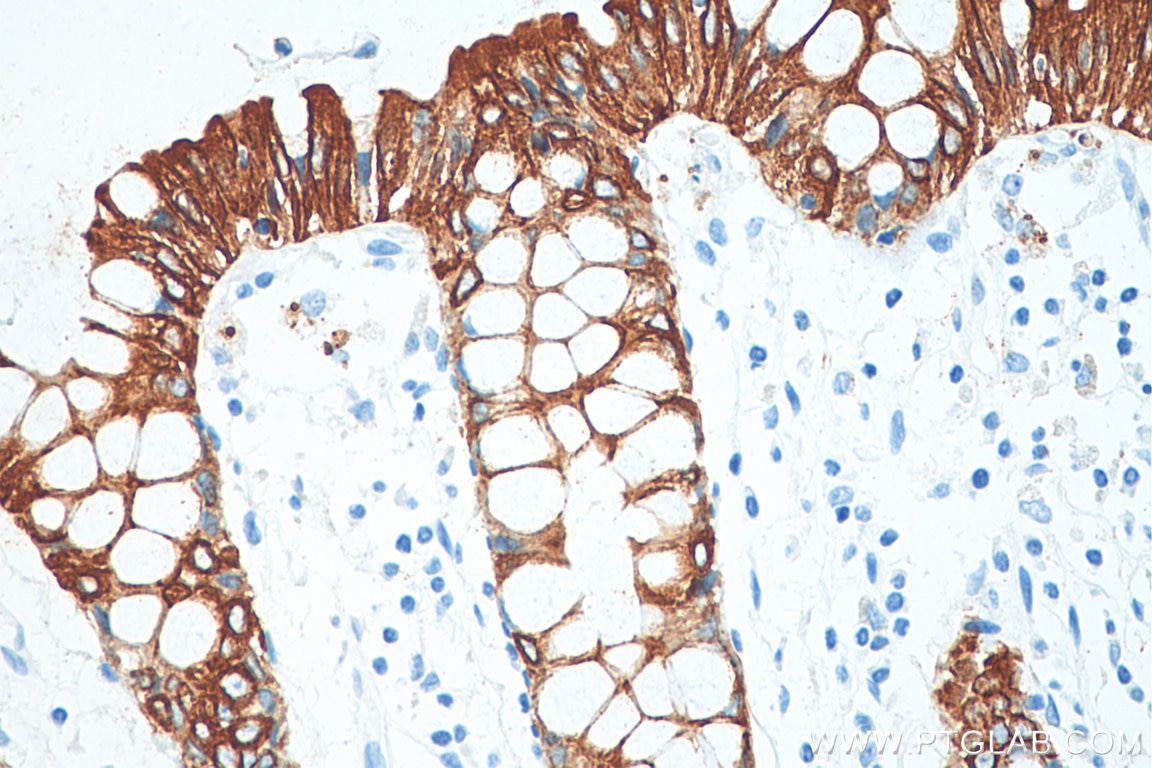
IHC staining of human colon using 10712-1-AP
Immunohistochemical analysis of paraffin-embedded human colon tissue slide using 10712-1-AP (Cytokeratin 19 antibody) at dilution of 1:6000 (under 40x lens). Heat mediated antigen retrieval with Tris-EDTA buffer (pH 9.0).
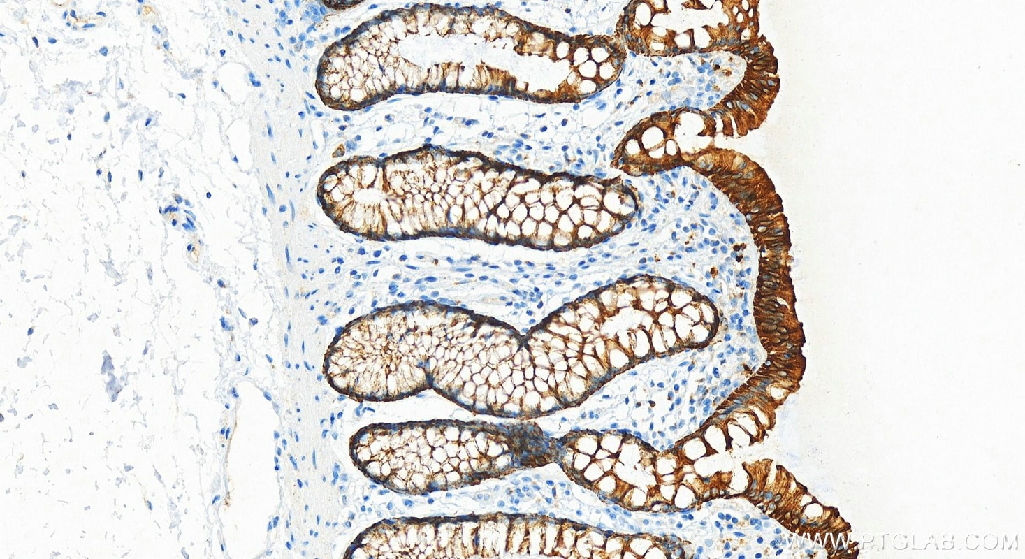
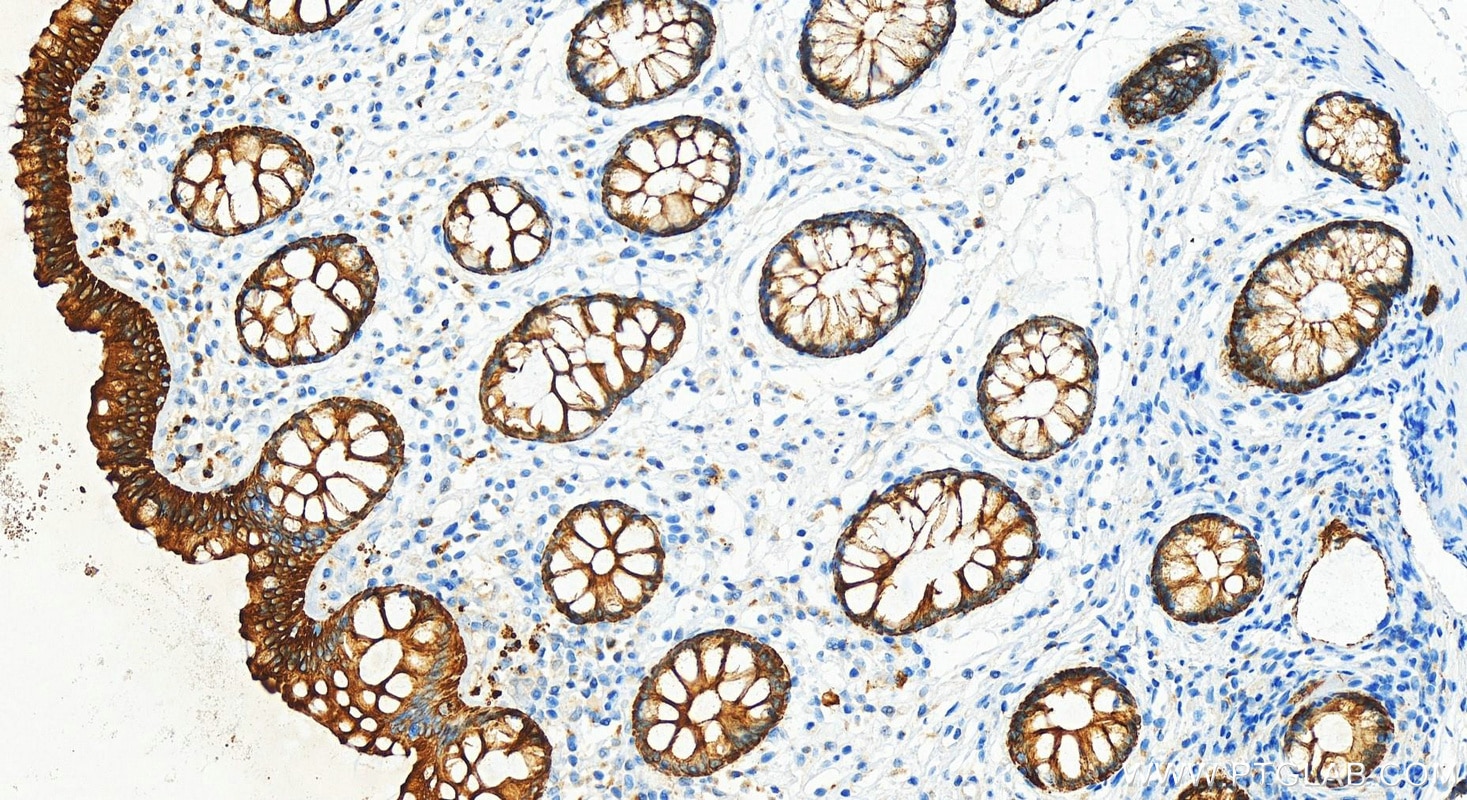
IHC staining of human colon using 10712-1-AP
Immunohistochemical analysis of paraffin-embedded human colon tissue slide using 10712-1-AP (Cytokeratin 19 antibody) at dilution of 1:20000 (under 20x lens). Heat mediated antigen retrieval with Tris-EDTA buffer (pH 9.0).
IHC staining of human colon using 10712-1-AP
Immunohistochemical analysis of paraffin-embedded human colon tissue slide using 10712-1-AP (Cytokeratin 19 antibody) at dilution of 1:20000 (under 20x lens). Heat mediated antigen retrieval with Tris-EDTA buffer (pH 9.0).
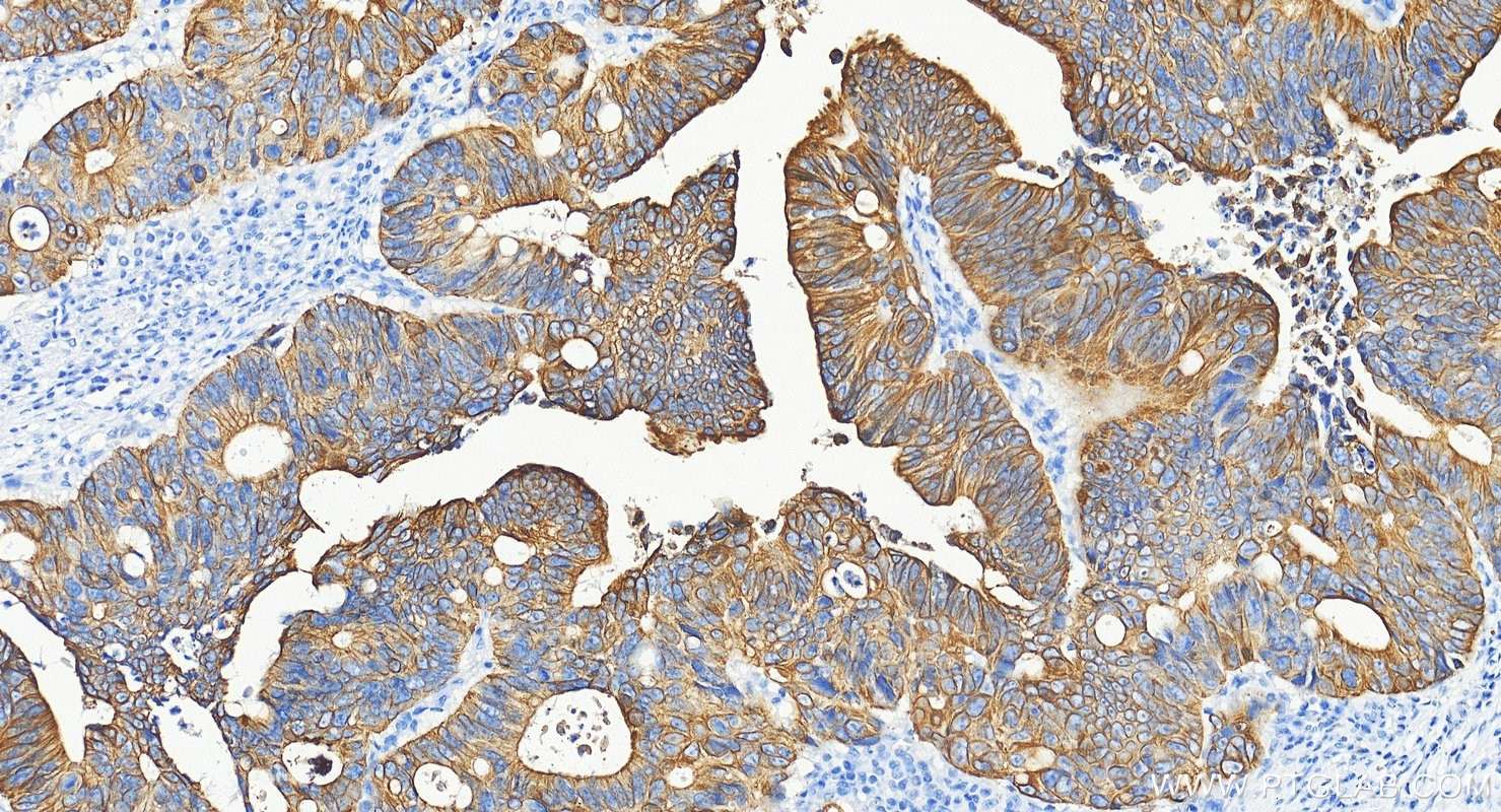
IHC staining of human colon cancer using 10712-1-AP
Immunohistochemical analysis of paraffin-embedded human colon cancer tissue slide using 10712-1-AP (Cytokeratin 19 antibody) at dilution of 1:20000 (under 20x lens). Heat mediated antigen retrieval with Tris-EDTA buffer (pH 9.0).
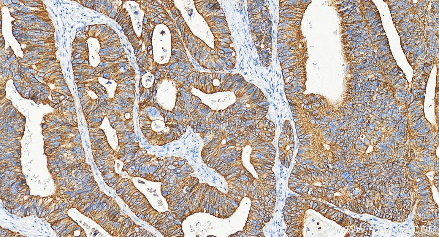
IHC staining of human colon cancer using 10712-1-AP
Immunohistochemical analysis of paraffin-embedded human colon cancer tissue slide using 10712-1-AP (Cytokeratin 19 antibody) at dilution of 1:20000 (under 20x lens). Heat mediated antigen retrieval with Tris-EDTA buffer (pH 9.0).
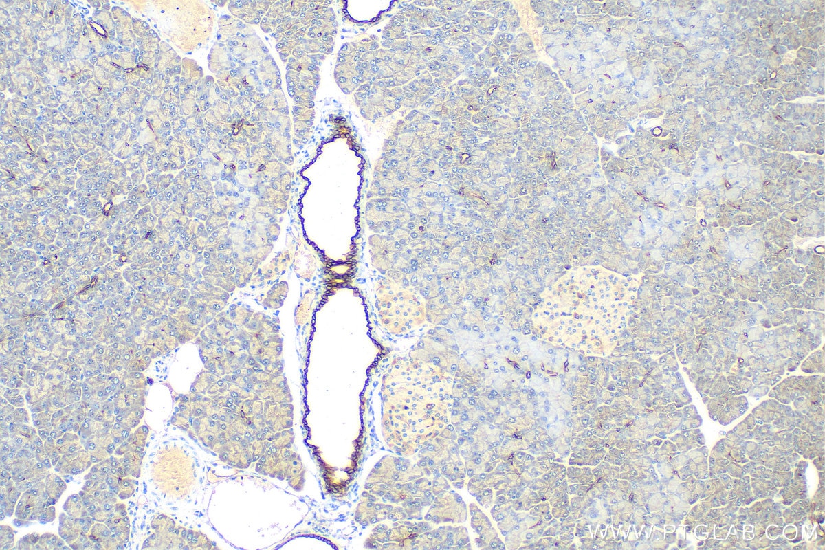
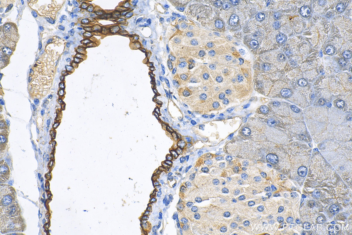
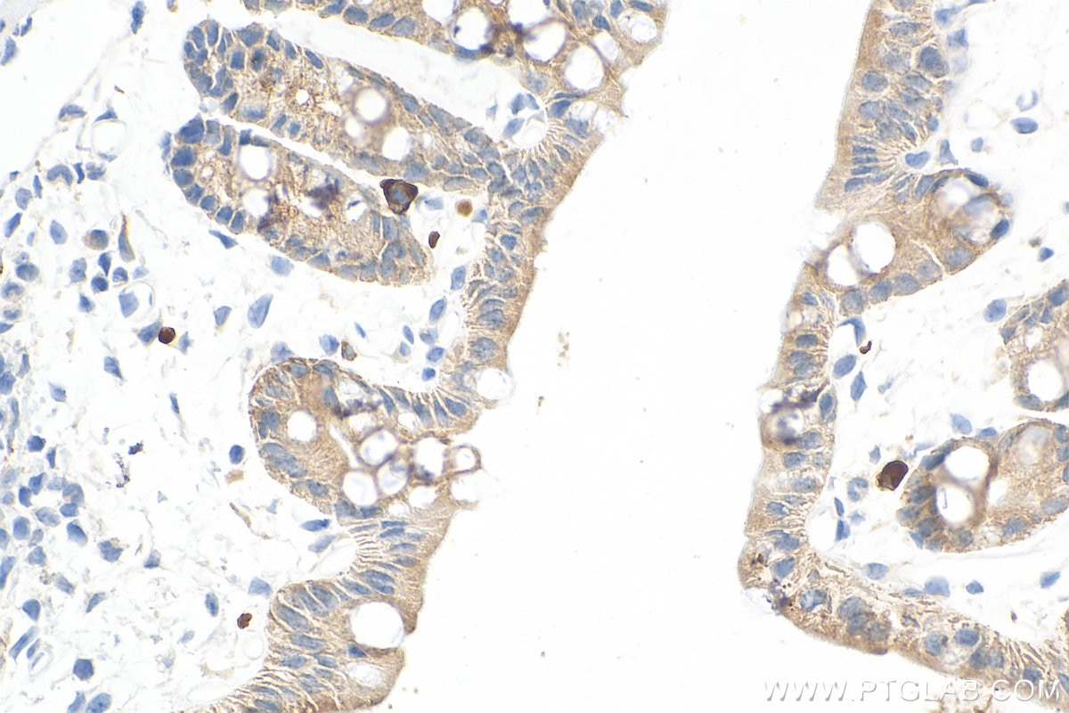
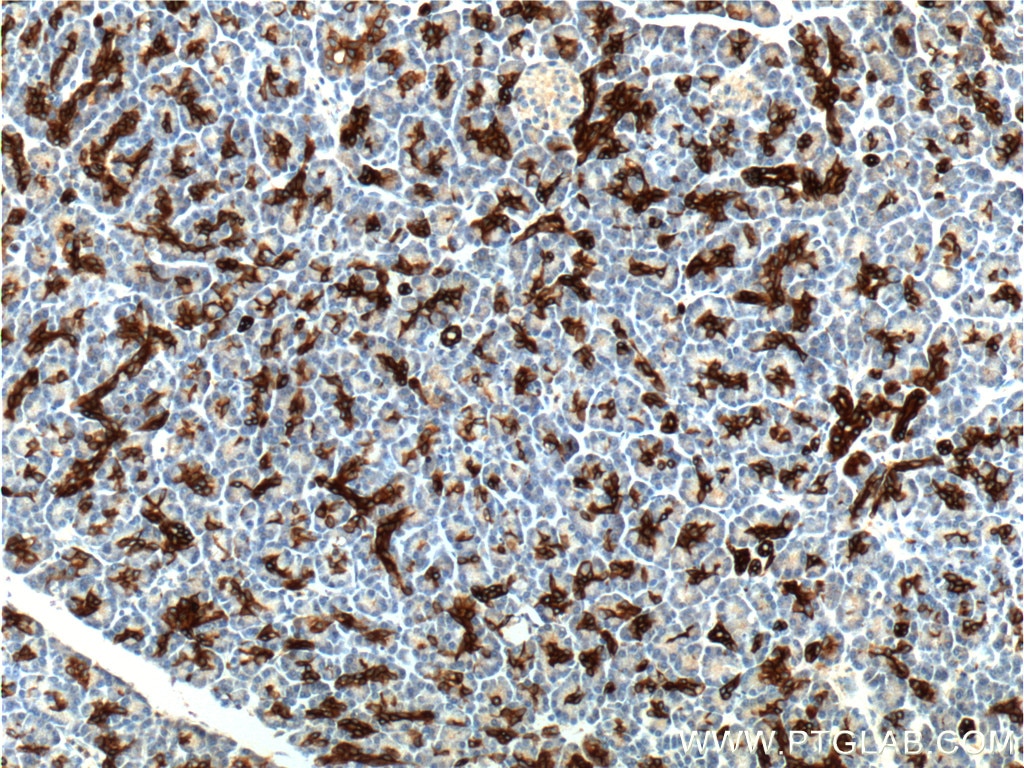
IHC staining of mouse pancreas using 10712-1-AP
Immunohistochemical analysis of paraffin-embedded mouse pancreas tissue slide using 10712-1-AP (Cytokeratin 19 antibody) at dilution of 1:1000 (under 10x lens). Heat mediated antigen retrieval with Tris-EDTA buffer (pH 9.0).
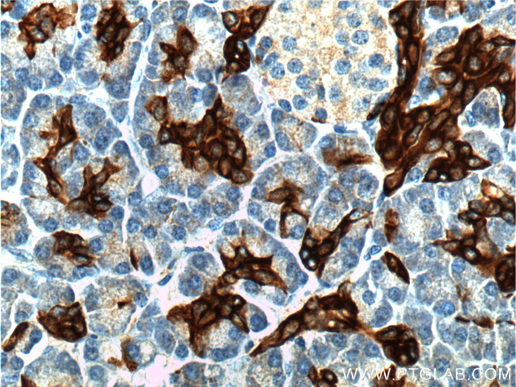
IHC staining of mouse pancreas using 10712-1-AP
Immunohistochemical analysis of paraffin-embedded mouse pancreas tissue slide using 10712-1-AP (Cytokeratin 19 antibody) at dilution of 1:1000 (under 40x lens). Heat mediated antigen retrieval with Tris-EDTA buffer (pH 9.0).
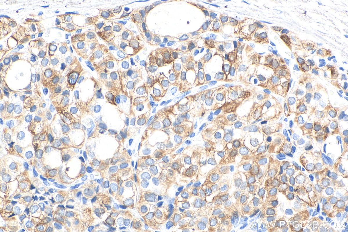
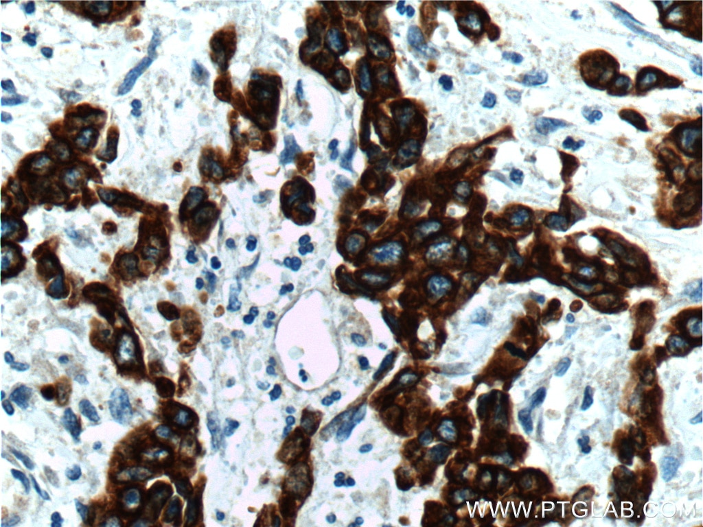
IHC staining of human thyroid cancer using 10712-1-AP
Immunohistochemical analysis of paraffin-embedded human thyroid cancer tissue slide using 10712-1-AP (Cytokeratin 19 antibody) at dilution of 1:10000 (under 40x lens). Heat mediated antigen retrieval with Tris-EDTA buffer (pH 9.0).
IHC staining of human thyroid cancer using 10712-1-AP
Immunohistochemical analysis of paraffin-embedded human thyroid cancer tissue slide using 10712-1-AP (Cytokeratin 19 antibody) at dilution of 1:10000 (under 10x lens). Heat mediated antigen retrieval with Tris-EDTA buffer (pH 9.0).
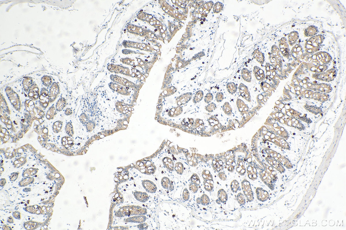
IHC staining of mouse colon using 10712-1-AP
Immunohistochemical analysis of paraffin-embedded mouse colon tissue slide using 10712-1-AP (Cytokeratin 19 antibody) at dilution of 1:10000 (under 10x lens). Heat mediated antigen retrieval with Tris-EDTA buffer (pH 9.0).
IHC staining of mouse colon using 10712-1-AP
Immunohistochemical analysis of paraffin-embedded mouse colon tissue slide using 10712-1-AP (Cytokeratin 19 antibody) at dilution of 1:10000 (under 40x lens). Heat mediated antigen retrieval with Tris-EDTA buffer (pH 9.0).
IHC staining of human stomach cancer using 10712-1-AP
Immunohistochemical analysis of paraffin-embedded human stomach cancer tissue slide using 10712-1-AP (Cytokeratin 19 antibody at dilution of 1:200 (under 10x lens).
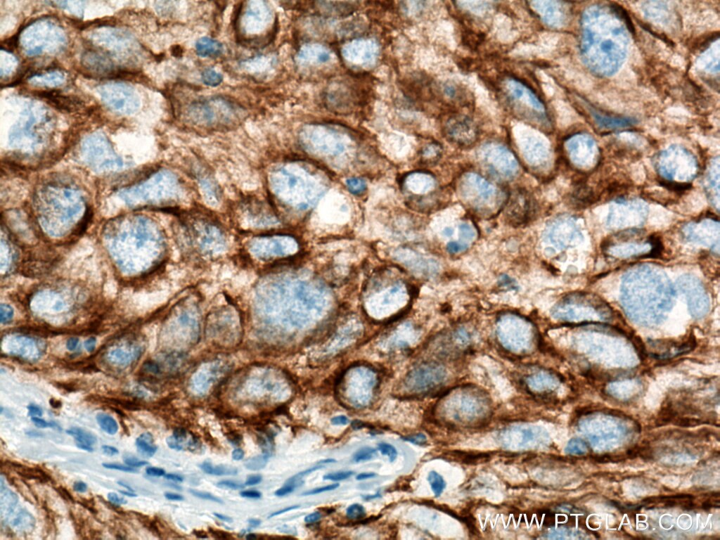
IHC staining of human stomach cancer using 10712-1-AP
Immunohistochemical analysis of paraffin-embedded human stomach cancer tissue slide using 10712-1-AP (Cytokeratin 19 antibody at dilution of 1:200 (under 40x lens).
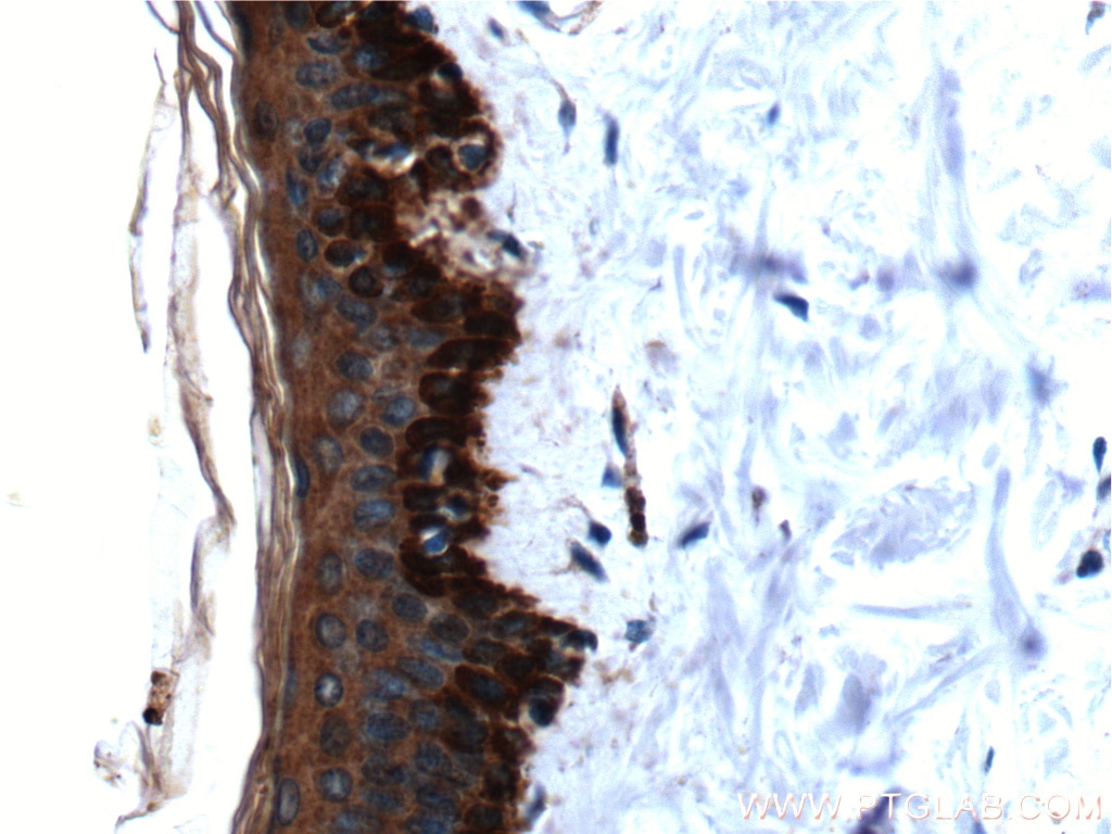
IHC staining of human skin using 10712-1-AP
Immunohistochemical analysis of paraffin-embedded human skin tissue slide using 10712-1-AP (Cytokeratin 19 antibody at dilution of 1:200 (under 10x lens).
IHC staining of human skin using 10712-1-AP
Immunohistochemical analysis of paraffin-embedded human skin tissue slide using 10712-1-AP (Cytokeratin 19 antibody at dilution of 1:200 (under 40x lens).
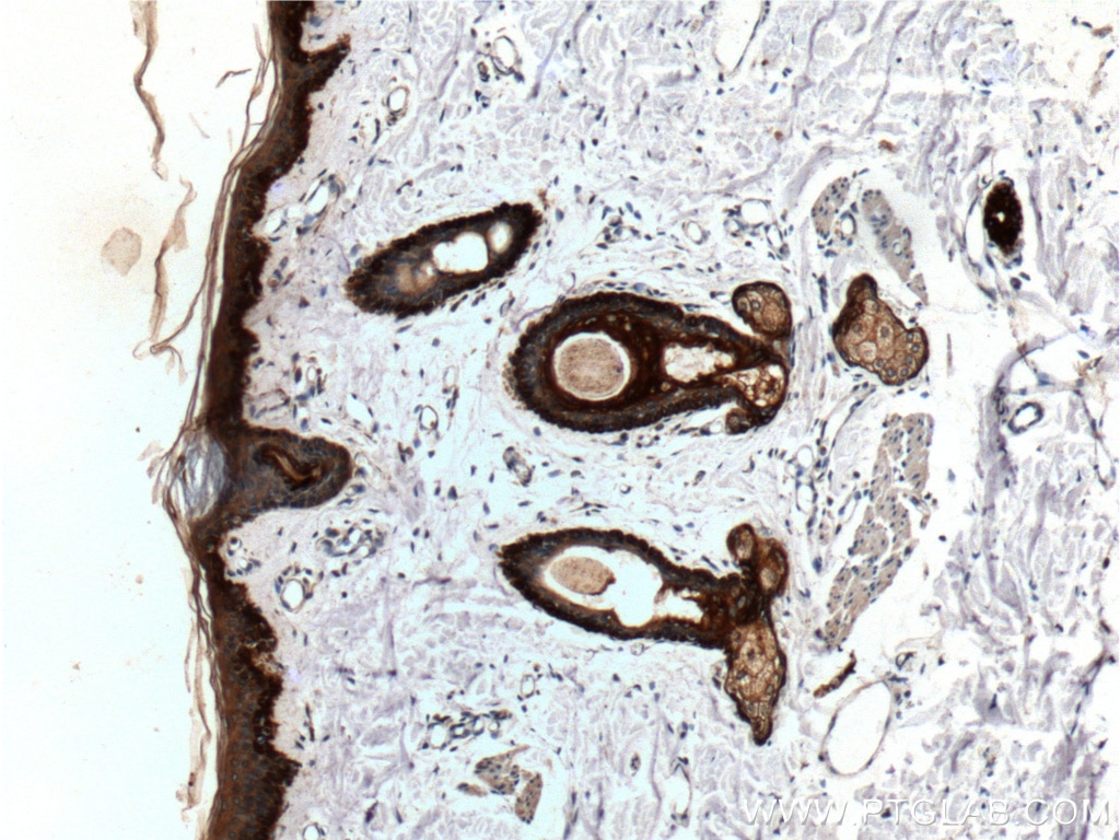
IHC staining of mouse skin using 10712-1-AP
Immunohistochemical analysis of paraffin-embedded mouse skin tissue slide using 10712-1-AP (Cytokeratin 19 antibody) at dilution of 1:3000 (under 10x lens). Heat mediated antigen retrieval with Tris-EDTA buffer (pH 9.0).
IHC staining of mouse skin using 10712-1-AP
Immunohistochemical analysis of paraffin-embedded mouse skin tissue slide using 10712-1-AP (Cytokeratin 19 antibody) at dilution of 1:3000 (under 40x lens). Heat mediated antigen retrieval with Tris-EDTA buffer (pH 9.0).
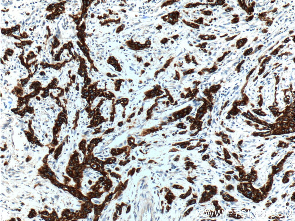
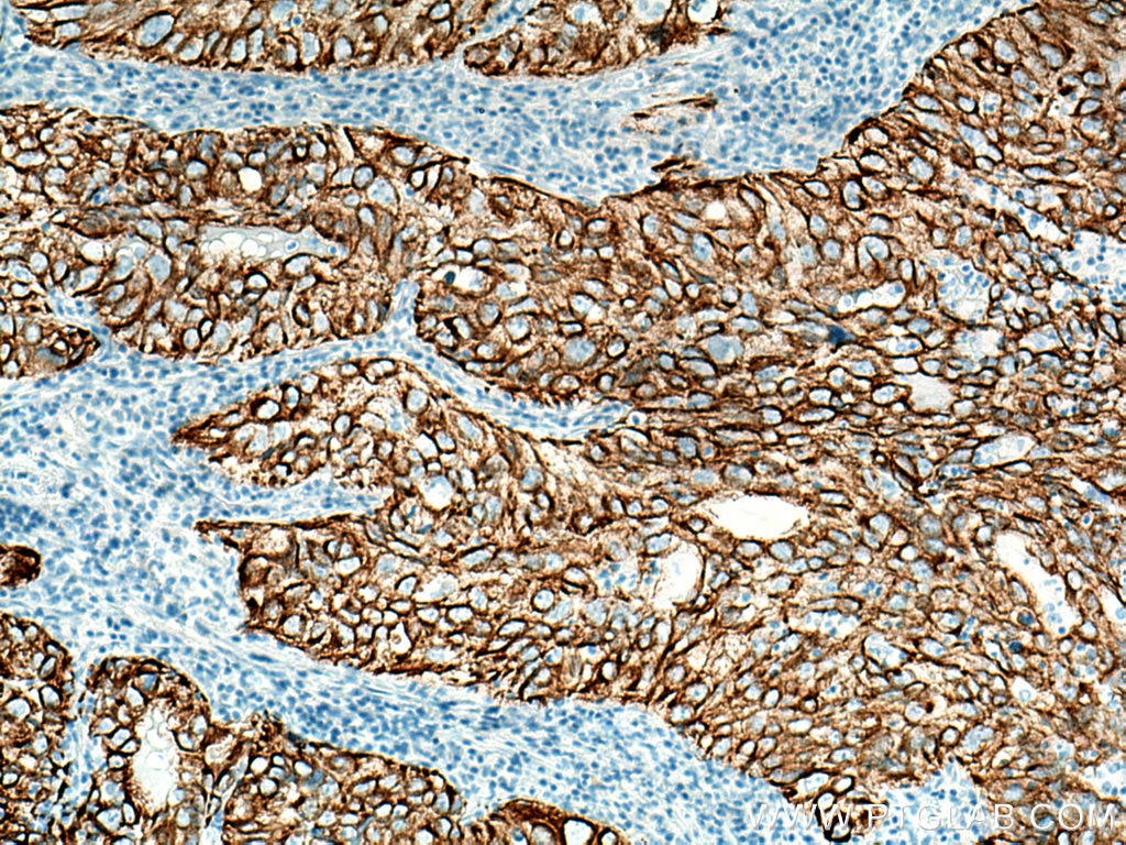
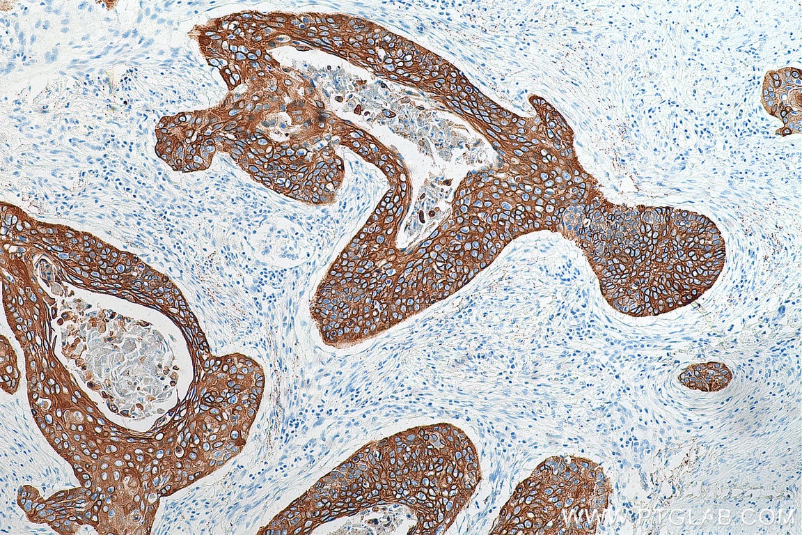
IHC staining of human lung cancer using 10712-1-AP
Immunohistochemical analysis of paraffin-embedded human lung cancer tissue slide using 10712-1-AP (Cytokeratin 19 antibody) at dilution of 1:5000 (under 10x lens). Heat mediated antigen retrieval with Tris-EDTA buffer (pH 9.0).
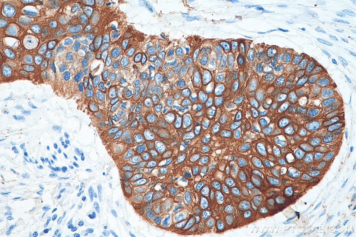
IHC staining of human lung cancer using 10712-1-AP
Immunohistochemical analysis of paraffin-embedded human lung cancer tissue slide using 10712-1-AP (Cytokeratin 19 antibody) at dilution of 1:5000 (under 40x lens). Heat mediated antigen retrieval with Tris-EDTA buffer (pH 9.0).
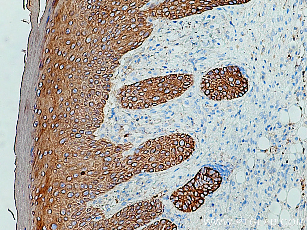
IHC staining of human oesophagus cancer using 10712-1-AP
Immunohistochemical analysis of paraffin-embedded human oesophagus cancer tissue slide using 10712-1-AP (Cytokeratin 19 antibody) at dilution of 1:6000 (under 10x lens). Heat mediated antigen retrieval with Tris-EDTA buffer (pH 9.0).
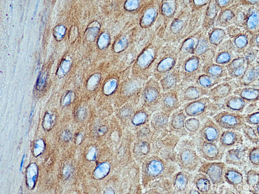
IHC staining of human oesophagus cancer using 10712-1-AP
Immunohistochemical analysis of paraffin-embedded human oesophagus cancer tissue slide using 10712-1-AP (Cytokeratin 19 antibody) at dilution of 1:6000 (under 40x lens). Heat mediated antigen retrieval with Tris-EDTA buffer (pH 9.0).
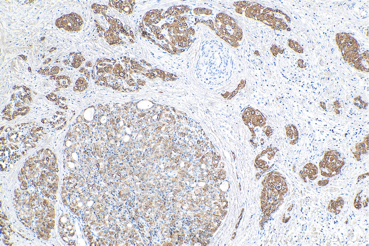
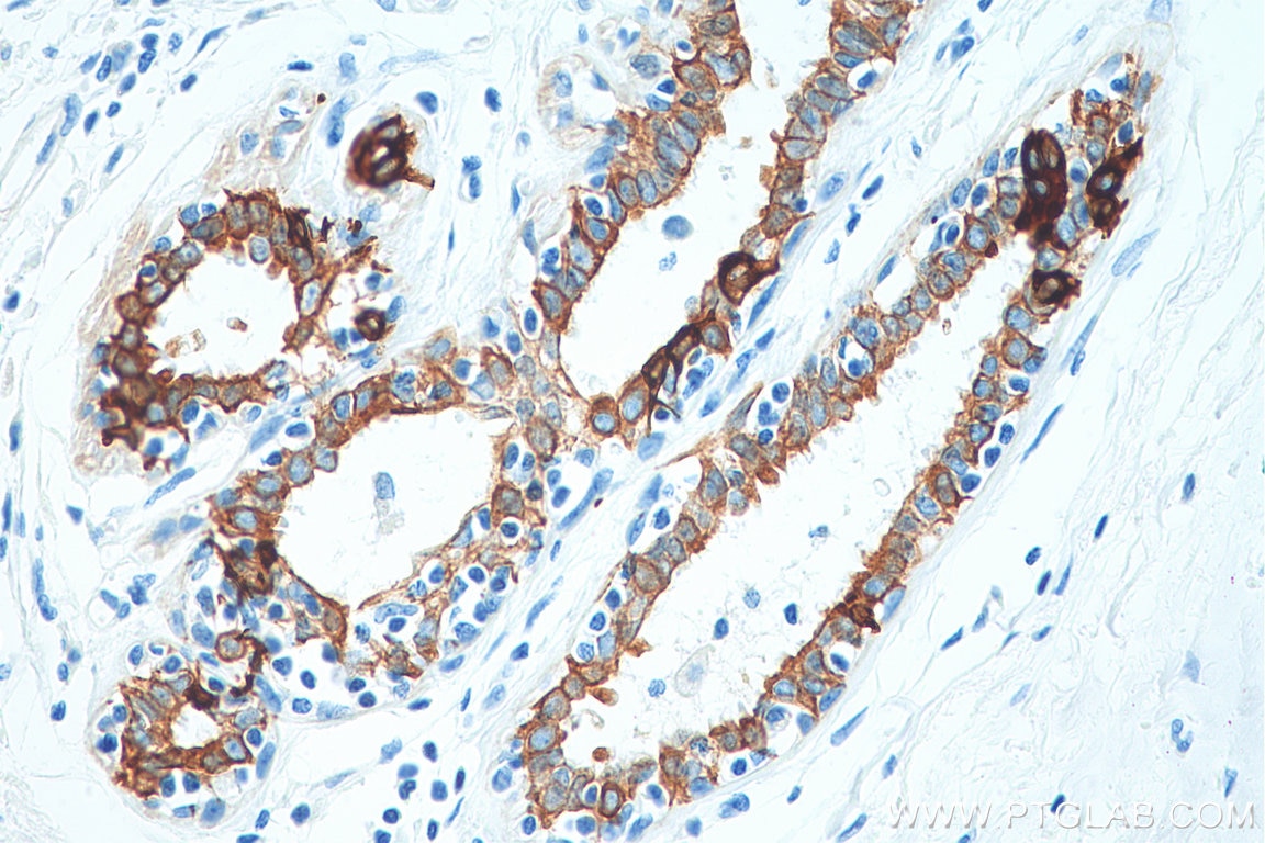
IHC staining of human breast cancer using 10712-1-AP
Immunohistochemical analysis of paraffin-embedded human breast cancer tissue slide using 10712-1-AP (Cytokeratin 19 antibody) at dilution of 1:6000 (under 10x lens). Heat mediated antigen retrieval with Tris-EDTA buffer (pH 9.0).
IHC staining of human breast cancer using 10712-1-AP
Immunohistochemical analysis of paraffin-embedded human breast cancer tissue slide using 10712-1-AP (Cytokeratin 19 antibody) at dilution of 1:6000 (under 40x lens). Heat mediated antigen retrieval with Tris-EDTA buffer (pH 9.0).
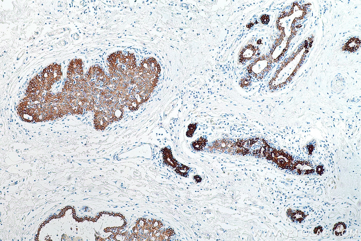
IHC staining of human pancreas using 10712-1-AP
Immunohistochemical analysis of paraffin-embedded human pancreas tissue slide using 10712-1-AP (Cytokeratin 19 antibody at dilution of 1:200 (under 10x lens).
IHC staining of human pancreas using 10712-1-AP
Immunohistochemical analysis of paraffin-embedded human pancreas tissue slide using 10712-1-AP (Cytokeratin 19 antibody at dilution of 1:200 (under 40x lens).
IF-P Figures
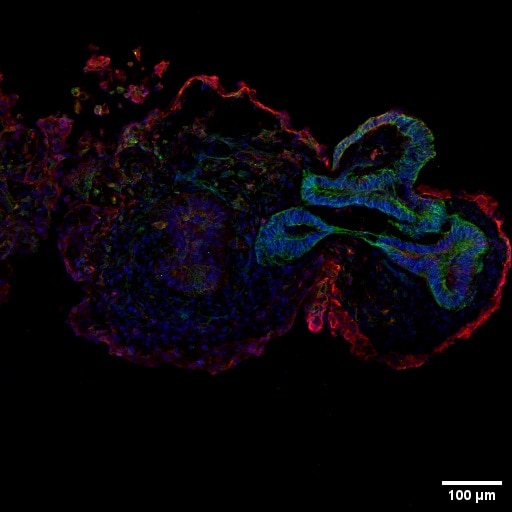
IF Staining of Retinal organoids using 10712-1-AP
Retinal organoids (day 60) generated from human induced pluripotent stem cells (iPSCs) and fixed with 4% PFA. Stained for Tubulin beta 3/TUJ1 using 66375-1-Ig at 1:500 dilution (green) and Cytokeratin 19 using 10712-1-AP at 1:200 (red). Nuclear stain DAPI (blue). Scale bar = 100 µm. Data generated by Alessandro Bellapianta at Johannes Kepler Universitat, Austria.
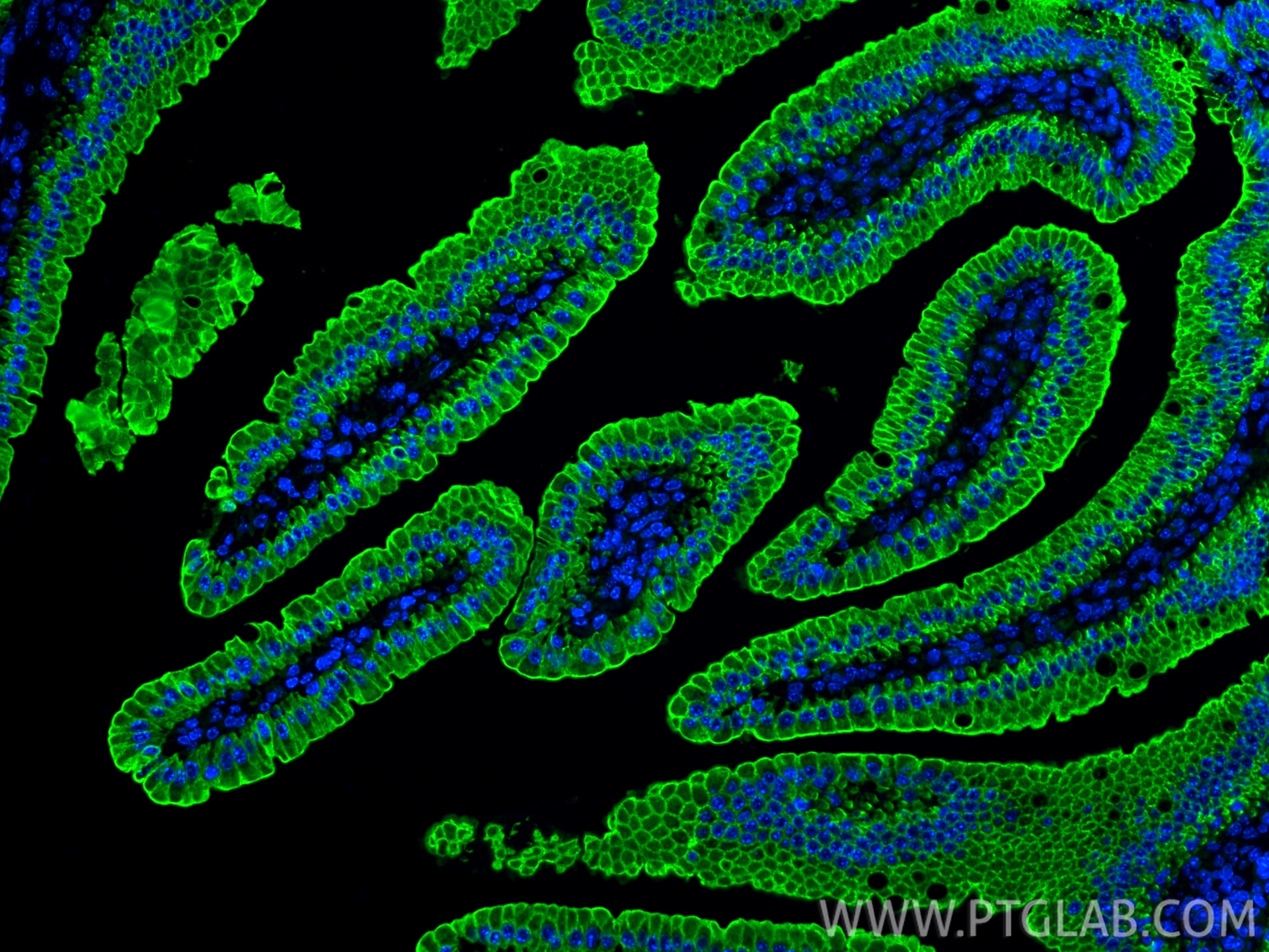
IF Staining of mouse small intestine using 10712-1-AP
Immunofluorescent analysis of (4% PFA) fixed paraffin-embedded mouse small intestine tissue using Cytokeratin 19 antibody (10712-1-AP) at dilution of 1:200 and CoraLite®488-Conjugated Goat Anti-Rabbit IgG(H+L) (SA00013-2). Heat mediated antigen retrieval with Tris-EDTA buffer (pH 9.0).
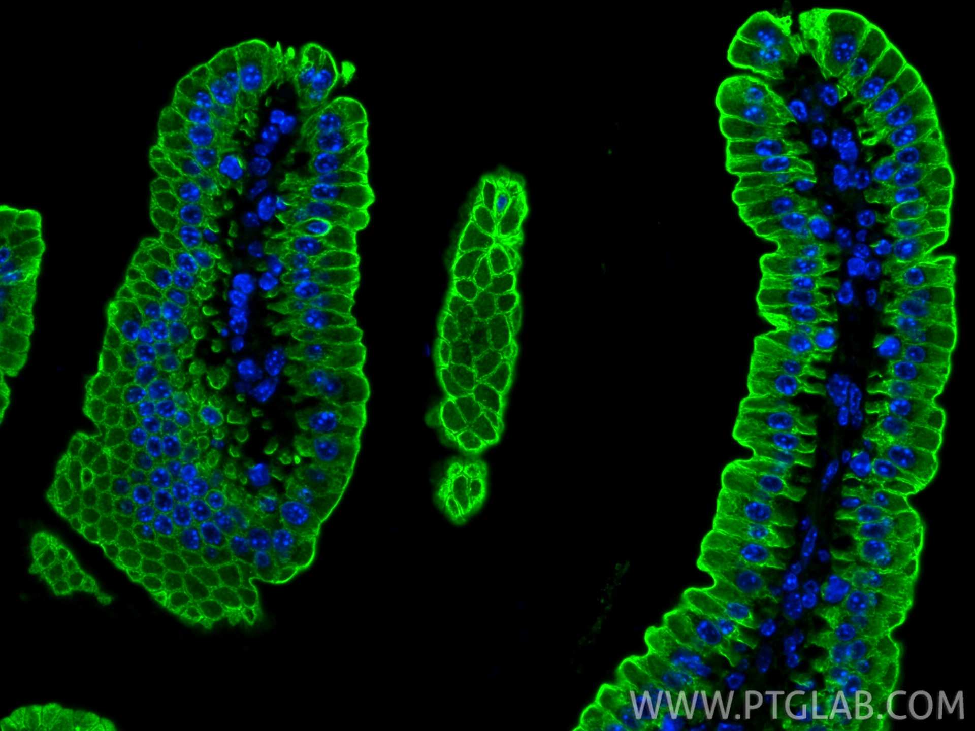
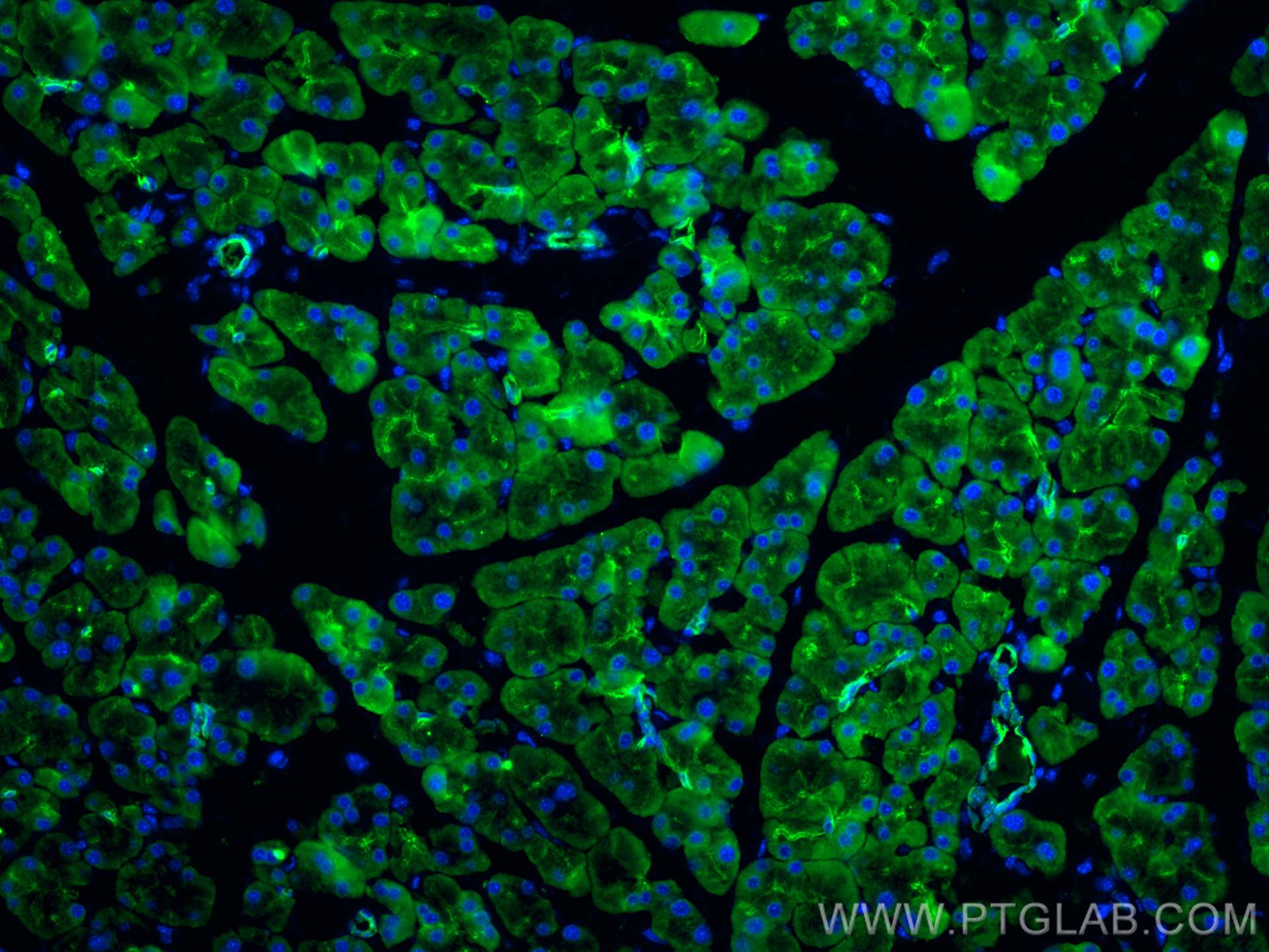
IF Staining of mouse pancreas using 10712-1-AP
Immunofluorescent analysis of (4% PFA) fixed paraffin-embedded mouse pancreas tissue using Cytokeratin 19 antibody (10712-1-AP) at dilution of 1:200 and CoraLite®488-Conjugated AffiniPure Goat Anti-Rabbit IgG(H+L) (SA00013-2). Heat mediated antigen retrieval with Tris-EDTA buffer (pH 9.0).
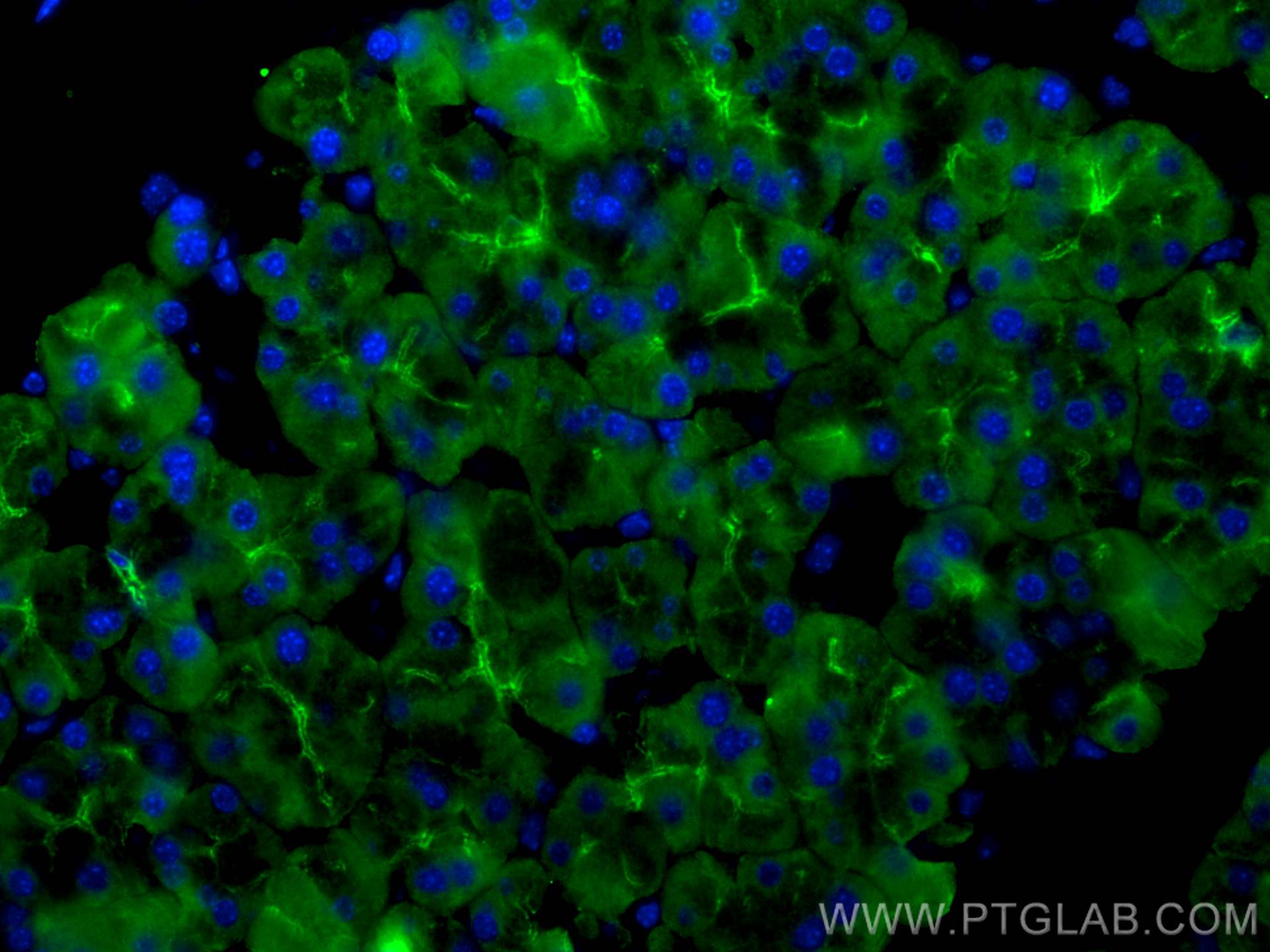
IF/ICC Figures
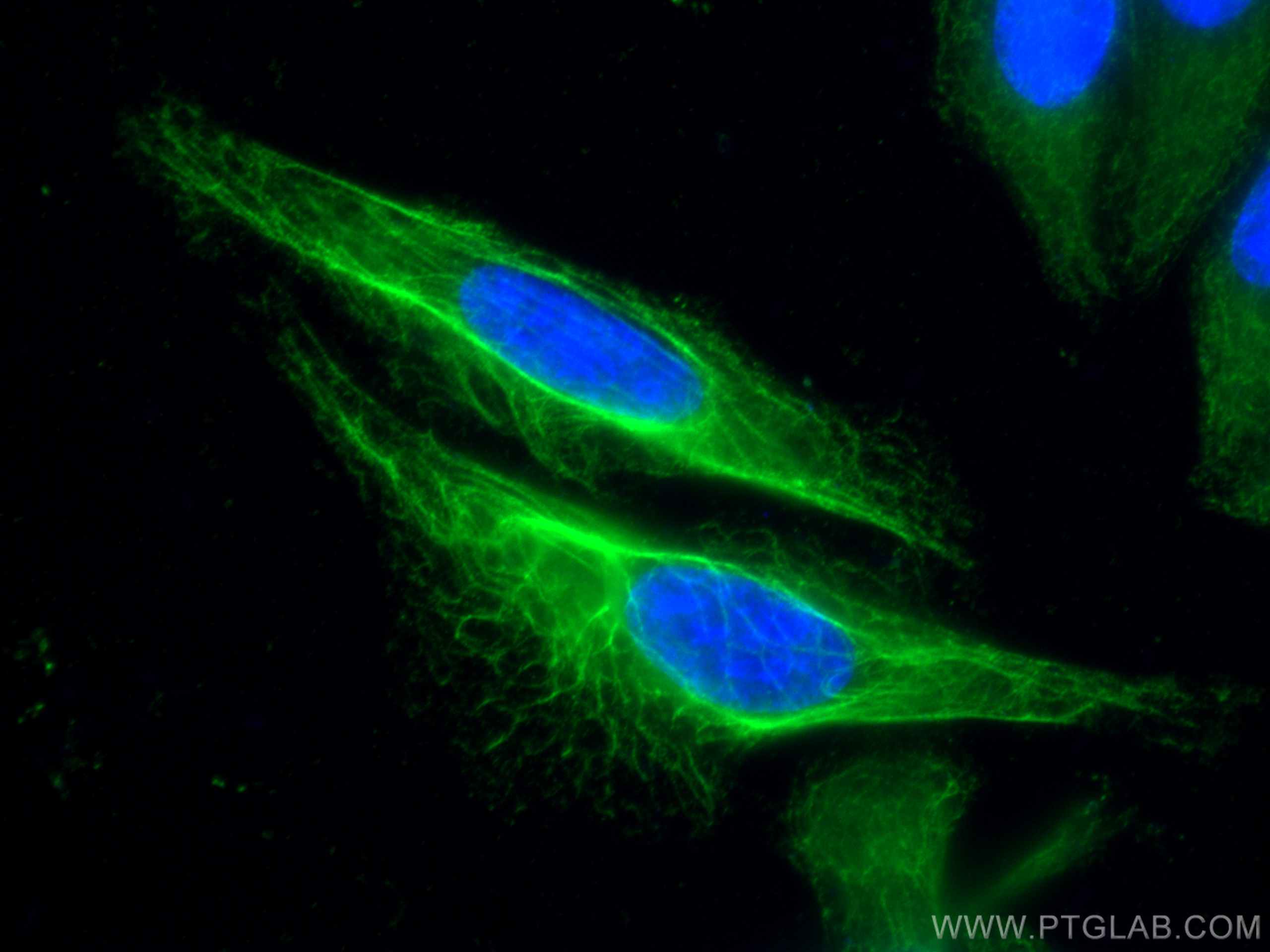
IF Staining of HeLa using 10712-1-AP
Immunofluorescent analysis of (-20°C Ethanol) fixed HeLa cells using Cytokeratin 19 antibody (10712-1-AP) at dilution of 1:200 and CoraLite®488-Conjugated AffiniPure Goat Anti-Rabbit IgG(H+L).
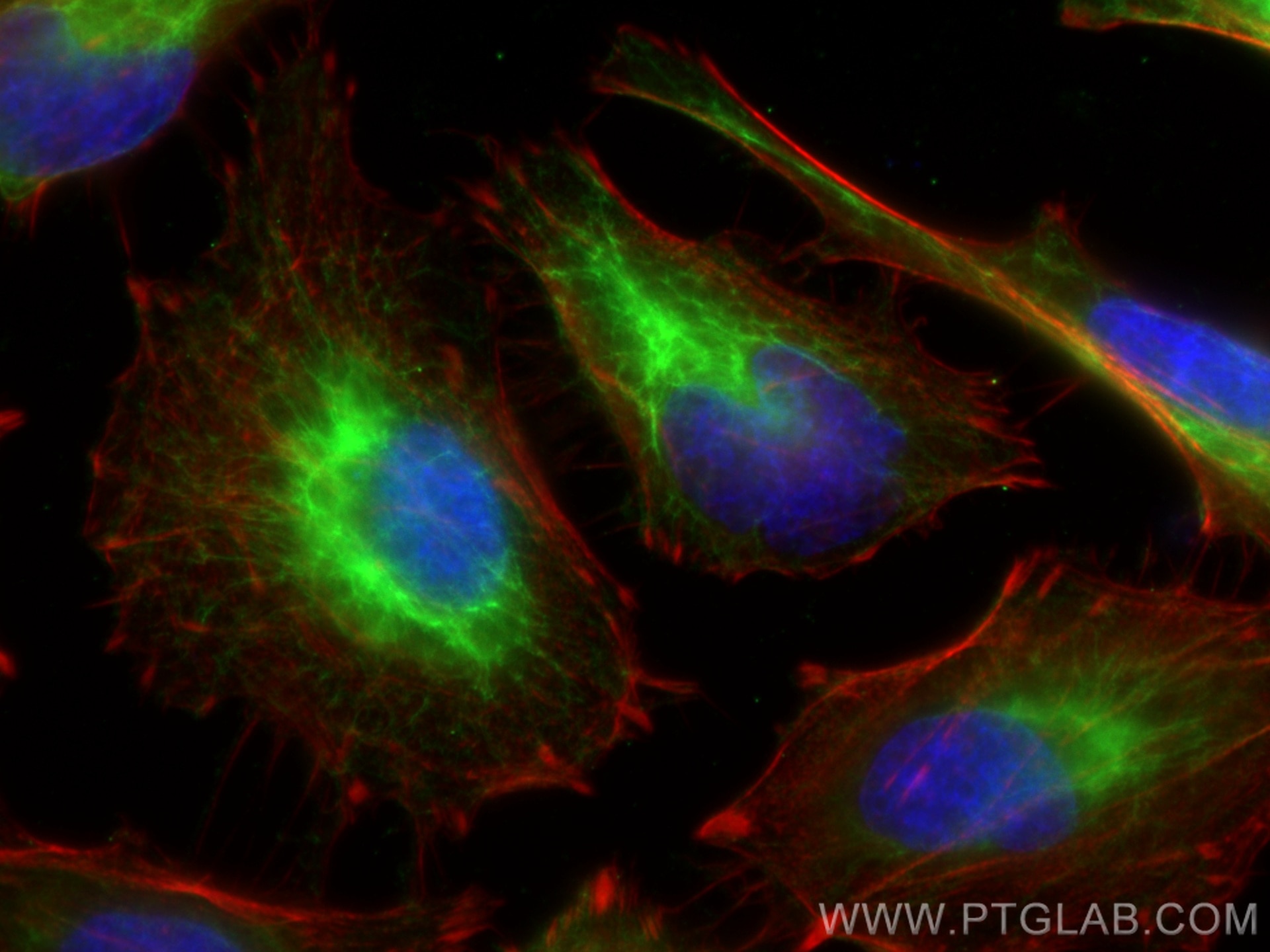
IF Staining of HeLa using 10712-1-AP
Immunofluorescent analysis of (-20°C Ethanol) fixed HeLa cells using Cytokeratin 19 antibody (10712-1-AP) at dilution of 1:400 and CoraLite®488-Conjugated AffiniPure Goat Anti-Rabbit IgG(H+L), CL594-Phalloidin (red).
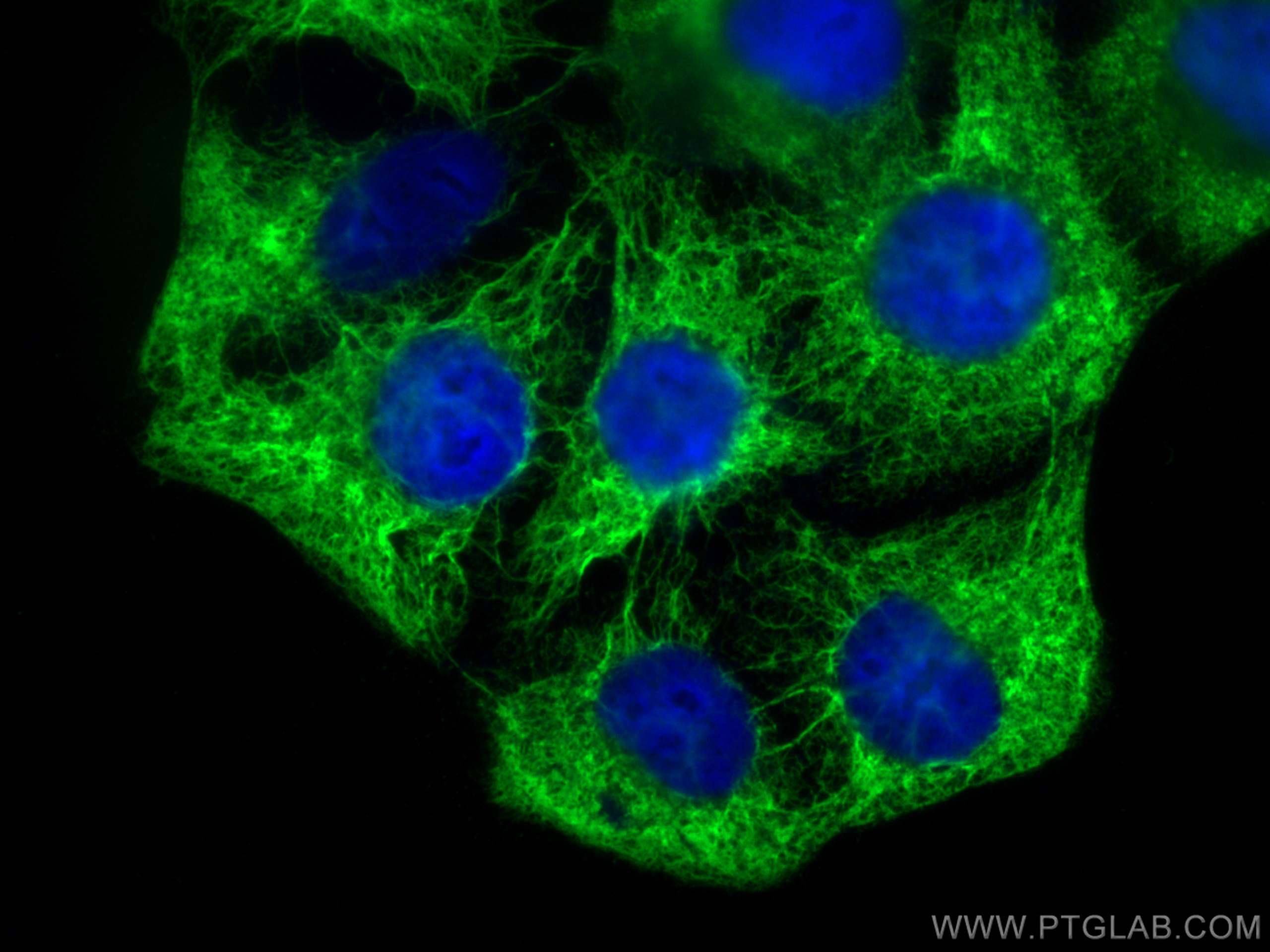
IF Staining of HaCaT using 10712-1-AP
Immunofluorescent analysis of (-20°C Methanol) fixed HaCaT cells using Cytokeratin 19 antibody (10712-1-AP) at dilution of 1:400 and CoraLite®488-Conjugated AffiniPure Goat Anti-Rabbit IgG(H+L).
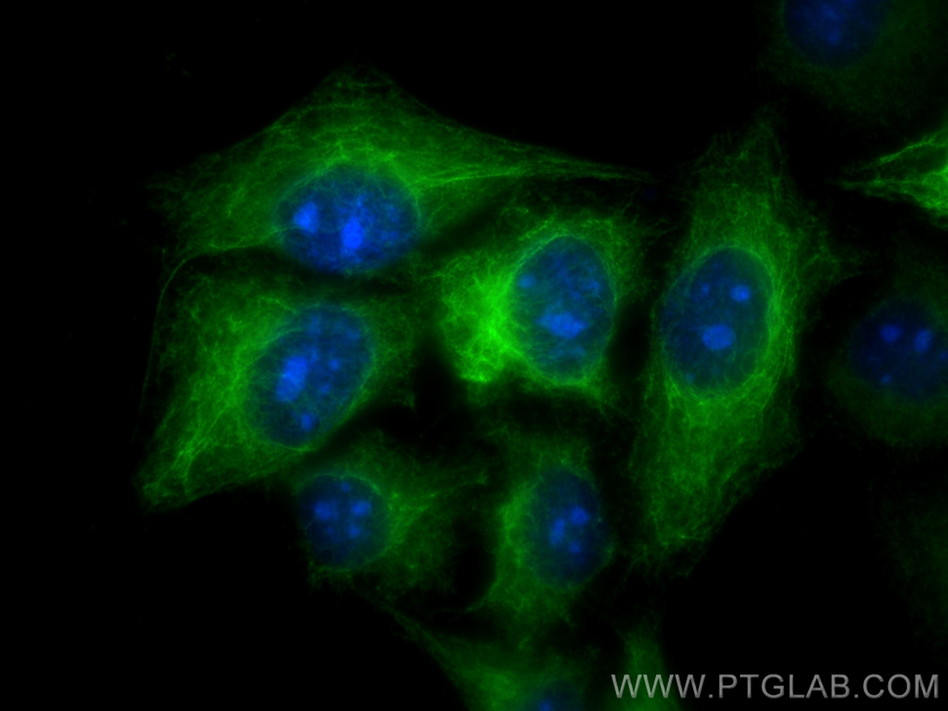
IF Staining of HepG2 using 10712-1-AP
Immunofluorescent analysis of (-20°C Ethanol) fixed HepG2 cells using Cytokeratin 19 antibody (10712-1-AP) at dilution of 1:400 and CoraLite®488-Conjugated AffiniPure Goat Anti-Rabbit IgG(H+L).