Tested Applications
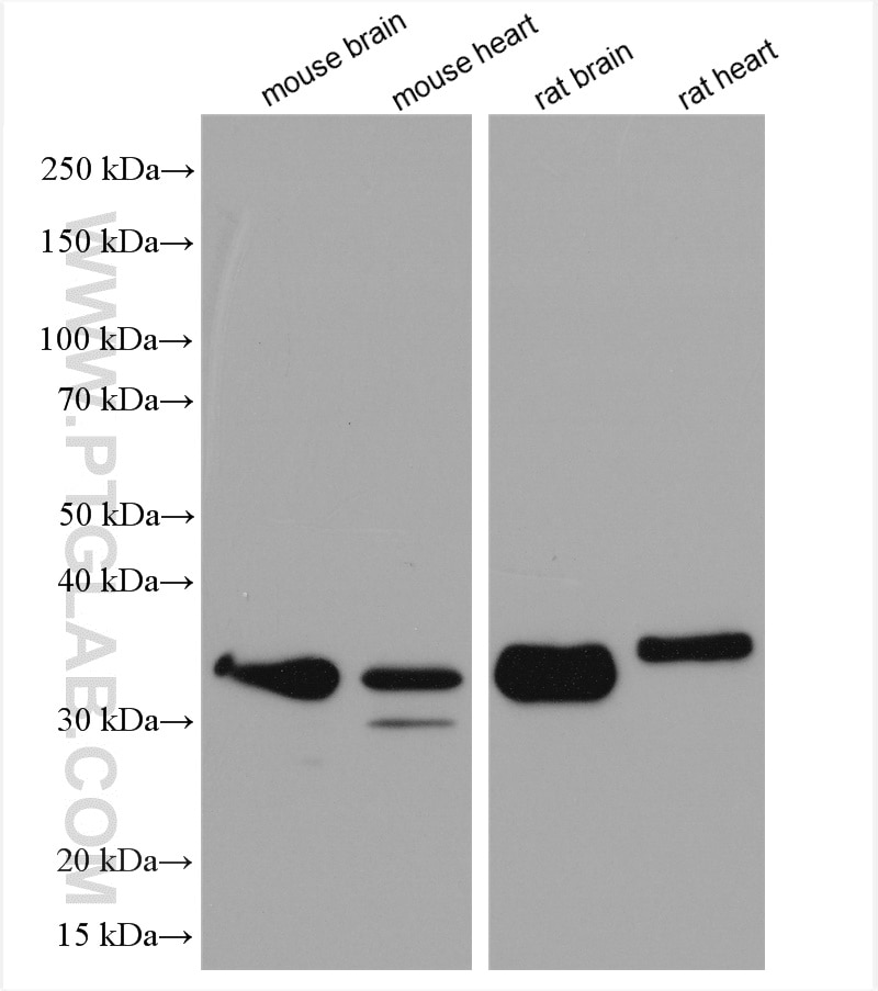
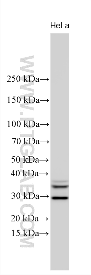
| Positive WB detected in | mouse brain tissue, HeLa cells, rat brain tissue, mouse heart tissue, rat heart tissue |
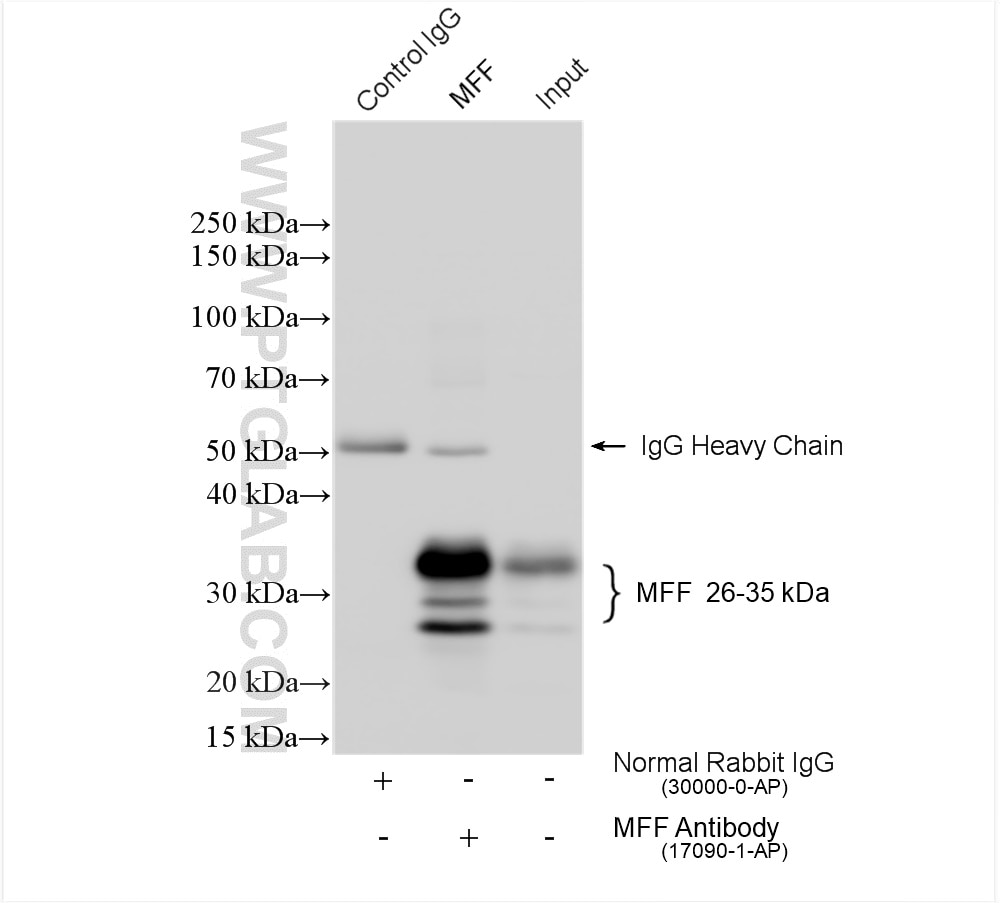
| Positive IP detected in | mouse brain tissue |
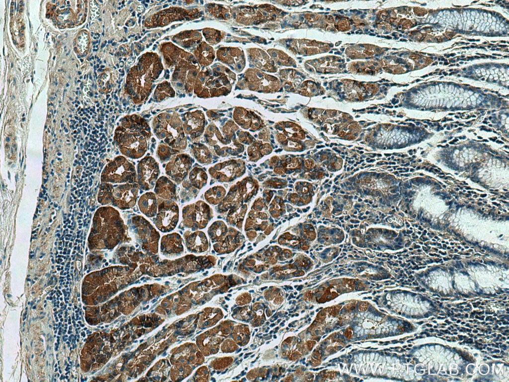
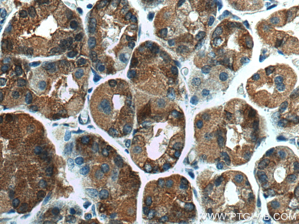
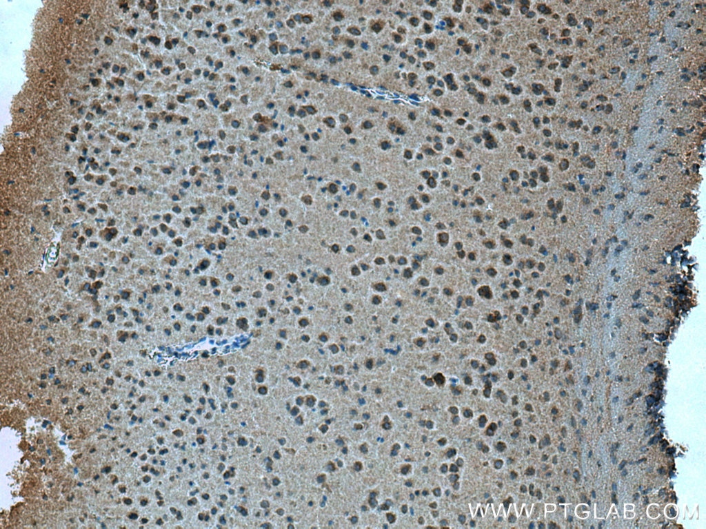
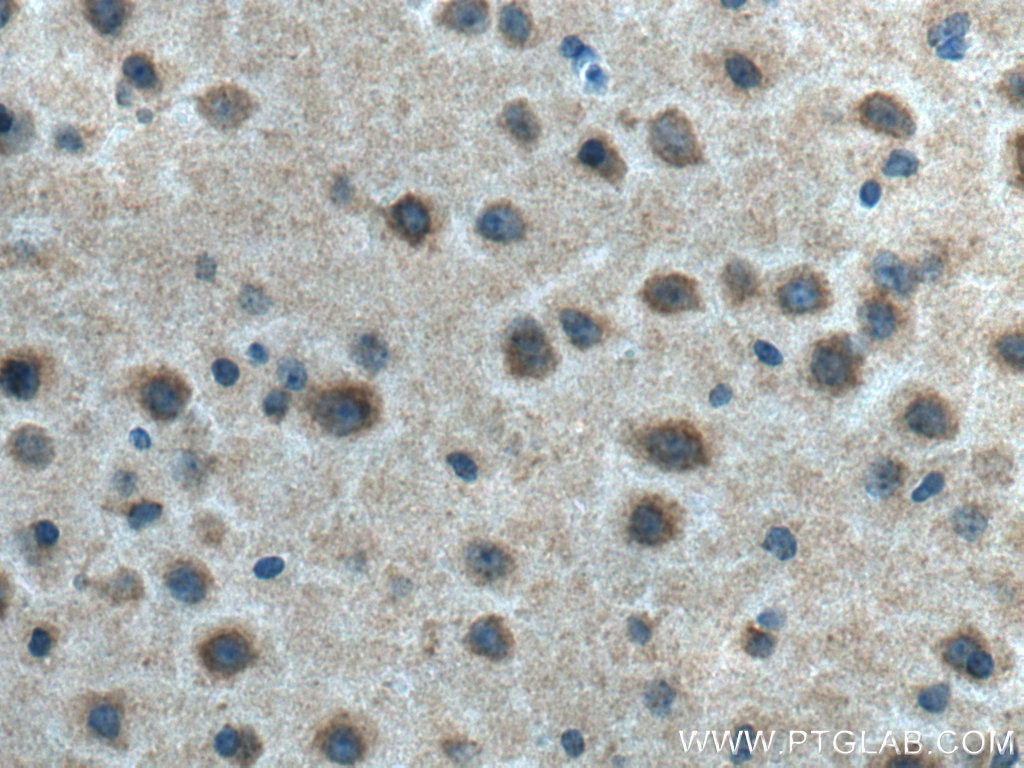
| Positive IHC detected in | human stomach tissue, mouse brain tissue Note: suggested antigen retrieval with TE buffer pH 9.0; (*) Alternatively, antigen retrieval may be performed with citrate buffer pH 6.0 |
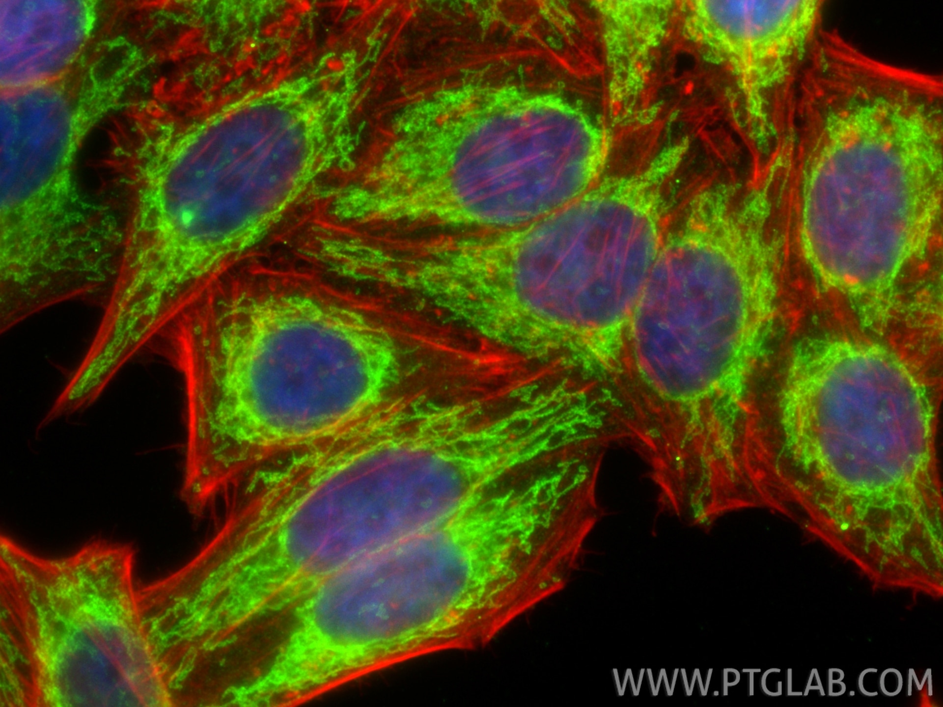
| Positive IF/ICC detected in | HepG2 cells |
Recommended dilution
| Application | Dilution |
|---|---|
| Western Blot (WB) | WB : 1:5000-1:50000 |
| Immunoprecipitation (IP) | IP : 0.5-4.0 ug for 1.0-3.0 mg of total protein lysate |
| Immunohistochemistry (IHC) | IHC : 1:500-1:2000 |
| Immunofluorescence (IF)/ICC | IF/ICC : 1:200-1:800 |
| It is recommended that this reagent should be titrated in each testing system to obtain optimal results. | |
| Sample-dependent, Check data in validation data gallery. | |
Published Applications
| KD/KO | See 20 publications below |
| WB | See 201 publications below |
| IHC | See 9 publications below |
| IF | See 29 publications below |
| IP | See 5 publications below |
Product Information
17090-1-AP targets MFF in WB, IHC, IF/ICC, IP, ELISA applications and shows reactivity with human, mouse, rat samples.
| Tested Reactivity | human, mouse, rat |
| Cited Reactivity | human, mouse, rat, pig, monkey, hamster, goat, squirrel |
| Host / Isotype | Rabbit / IgG |
| Class | Polyclonal |
| Type | Antibody |
| Immunogen |
CatNo: Ag9990 Product name: Recombinant human MFF protein Source: e coli.-derived, PGEX-4T Tag: GST Domain: 1-238 aa of BC000797 Sequence: MAEISRIQYEMEYTEGISQRMRVPEKLKVAPPNADLEQGFQEGVPNASVIMQVPERIVVAGNNEDVSFSRPADLDLIQSTPFKPLALKTPPRVLTLSERPLDFLDLERPPTTPQNEEIRAVGRLKRERSMSENAVRQNGQLVRNDSLPVLRGGSAAATSNPHHDNVRYGISNIDTTIEGTSDDLTVVDAASLRRQIIKLNRRLQLLEEENKERAKREMVMYSITVAFWLLNSWLWFRR Predict reactive species |
| Full Name | mitochondrial fission factor |
| Calculated Molecular Weight | 38 kDa |
| Observed Molecular Weight | 27-38 kDa |
| GenBank Accession Number | BC000797 |
| Gene Symbol | MFF |
| Gene ID (NCBI) | 56947 |
| RRID | AB_2142463 |
| Conjugate | Unconjugated |
| Form | Liquid |
| Purification Method | Antigen affinity purification |
| UNIPROT ID | Q9GZY8 |
| Storage Buffer | PBS with 0.02% sodium azide and 50% glycerol, pH 7.3. |
| Storage Conditions | Store at -20°C. Stable for one year after shipment. Aliquoting is unnecessary for -20oC storage. 20ul sizes contain 0.1% BSA. |
Background Information
MFF (mitochondrial fission factor) is a mitochondrial outer membrane protein that is involved in mitochondrial localization of Drp1 and mitochondrial fission. Multiple isoforms of MFF exist due to the alternative splicing. This antibody recognizes the endogenous MFF protein around 26-29 kDa and 35-38 kDa.
Protocols
| Product Specific Protocols | |
|---|---|
| IF protocol for MFF antibody 17090-1-AP | Download protocol |
| IHC protocol for MFF antibody 17090-1-AP | Download protocol |
| IP protocol for MFF antibody 17090-1-AP | Download protocol |
| WB protocol for MFF antibody 17090-1-AP | Download protocol |
| Standard Protocols | |
|---|---|
| Click here to view our Standard Protocols |
Publications
| Species | Application | Title |
|---|---|---|
Science Golgi-derived PI(4)P-containing vesicles drive late steps of mitochondrial division. | ||
Signal Transduct Target Ther Targeting CRL4 suppresses chemoresistant ovarian cancer growth by inducing mitophagy | ||
Cell CerS6-Derived Sphingolipids Interact with Mff and Promote Mitochondrial Fragmentation in Obesity. |
Reviews
The reviews below have been submitted by verified Proteintech customers who received an incentive for providing their feedback.
FH Parmita (Verified Customer) (07-23-2025) | working very good
|
FH Chun (Verified Customer) (09-07-2020) | This antibody is good for IP, and there are non-specific band(s) detected when it is used for immunoblotting.
|
FH JAE HO (Verified Customer) (04-22-2019) | I used couple of antibodies for MFF proteins. Only this antibody was working nicely. I satisfied its higher titer and specificity. I have tested and used this antibody for WB and IP.
|