

Tested Applications
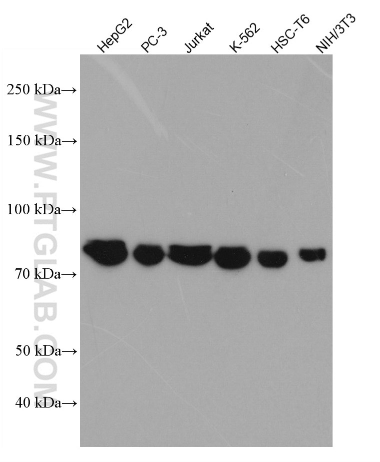
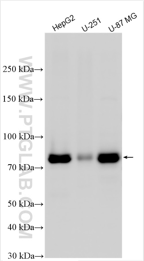
| Positive WB detected in | HepG2 cells, PC-3 cells, Jurkat cells, K-562 cells, HSC-T6 cells, NIH/3T3 cells, U-251 cells, U-87 MG cells |
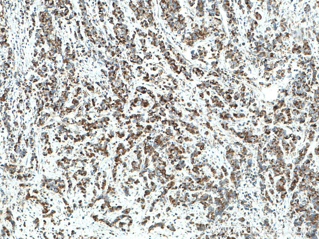
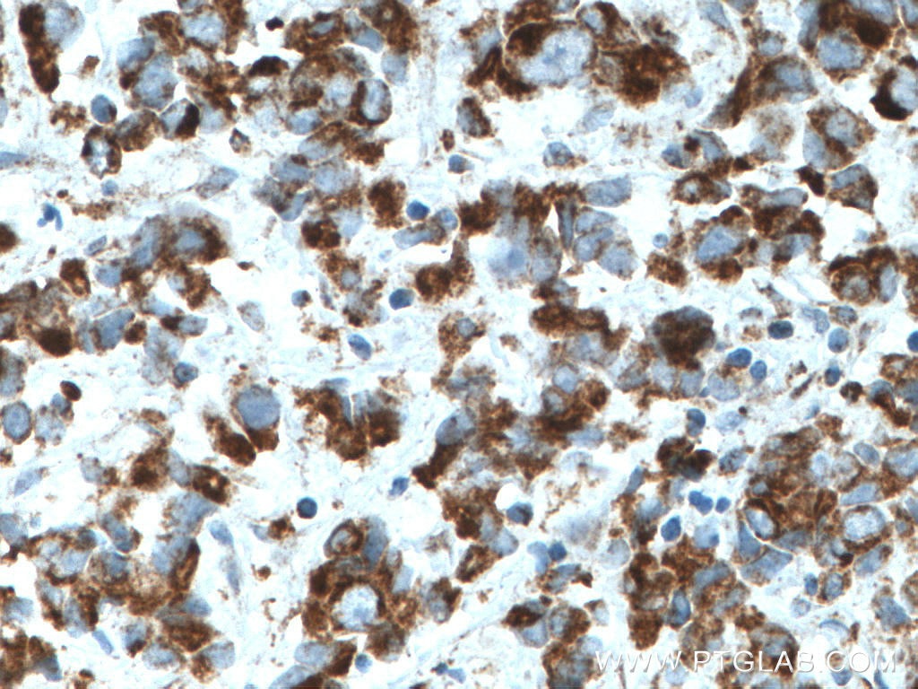
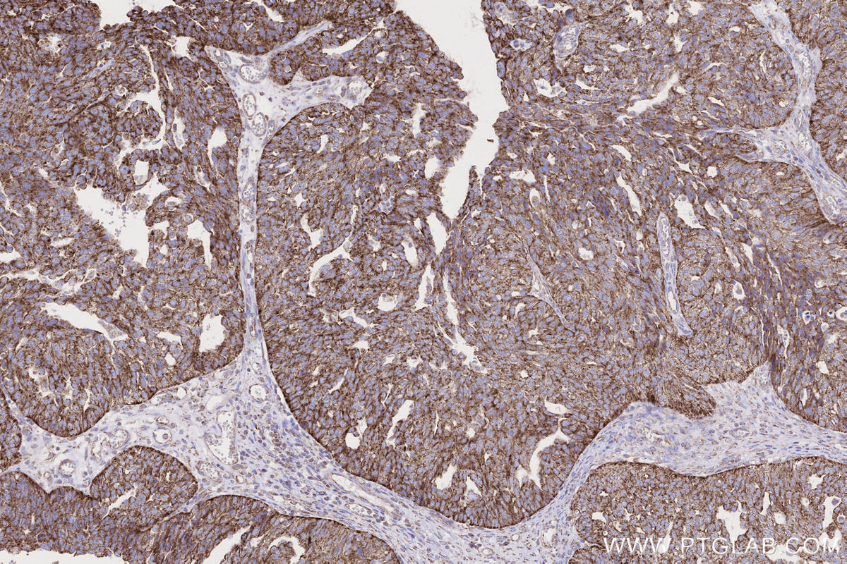
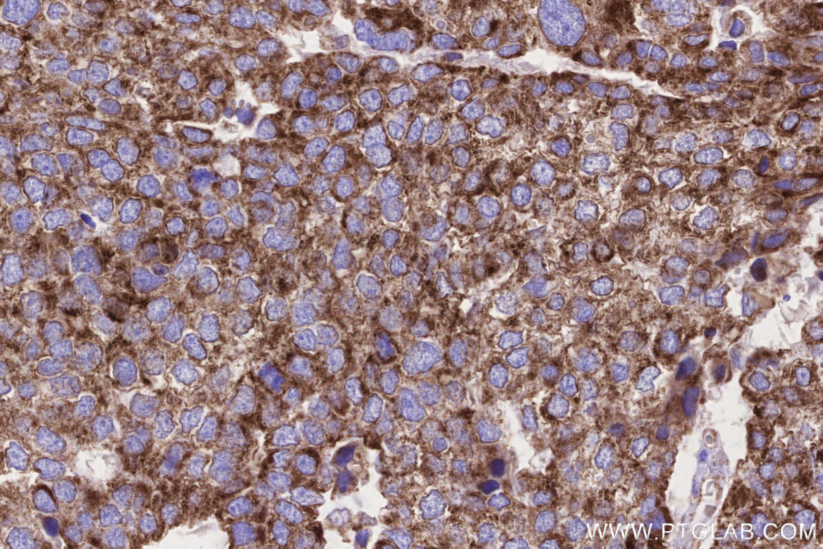
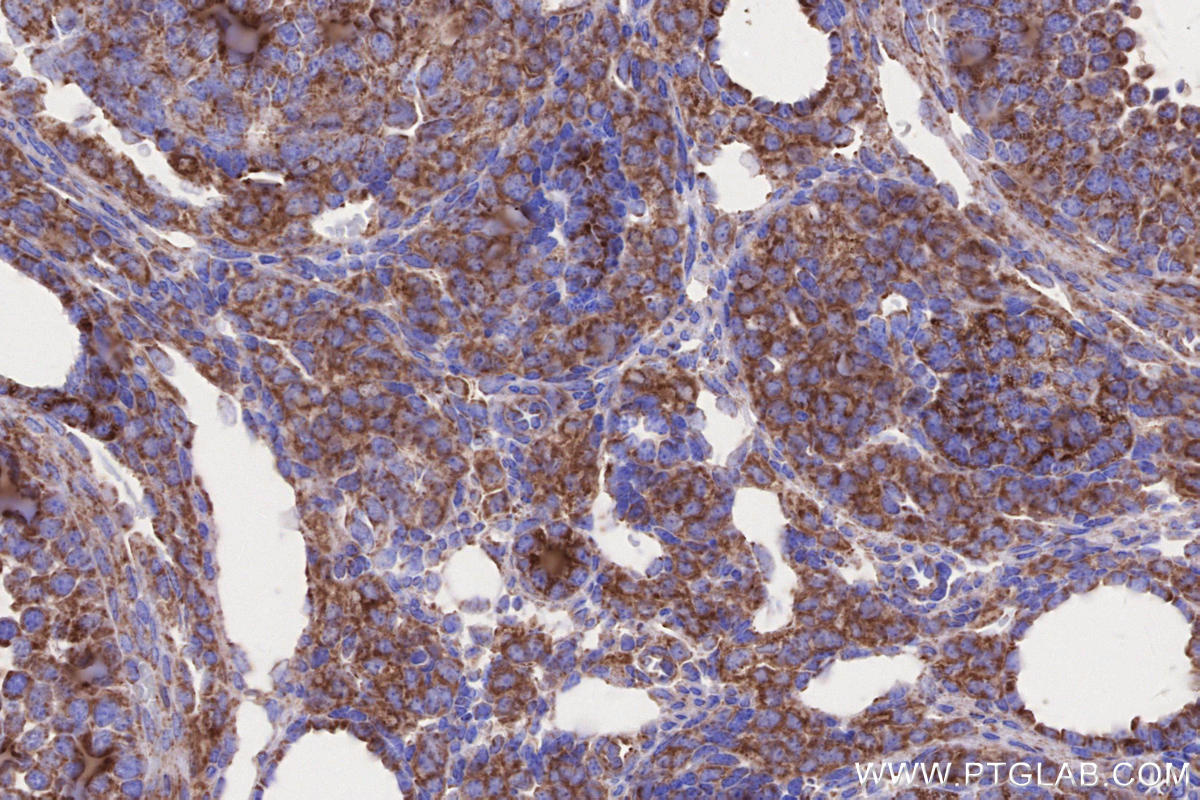
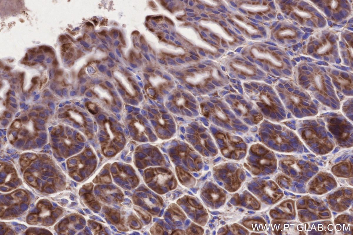
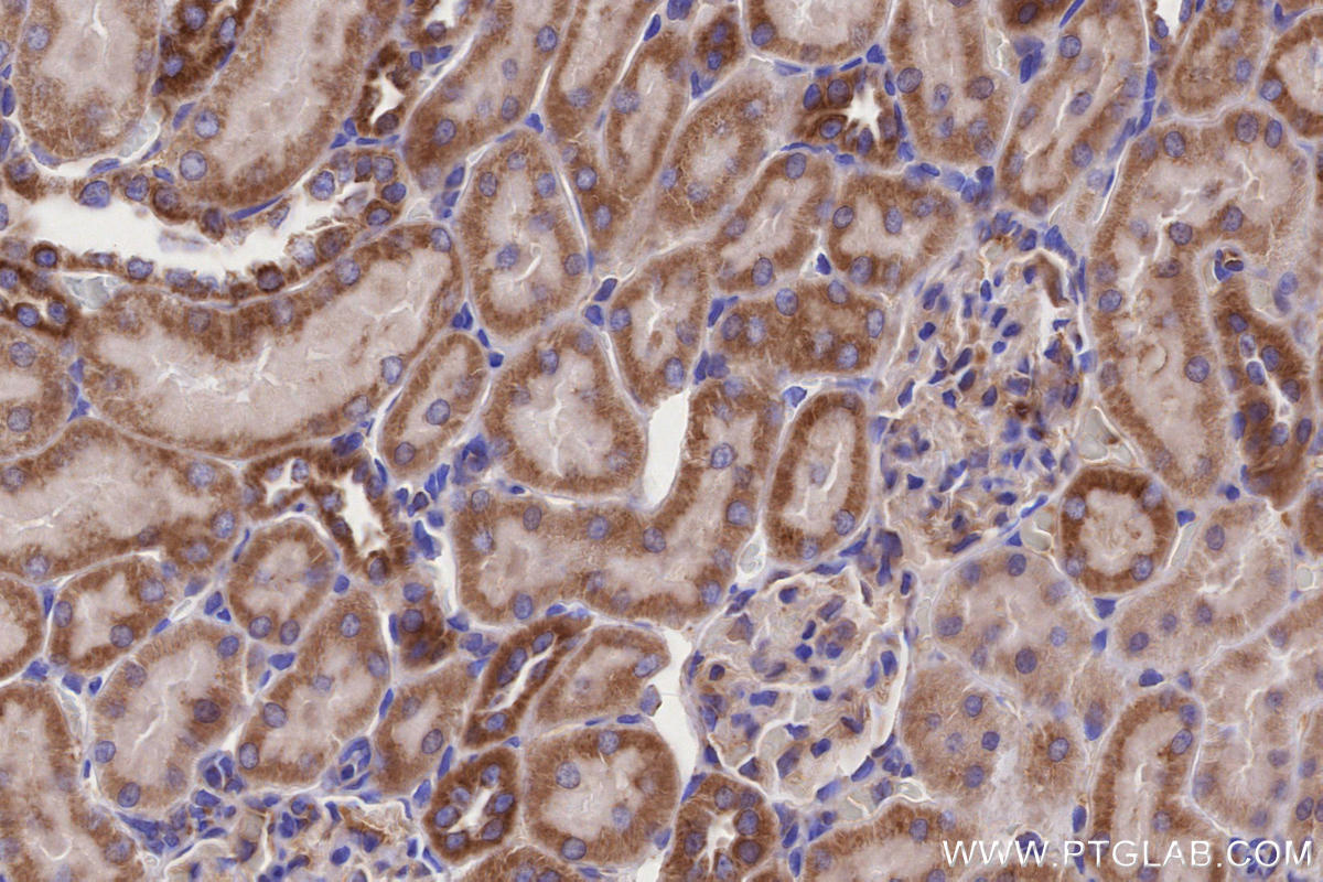
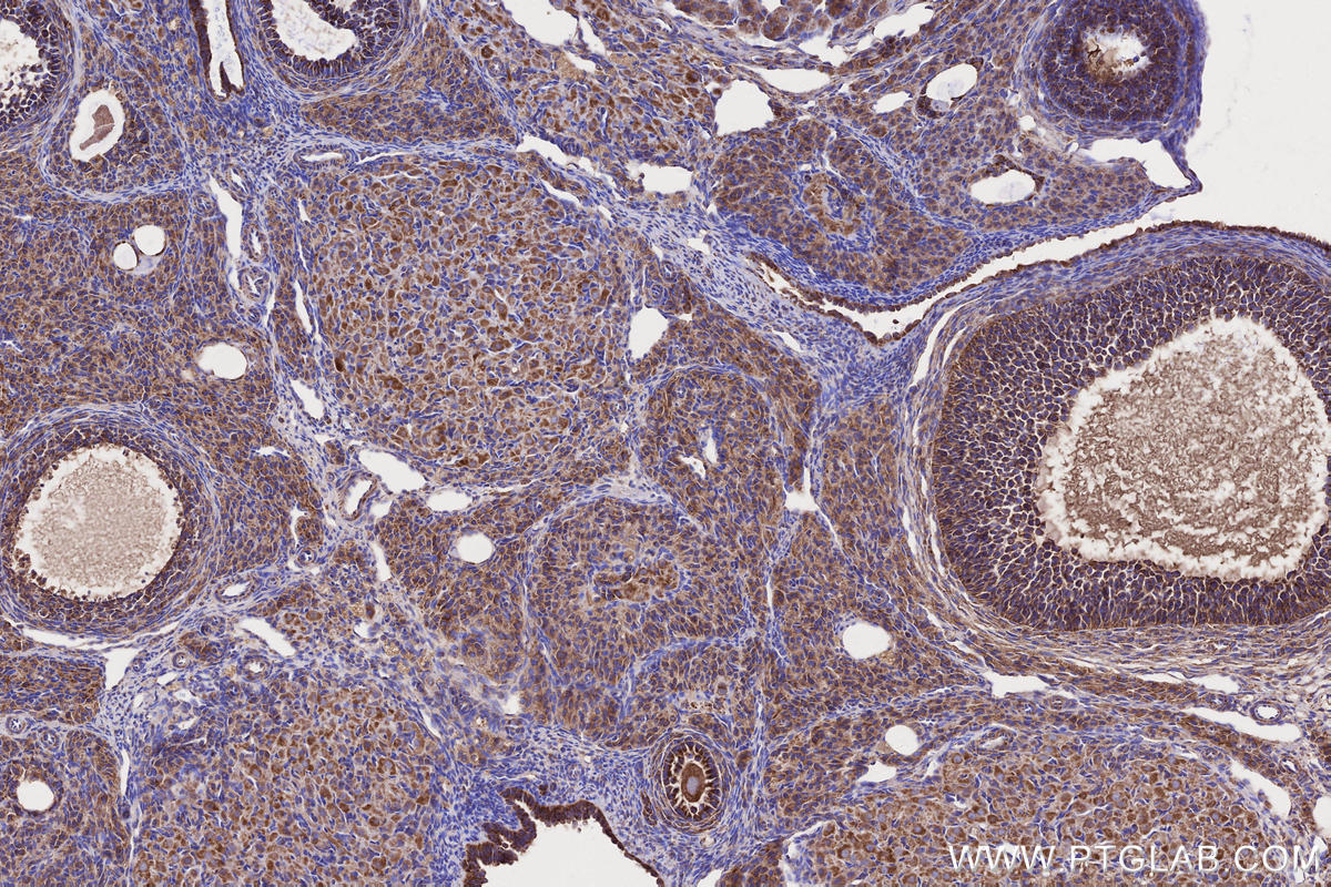
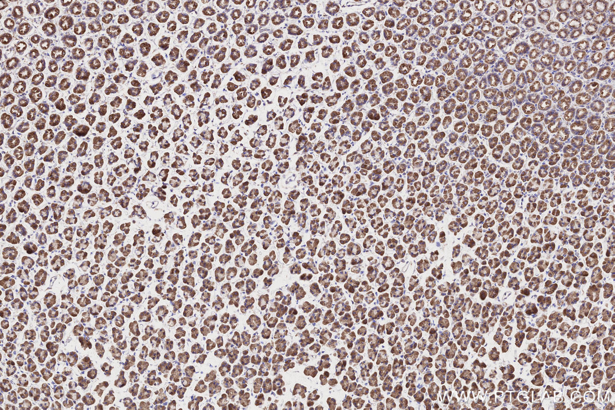
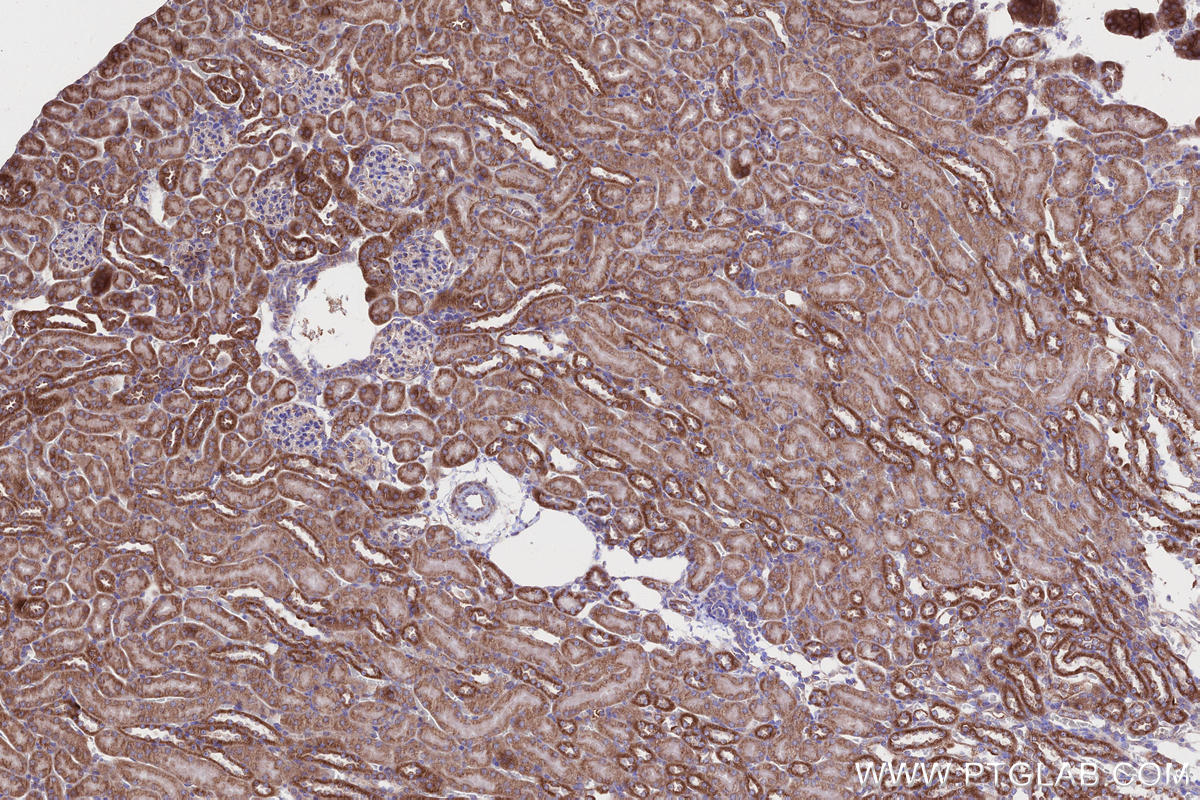
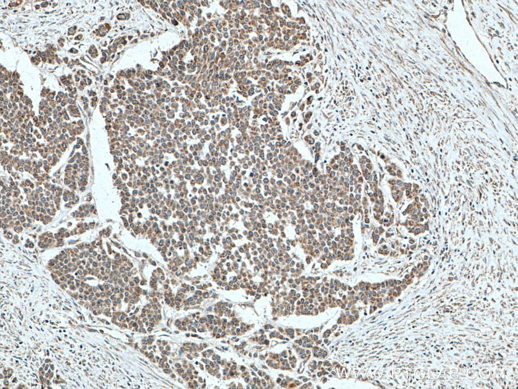
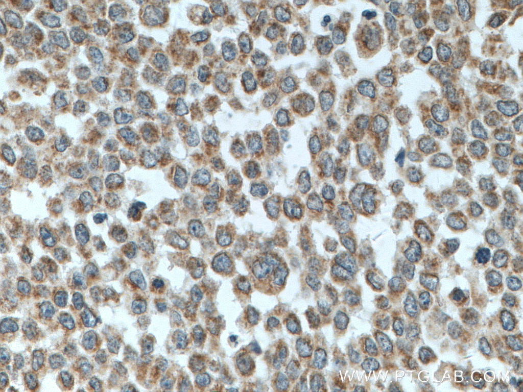
| Positive IHC detected in | human prostate cancer tissue, human colon cancer tissue, human ovary cancer tissue, mouse ovary tissue, mouse stomach tissue, rat stomach tissue, human stomach cancer tissue, mouse kidney tissue, rat ovary tissue, rat kidney tissue Note: suggested antigen retrieval with TE buffer pH 9.0; (*) Alternatively, antigen retrieval may be performed with citrate buffer pH 6.0 |
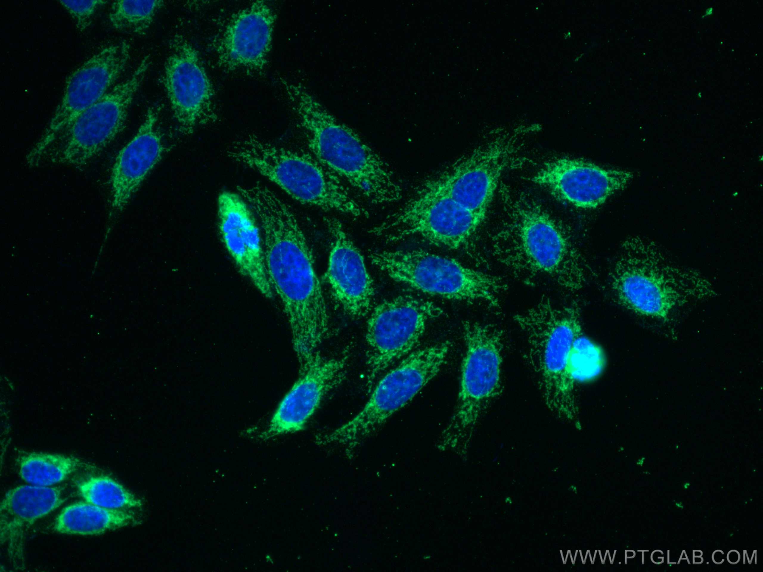
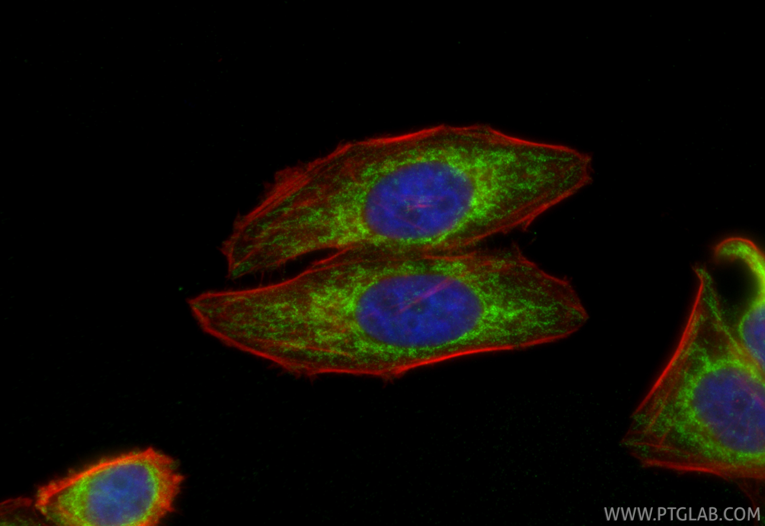
| Positive IF/ICC detected in | HepG2 cells |
Recommended dilution
| Application | Dilution |
|---|---|
| Western Blot (WB) | WB : 1:2000-1:10000 |
| Immunohistochemistry (IHC) | IHC : 1:500-1:2000 |
| Immunofluorescence (IF)/ICC | IF/ICC : 1:400-1:1600 |
| It is recommended that this reagent should be titrated in each testing system to obtain optimal results. | |
| Sample-dependent, Check data in validation data gallery. | |
Product Information
66776-1-Ig targets MFN1 in WB, IHC, IF/ICC, ELISA applications and shows reactivity with human, mouse, rat samples.
| Tested Reactivity | human, mouse, rat |
| Cited Reactivity | human, mouse, rat, pig |
| Host / Isotype | Mouse / IgG2a |
| Class | Monoclonal |
| Type | Antibody |
| Immunogen |
CatNo: Ag4890 Product name: Recombinant human MFN1 protein Source: e coli.-derived, PET28a Tag: 6*His Domain: 27-351 aa of BC040557 Sequence: EFVTEGSHFVEATYKNPELDRIATEDDLVEMQGYKDKLSIIGEVLSRRHMKVAFFGRTSSGKSSVINAMLWDKVLPSGIGHITNCFLSVEGTDGDKAYLMTEGSDEKKSVKTVNQLAHALHMDKDLKAGCLVRVFWPKAKCALLRDDLVLVDSPGTDVTTELDSWIDKFCLDADVFVLVANSESTLMNTEKHFFHKVNERLSKPNIFILNNRWDASASEPEYMEDVRRQHMERCLHFLVEELKVVNALEAQNRIFFVSAKEVLSARKQKAQGMPESGVALAEGFHARLQEFQNFEQIFEECISQSAVKTKFEQHTIRAKQILATV Predict reactive species |
| Full Name | mitofusin 1 |
| Calculated Molecular Weight | 741 aa, 84 kDa |
| Observed Molecular Weight | 84 kDa |
| GenBank Accession Number | BC040557 |
| Gene Symbol | MFN1 |
| Gene ID (NCBI) | 55669 |
| RRID | AB_2882122 |
| Conjugate | Unconjugated |
| Form | Liquid |
| Purification Method | Protein A purification |
| UNIPROT ID | Q8IWA4 |
| Storage Buffer | PBS with 0.02% sodium azide and 50% glycerol, pH 7.3. |
| Storage Conditions | Store at -20°C. Stable for one year after shipment. Aliquoting is unnecessary for -20oC storage. 20ul sizes contain 0.1% BSA. |
Background Information
Mitofusin-1 (MFN1) is a mediator of mitochondrial fusion. This protein and mitofusin 2 are homologs of the Drosophila protein fuzzy onion (Fzo). Mitofusins are large predicted GTPases located in outer mitochondrial membrane. They are essential for outer membrane fusion by interacting with each other to facilitate mitochondrial targeting. The mitofusins are the first known protein mediator of mitochondrial fusion, and mediate developmentally regulated post-meiotic fusion of mitochondria. Mitofusin 1 and mitofusin 2 are ubiquitinated in a PINK1/parkin-dependent manner upon induction of mitophagy(PMID: 20871098).
Protocols
| Product Specific Protocols | |
|---|---|
| IF protocol for MFN1 antibody 66776-1-Ig | Download protocol |
| IHC protocol for MFN1 antibody 66776-1-Ig | Download protocol |
| WB protocol for MFN1 antibody 66776-1-Ig | Download protocol |
| Standard Protocols | |
|---|---|
| Click here to view our Standard Protocols |
Publications
What published studies show
MFN1 Monoclonal antibody (3F11C11), catalog no. 66776-1-Ig, has 51 citations. Publications appear in high-impact journals including Nature Communications, Redox Biology, Advanced Science, Cell Death & Disease, FASEB Journal, Cell Reports, and Development. Research fields span mitophagy, mitochondrial fusion/dynamics, oxidative stress, neurodegeneration (Alzheimer's, Parkinson's), cardiovascular disease, reproductive biology (oocyte maturation), cancer, diabetic kidney disease, and inflammation. Applications cited include WB, IHC, and IF across human, mouse, rat, and pig species.
| Species | Application | Title |
|---|---|---|
Nat Commun BNIP3L/NIX-mediated mitophagy protects against glucocorticoid-induced synapse defects. | ||
Redox Biol Novel mitophagy activator TJ0113 restricts cytosolic mtDNA to suppress ZBP1-driven inflammation in acute lung injury. | ||
Redox Biol Nogo-B mediates endothelial oxidative stress and inflammation to promote coronary atherosclerosis in pressure-overloaded mouse hearts | ||
Adv Sci (Weinh) HIGD1A Alleviates Oxidative Stress Related Ovarian Hypofunction by Enhancing Granulosa Cell Functions via NF-κB/SOD2 Signaling Pathway | ||
Cell Death Dis Crotonylated BEX2 interacts with NDP52 and enhances mitophagy to modulate chemotherapeutic agent-induced apoptosis in non-small-cell lung cancer cells | ||
Int J Biol Sci Targeting ALG3/FOXD1/BNIP3 Axis Prevents Mitophagy and Gemcitabine Resistance of Nasopharyngeal Carcinoma |
Reviews
What customers say
Both reviewers used this antibody for Western Blot and reported positive results. One user (5/5) described it as "Excellent" when applied to T cells. Another (4/5) noted it performed very strongly at a 1:2000 dilution with 20 µg of protein from HeLa cells. Overall, the antibody is well-received for WB applications across different cell types.
The reviews below have been submitted by verified Proteintech customers who received an incentive for providing their feedback.
Pierre (Verified Customer) (09-26-2025) | Excellent
|
benjamin (Verified Customer) (01-31-2019) | Very strong for WB with 20ug of protein using heLa cells
|

















