Tested Applications
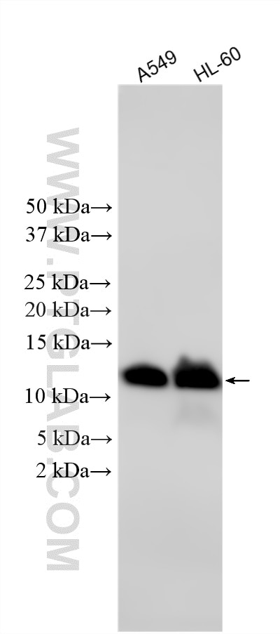
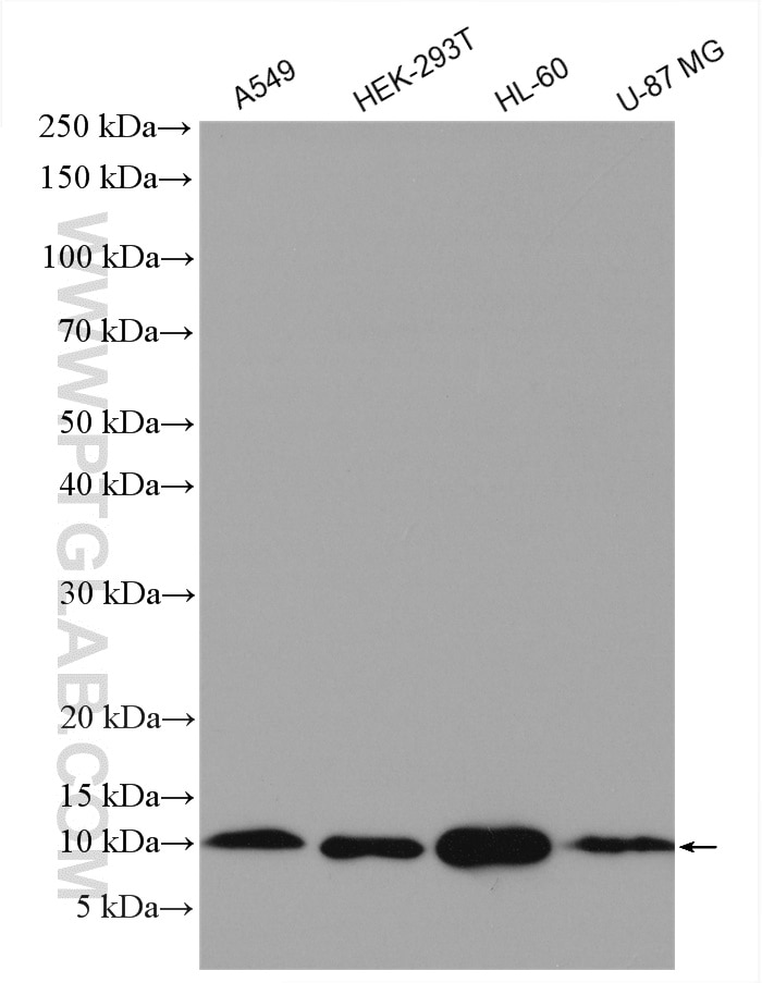
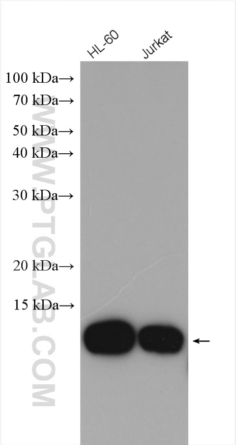
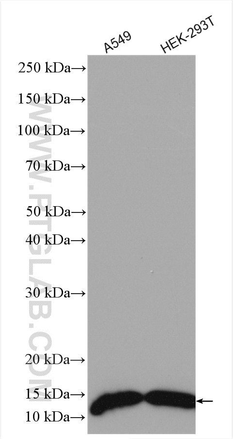
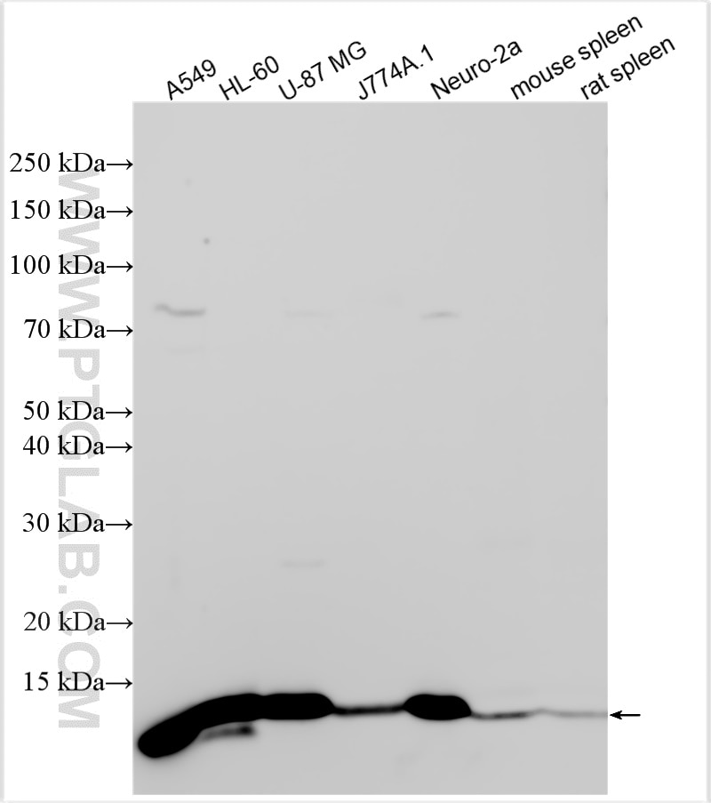
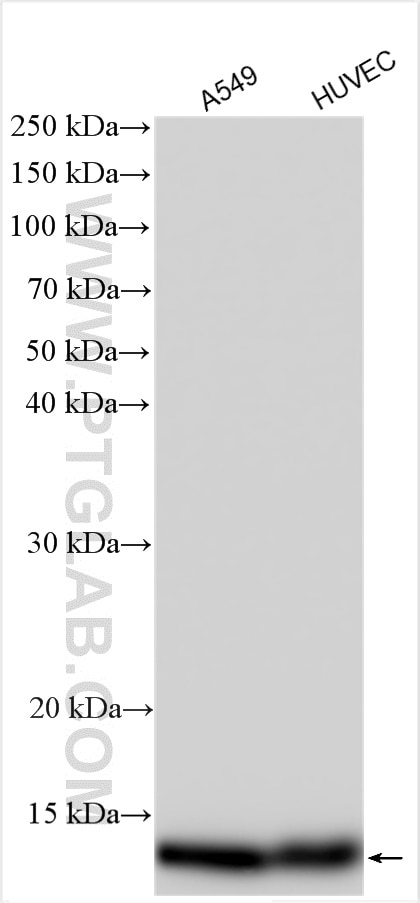
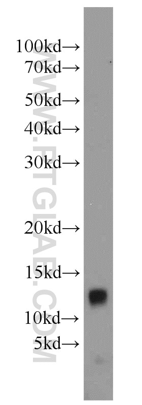
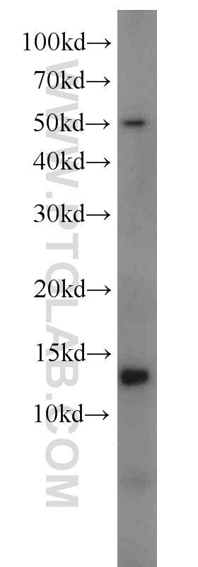
| Positive WB detected in | A549 cells, HL-60 cells, U-937 cells, Y79 cells, U-87 MG cells, HEK-293T cells, HUVEC cells, J774A.1 cells, Neuro-2a cells, mouse spleen tissue, rat spleen tissue, Jurkat cells |
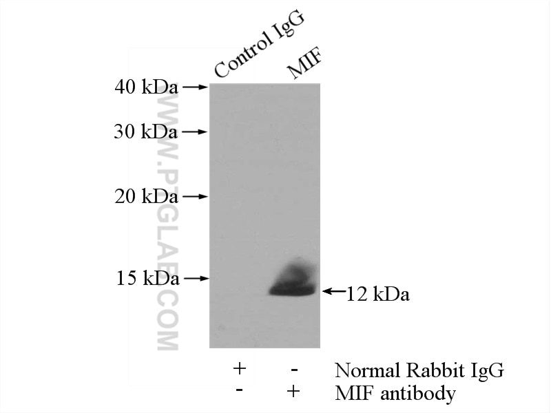
| Positive IP detected in | mouse spleen tissue |
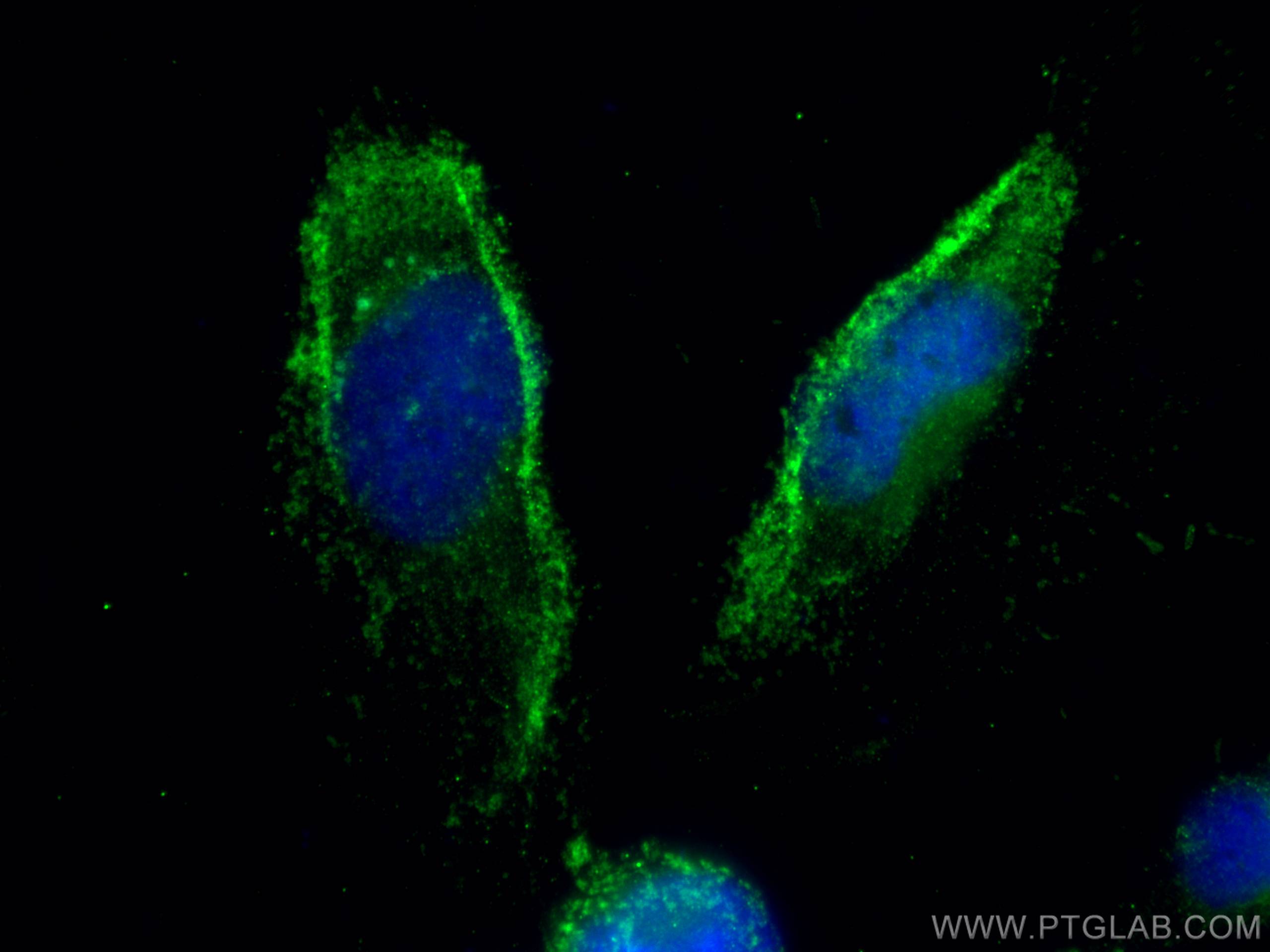
| Positive IF/ICC detected in | U-251 cells |
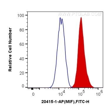
| Positive FC (Intra) detected in | THP-1 cells |
Recommended dilution
| Application | Dilution |
|---|---|
| Western Blot (WB) | WB : 1:5000-1:50000 |
| Immunoprecipitation (IP) | IP : 0.5-4.0 ug for 1.0-3.0 mg of total protein lysate |
| Immunofluorescence (IF)/ICC | IF/ICC : 1:50-1:500 |
| Flow Cytometry (FC) (INTRA) | FC (INTRA) : 0.40 ug per 10^6 cells in a 100 µl suspension |
| It is recommended that this reagent should be titrated in each testing system to obtain optimal results. | |
| Sample-dependent, Check data in validation data gallery. | |
Published Applications
| KD/KO | See 6 publications below |
| WB | See 21 publications below |
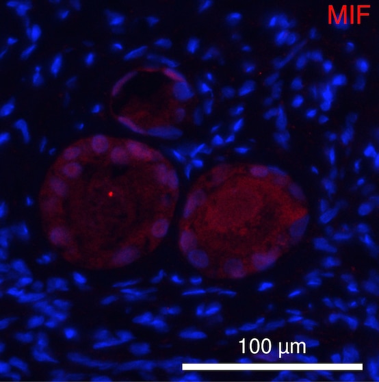
| IF | See 15 publications below |
Product Information
20415-1-AP targets MIF in WB, IF/ICC, FC (Intra), IP, ELISA applications and shows reactivity with human, mouse, rat samples.
| Tested Reactivity | human, mouse, rat |
| Cited Reactivity | human, mouse, rat |
| Host / Isotype | Rabbit / IgG |
| Class | Polyclonal |
| Type | Antibody |
| Immunogen |
CatNo: Ag14058 Product name: Recombinant human MIF protein Source: e coli.-derived, PGEX-4T Tag: GST Domain: 51-115 aa of BC000447 Sequence: GGSSEPCALCSLHSIGKIGGAQNRSYSKLLCGLLAERLRISPDRVYINYYDMNAANVGWNNSTFA Predict reactive species |
| Full Name | macrophage migration inhibitory factor (glycosylation-inhibiting factor) |
| Calculated Molecular Weight | 115 aa, 12 kDa |
| Observed Molecular Weight | 12 kDa |
| GenBank Accession Number | BC000447 |
| Gene Symbol | MIF |
| Gene ID (NCBI) | 4282 |
| RRID | AB_10694820 |
| Conjugate | Unconjugated |
| Form | Liquid |
| Purification Method | Antigen affinity purification |
| UNIPROT ID | P14174 |
| Storage Buffer | PBS with 0.02% sodium azide and 50% glycerol, pH 7.3. |
| Storage Conditions | Store at -20°C. Stable for one year after shipment. Aliquoting is unnecessary for -20oC storage. 20ul sizes contain 0.1% BSA. |
Background Information
MIF is a pleiotropic cytokine that contributes to the pathogenesis of many autoimmune diseases through its upstream immunoregulatory function and its polymorphic genetic locus. MIF is a highly conserved protein of 12.5 kDa, with evolutionarily ancient homologues in plants, protozoans, nematodes, and invertebrates.
Protocols
| Product Specific Protocols | |
|---|---|
| FC protocol for MIF antibody 20415-1-AP | Download protocol |
| IF protocol for MIF antibody 20415-1-AP | Download protocol |
| IP protocol for MIF antibody 20415-1-AP | Download protocol |
| WB protocol for MIF antibody 20415-1-AP | Download protocol |
| Standard Protocols | |
|---|---|
| Click here to view our Standard Protocols |
Publications
| Species | Application | Title |
|---|---|---|
Stem Cell Res Ther DPSCs regulate epithelial-T cell interactions in oral submucous fibrosis | ||
Am J Pathol CDR1as Deficiency Prevents Photoreceptor Degeneration by Regulating miR-7a-5p/α-syn/Parthanatos Pathway in Retinal Detachment | ||
Cancer Lett Macrophage migration inhibitory factor promotes tumor aggressiveness of esophageal squamous cell carcinoma via activation of Akt and inactivation of GSK3β.
| ||
Cell Prolif TSP50 promotes hepatocyte proliferation and tumour formation by activating glucose-6-phosphate dehydrogenase (G6PD). | ||
Front Endocrinol (Lausanne) Exercise prevents fatal stress-induced myocardial injury in obese mice | ||
Food Chem Toxicol The crosstalk between M1 macrophage polarization and energy metabolism disorder contributes to polystyrene nanoplastics-triggered testicular inflammation |
Reviews
The reviews below have been submitted by verified Proteintech customers who received an incentive for providing their feedback.
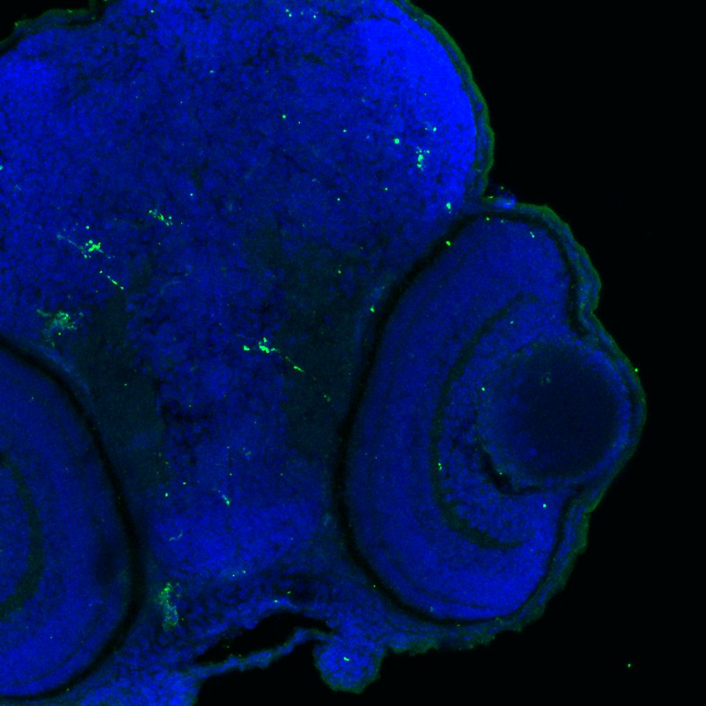
FH Tianyi (Verified Customer) (01-12-2024) | Antigen retrieval in TE buffer, incubated with AF594 for 2 h at RT
 |
FH sarah (Verified Customer) (10-27-2023) | antibody worked great for WB
|
FH Sarah (Verified Customer) (01-24-2023) | This antibody worked well despite low protein concentration.
|
FH Emma (Verified Customer) (03-15-2022) | Nice antibody have used overnight @ 1:1000 on cell lysates and conditioned media. Produces a band at the correct size.
|
FH Ryan (Verified Customer) (02-27-2019) | Tissue was fixed in PFA with no additional antigen retrieval. Co-localisation with microglia based on known markers (not shown).
 |